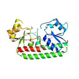
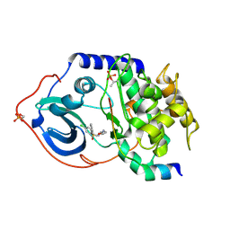
6QAH
 
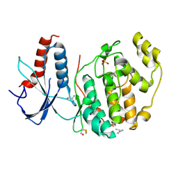 | | ERK2 mini-fragment binding | | 分子名称: | 1-azanylpropylideneazanium, Mitogen-activated protein kinase 1, SULFATE ION | | 著者 | O'Reilly, M, Cleasby, A, Davies, T.G, Hall, R, Ludlow, F, Murray, C.W, Tisi, D, Jhoti, H. | | 登録日 | 2018-12-19 | | 公開日 | 2019-06-26 | | 実験手法 | X-RAY DIFFRACTION (1.58 Å) | | 主引用文献 | Crystallographic screening using ultra-low-molecular-weight ligands to guide drug design.
Drug Discov Today, 24, 2019
|
|
4OEB
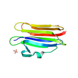 | | Structure of membrane binding protein pleurotolysin A from Pleurotus ostreatus | | 分子名称: | Pleurotolysin A, SULFATE ION | | 著者 | Dunstone, M.A, Caradoc-Davies, T.T, Whisstock, J.C, Law, R.H.P. | | 登録日 | 2014-01-12 | | 公開日 | 2015-02-18 | | 実験手法 | X-RAY DIFFRACTION (1.85 Å) | | 主引用文献 | Conformational Changes during Pore Formation by the Perforin-Related Protein Pleurotolysin.
Plos Biol., 13, 2015
|
|
4OEJ
 | | Structure of membrane binding protein pleurotolysin B from Pleurotus ostreatus | | 分子名称: | ACETATE ION, CHLORIDE ION, GLYCEROL, ... | | 著者 | Dunstone, M.A, Caradoc-Davies, T.T, Whisstock, J.C, Law, R.H.P. | | 登録日 | 2014-01-13 | | 公開日 | 2015-02-18 | | 最終更新日 | 2024-02-28 | | 実験手法 | X-RAY DIFFRACTION (2.2 Å) | | 主引用文献 | Conformational Changes during Pore Formation by the Perforin-Related Protein Pleurotolysin.
Plos Biol., 13, 2015
|
|
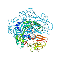
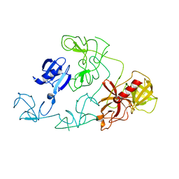
4DUR
 | | The X-ray Crystal Structure of Full-Length type II Human Plasminogen | | 分子名称: | ACETATE ION, BICARBONATE ION, CHLORIDE ION, ... | | 著者 | Law, R.H.P, Caradoc-Davies, T, Whisstock, J.C. | | 登録日 | 2012-02-22 | | 公開日 | 2012-03-28 | | 最終更新日 | 2023-11-08 | | 実験手法 | X-RAY DIFFRACTION (2.45 Å) | | 主引用文献 | The X-ray crystal structure of full-length human plasminogen
Cell Rep, 1, 2012
|
|
4DUU
 | |
1RXU
 | | E. coli uridine phosphorylase: thymidine phosphate complex | | 分子名称: | PHOSPHATE ION, POTASSIUM ION, THYMIDINE, ... | | 著者 | Caradoc-Davies, T.T, Cutfield, S.M, Lamont, I.L, Cutfield, J.F. | | 登録日 | 2003-12-18 | | 公開日 | 2004-04-13 | | 最終更新日 | 2024-04-03 | | 実験手法 | X-RAY DIFFRACTION (3.1 Å) | | 主引用文献 | Crystal structures of escherichia coli uridine phosphorylase in two native and three complexed forms reveal basis of substrate specificity, induced conformational changes and influence of potassium
J.Mol.Biol., 337, 2004
|
|
1RXC
 | | E. COLI uridine phosphorylase: 5-fluorouracil ribose-1-phosphate complex | | 分子名称: | 1-O-phosphono-alpha-D-ribofuranose, 5-FLUOROURACIL, 5-FLUOROURIDINE, ... | | 著者 | Caradoc-Davies, T.T, Cutfield, S.M, Lamont, I.L, Cutfield, J.F. | | 登録日 | 2003-12-18 | | 公開日 | 2004-04-13 | | 最終更新日 | 2024-03-13 | | 実験手法 | X-RAY DIFFRACTION (2.35 Å) | | 主引用文献 | Crystal structures of escherichia coli uridine phosphorylase in two native and three complexed forms reveal basis of substrate specificity, induced conformational changes and influence of potassium
J.Mol.Biol., 337, 2004
|
|
1RXS
 | | E. coli uridine phosphorylase: 2'-deoxyuridine phosphate complex | | 分子名称: | 2'-DEOXYURIDINE, META VANADATE, PHOSPHATE ION, ... | | 著者 | Caradoc-Davies, T.T, Cutfield, S.M, Lamont, I.L, Cutfield, J.F. | | 登録日 | 2003-12-18 | | 公開日 | 2004-04-13 | | 最終更新日 | 2024-04-03 | | 実験手法 | X-RAY DIFFRACTION (2.8 Å) | | 主引用文献 | Crystal structures of escherichia coli uridine phosphorylase in two native and three complexed forms reveal basis of substrate specificity, induced conformational changes and influence of potassium
J.Mol.Biol., 337, 2004
|
|
4P48
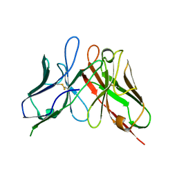 | | The structure of a chicken anti-cardiac Troponin I scFv | | 分子名称: | Antibody scFv 180 | | 著者 | Conroy, P.J, Law, R.H.P, Gillgunn, S, Hearty, S, Caradoc-Davies, T.T, Llyod, G, O'Kennedy, R.J, Whisstock, J.C. | | 登録日 | 2014-03-12 | | 公開日 | 2014-04-23 | | 最終更新日 | 2023-12-27 | | 実験手法 | X-RAY DIFFRACTION (1.35 Å) | | 主引用文献 | Reconciling the structural attributes of avian antibodies.
J.Biol.Chem., 289, 2014
|
|
4P49
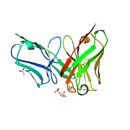 | | The structure of a chicken anti-prostate specific antigen scFv | | 分子名称: | ACETATE ION, Antibody scFv B8, SULFATE ION | | 著者 | Conroy, P.J, Law, R.H.P, Gilgunn, S, Hearty, S, Llyod, G, Caradoc-Davies, T.T, O'Kennedy, R.J, Whisstock, J.C. | | 登録日 | 2014-03-12 | | 公開日 | 2014-04-23 | | 最終更新日 | 2023-12-27 | | 実験手法 | X-RAY DIFFRACTION (1.4 Å) | | 主引用文献 | Reconciling the structural attributes of avian antibodies.
J.Biol.Chem., 289, 2014
|
|
1RXY
 | | E. coli uridine phosphorylase: type-B native | | 分子名称: | ACETATE ION, GLYCEROL, POTASSIUM ION, ... | | 著者 | Caradoc-Davies, T.T, Cutfield, S.M, Lamont, I.L, Cutfield, J.F. | | 登録日 | 2003-12-18 | | 公開日 | 2004-04-13 | | 最終更新日 | 2023-10-25 | | 実験手法 | X-RAY DIFFRACTION (1.7 Å) | | 主引用文献 | Crystal structures of escherichia coli uridine phosphorylase in two native and three complexed forms reveal basis of substrate specificity, induced conformational changes and influence of potassium
J.Mol.Biol., 337, 2004
|
|
1T0U
 | | Crystal structure of E.coli uridine phosphorylase at 2.2 A resolution (Type-A Native) | | 分子名称: | Uridine phosphorylase | | 著者 | Caradoc-Davies, T.T, Cutfield, S.M, Lamont, I.L, Cutfield, J.F. | | 登録日 | 2004-04-13 | | 公開日 | 2004-04-27 | | 最終更新日 | 2024-03-13 | | 実験手法 | X-RAY DIFFRACTION (2.2 Å) | | 主引用文献 | Crystal structures of escherichia coli uridine phosphorylase in two native and three complexed forms reveal basis of substrate specificity, induced conformational changes and influence of potassium
J.Mol.Biol., 337, 2004
|
|
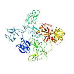
6O1Z
 | | Structure of pCW3 conjugation coupling protein TcpA hexagonal crystal form | | 分子名称: | DNA translocase coupling protein | | 著者 | Traore, D.A.K, Ahktar, N, Torres, V.T, Adams, V, Coulibaly, F, Panjikar, S, Caradoc-Davies, T.T, Rood, J.I, Whisstock, J.C. | | 登録日 | 2019-02-22 | | 公開日 | 2020-03-04 | | 最終更新日 | 2023-10-11 | | 実験手法 | X-RAY DIFFRACTION (3.1 Å) | | 主引用文献 | Structure of pCW3 conjugation coupling protein TcpA
To be published
|
|
6O1Y
 | | Structure of pCW3 conjugation coupling protein TcpA monomeric form with ATP | | 分子名称: | ADENOSINE-5'-TRIPHOSPHATE, DNA translocase coupling protein, beta-D-glucopyranose | | 著者 | Traore, D.A.K, Ahktar, N, Torres, V.T, Adams, V, Coulibaly, F, Panjikar, S, Caradoc-Davies, T.T, Rood, J.I, Whisstock, J.C. | | 登録日 | 2019-02-22 | | 公開日 | 2020-02-26 | | 最終更新日 | 2023-10-11 | | 実験手法 | X-RAY DIFFRACTION (2.7 Å) | | 主引用文献 | Structure of pCW3 conjugation coupling protein TcpA
To be published
|
|
6O1W
 | | Structure of pCW3 conjugation coupling protein TcpA monomer orthorhombic crystal form | | 分子名称: | DNA translocase coupling protein, beta-D-glucopyranose | | 著者 | Traore, D.A.K, Ahktar, N, Torres, V.T, Adams, V, Coulibaly, F, Panjikar, S, Caradoc-Davies, T.T, Rood, J.I, Whisstock, J.C. | | 登録日 | 2019-02-22 | | 公開日 | 2020-02-26 | | 最終更新日 | 2020-07-29 | | 実験手法 | X-RAY DIFFRACTION (2.2 Å) | | 主引用文献 | Structure of pCW3 conjugation coupling protein TcpA
To be published
|
|
6O1X
 | | Structure of pCW3 conjugation coupling protein TcpA monomer form with ATPgS | | 分子名称: | DNA translocase coupling protein, PHOSPHOTHIOPHOSPHORIC ACID-ADENYLATE ESTER, beta-D-glucopyranose | | 著者 | Traore, D.A.K, Ahktar, N, Torres, V.T, Adams, V, Coulibaly, F, Panjikar, S, Caradoc-Davies, T.T, Rood, J.I, Whisstock, J.C. | | 登録日 | 2019-02-22 | | 公開日 | 2020-02-26 | | 最終更新日 | 2023-10-11 | | 実験手法 | X-RAY DIFFRACTION (2.46 Å) | | 主引用文献 | Structure of pCW3 conjugation coupling protein TcpA
To be published
|
|
3GI1
 | | Crystal Structure of the laminin-binding protein Lbp of Streptococcus pyogenes | | 分子名称: | Laminin-binding protein of group A streptococci, ZINC ION | | 著者 | Linke, C, Caradoc-Davies, T.T, Young, P.G, Proft, T, Baker, E.N. | | 登録日 | 2009-03-04 | | 公開日 | 2009-08-04 | | 最終更新日 | 2023-11-01 | | 実験手法 | X-RAY DIFFRACTION (2.45 Å) | | 主引用文献 | The laminin-binding protein Lbp from Streptococcus pyogenes is a zinc receptor
J.Bacteriol., 191, 2009
|
|
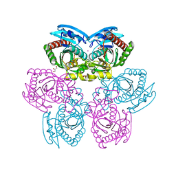
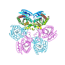
2HUO
 | | Crystal structure of mouse myo-inositol oxygenase in complex with substrate | | 分子名称: | 1,2,3,4,5,6-HEXAHYDROXY-CYCLOHEXANE, FE (III) ION, FORMIC ACID, ... | | 著者 | Brown, P.M, Caradoc-Davies, T.T, Dickson, J.M.J, Cooper, G.J.S, Loomes, K.M, Baker, E.N. | | 登録日 | 2006-07-27 | | 公開日 | 2006-09-26 | | 最終更新日 | 2024-02-14 | | 実験手法 | X-RAY DIFFRACTION (2 Å) | | 主引用文献 | Crystal structure of a substrate complex of myo-inositol oxygenase, a di-iron oxygenase with a key role in inositol metabolism.
Proc.Natl.Acad.Sci.Usa, 103, 2006
|
|
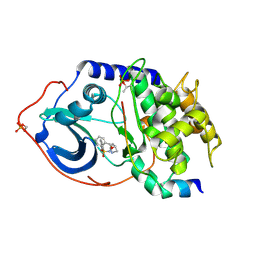
6Q7S
 | | ERK2 mini-fragment binding | | 分子名称: | Mitogen-activated protein kinase 1, PHENOL, SULFATE ION | | 著者 | O'Reilly, M, Cleasby, A, Davies, T.G, Hall, R, Ludlow, F, Murray, C.W, Tisi, D, Jhoti, H. | | 登録日 | 2018-12-13 | | 公開日 | 2019-06-26 | | 実験手法 | X-RAY DIFFRACTION (1.73 Å) | | 主引用文献 | Crystallographic screening using ultra-low-molecular-weight ligands to guide drug design.
Drug Discov Today, 24, 2019
|
|
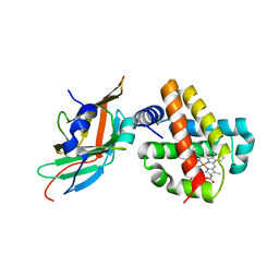
6Q7T
 | | ERK2 mini-fragment binding | | 分子名称: | 1,2-oxazol-3-amine, Mitogen-activated protein kinase 1, SULFATE ION | | 著者 | O'Reilly, M, Cleasby, A, Davies, T.G, Hall, R, Ludlow, F, Murray, C.W, Tisi, D, Jhoti, H. | | 登録日 | 2018-12-13 | | 公開日 | 2019-06-26 | | 実験手法 | X-RAY DIFFRACTION (1.6 Å) | | 主引用文献 | Crystallographic screening using ultra-low-molecular-weight ligands to guide drug design.
Drug Discov Today, 24, 2019
|
|
2C1A
 | | Structure of cAMP-dependent protein kinase complexed with Isoquinoline-5-sulfonic acid (2-(2-(4-chlorobenzyloxy)ethylamino) ethyl)amide | | 分子名称: | CAMP-DEPENDENT PROTEIN KINASE, CAMP-DEPENDENT PROTEIN KINASE INHIBITOR, ISOQUINOLINE-5-SULFONIC ACID (2-(2-(4-CHLOROBENZYLOXY)ETHYLAMINO)ETHYL)AMIDE | | 著者 | Collins, I, Caldwell, J, Fonseca, T, Donald, A, Bavetsias, V, Hunter, L.J, Garrett, M.D, Rowlands, M.G, Aherne, G.W, Davies, T.G, Berdini, V, Woodhead, S.J, Seavers, L.C.A, Wyatt, P.G, Workman, P, McDonald, E. | | 登録日 | 2005-09-12 | | 公開日 | 2005-11-02 | | 最終更新日 | 2023-12-13 | | 実験手法 | X-RAY DIFFRACTION (1.95 Å) | | 主引用文献 | Structure-based design of isoquinoline-5-sulfonamide inhibitors of protein kinase B.
Bioorg. Med. Chem., 14, 2006
|
|
2C1B
 | | Structure of cAMP-dependent protein kinase complexed with (4R,2S)-5'-(4-(4-Chlorobenzyloxy)pyrrolidin-2-ylmethanesulfonyl)isoquinoline | | 分子名称: | (4R,2S)-5'-(4-(4-CHLOROBENZYLOXY)PYRROLIDIN-2-YLMETHANESULFONYL)ISOQUINOLINE, CAMP-DEPENDENT PROTEIN KINASE, CAMP-DEPENDENT PROTEIN KINASE INHIBITOR | | 著者 | Collins, I, Caldwell, J, Fonseca, T, Donald, A, Bavetsias, V, Hunter, L.J, Garrett, M.D, Rowlands, M.G, Aherne, G.W, Davies, T.G, Berdini, V, Woodhead, S.J, Seavers, L.C.A, Wyatt, P.G, Workman, P, McDonald, E. | | 登録日 | 2005-09-12 | | 公開日 | 2005-11-02 | | 最終更新日 | 2018-02-28 | | 実験手法 | X-RAY DIFFRACTION (2 Å) | | 主引用文献 | Structure-based design of isoquinoline-5-sulfonamide inhibitors of protein kinase B.
Bioorg. Med. Chem., 14, 2006
|
|
3S48
 | | Human Alpha-Haemoglobin Complexed with the First NEAT Domain of IsdH from Staphylococcus aureus | | 分子名称: | Hemoglobin subunit alpha, Iron-regulated surface determinant protein H, PROTOPORPHYRIN IX CONTAINING FE | | 著者 | Kumar, K.K, Jacques, D.A, Caradoc-Davies, T.T, Guss, J.M, Gell, D.A. | | 登録日 | 2011-05-19 | | 公開日 | 2012-05-23 | | 最終更新日 | 2023-11-01 | | 実験手法 | X-RAY DIFFRACTION (3.05 Å) | | 主引用文献 | The structure of alpha-haemoglobin in complex with a haemoglobin-binding domain from Staphylococcus aureus reveals the elusive alpha-haemoglobin dimerization interface
ACTA CRYSTALLOGR.,SECT.F, 70, 2014
|
|
1H0W
 | | Human cyclin dependent protein kinase 2 in complex with the inhibitor 2-Amino-6-[cyclohex-3-enyl]methoxypurine | | 分子名称: | 1-AMINO-6-CYCLOHEX-3-ENYLMETHYLOXYPURINE, CELL DIVISION PROTEIN KINASE 2 | | 著者 | Gibson, A.E, Arris, C.E, Bentley, J, Boyle, F.T, Curtin, N.J, Davies, T.G, Endicott, J.A, Golding, B.T, Grant, S, Griffin, R.J, Jewsbury, P, Johnson, L.N, Mesguiche, V, Newell, D.R, Noble, M.E.M, Tucker, J.A, Whitfield, H.J. | | 登録日 | 2002-06-27 | | 公開日 | 2003-06-27 | | 最終更新日 | 2023-12-13 | | 実験手法 | X-RAY DIFFRACTION (2.1 Å) | | 主引用文献 | Probing the ATP Ribose-Binding Domain of Cyclin-Dependent Kinases 1 and 2 with O(6)-Substituted Guanine Derivatives
J.Med.Chem., 45, 2002
|
|
1H0V
 | | Human cyclin dependent protein kinase 2 in complex with the inhibitor 2-Amino-6-[(R)-pyrrolidino-5'-yl]methoxypurine | | 分子名称: | 5-{[(2-AMINO-9H-PURIN-6-YL)OXY]METHYL}-2-PYRROLIDINONE, CELL DIVISION PROTEIN KINASE 2 | | 著者 | Gibson, A.E, Arris, C.E, Bentley, J, Boyle, F.T, Curtin, N.J, Davies, T.G, Endicott, J.A, Golding, B.T, Grant, S, Griffin, R.J, Jewsbury, P, Johnson, L.N, Mesguiche, V, Newell, D.R, Noble, M.E.M, Tucker, J.A, Whitfield, H.J. | | 登録日 | 2002-06-27 | | 公開日 | 2003-06-27 | | 最終更新日 | 2023-12-13 | | 実験手法 | X-RAY DIFFRACTION (1.9 Å) | | 主引用文献 | Probing the ATP Ribose-Binding Domain of Cyclin-Dependent Kinases 1 and 2 with O(6)-Substituted Guanine Derivatives
J.Med.Chem., 45, 2002
|
|


