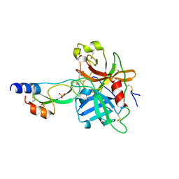
1W8U
 
 | | CBM29-2 mutant D83A complexed with mannohexaose: Probing the Mechanism of Ligand Recognition by Family 29 Carbohydrate Binding Modules | | 分子名称: | NON CATALYTIC PROTEIN 1, beta-D-mannopyranose-(1-4)-beta-D-mannopyranose-(1-4)-beta-D-mannopyranose-(1-4)-beta-D-mannopyranose-(1-4)-beta-D-mannopyranose | | 著者 | Flint, J, Bolam, D.N, Nurizzo, D, Taylor, E.J, Williamson, M.P, Walters, C, Davies, G.J, Gilbert, H.J. | | 登録日 | 2004-09-28 | | 公開日 | 2005-03-22 | | 最終更新日 | 2023-12-13 | | 実験手法 | X-RAY DIFFRACTION (1.3 Å) | | 主引用文献 | Probing the Mechanism of Ligand Recognition in Family 29 Carbohydrate-Binding Modules
J.Biol.Chem., 280, 2005
|
|
7QII
 | |
7QIH
 | |
1W9B
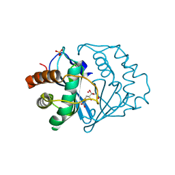
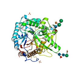 | | S. alba myrosinase in complex with carba-glucotropaeolin | | 分子名称: | 2-acetamido-2-deoxy-beta-D-glucopyranose, 2-acetamido-2-deoxy-beta-D-glucopyranose-(1-4)-2-acetamido-2-deoxy-beta-D-glucopyranose, CARBA-GLUCOTROPAEOLIN, ... | | 著者 | Bourderioux, A, Lefoix, M, Gueyrard, D, Tatibouet, A, Cottaz, S, Arzt, S, Burmeister, W.P, Rollin, P. | | 登録日 | 2004-10-08 | | 公開日 | 2005-05-19 | | 最終更新日 | 2023-12-13 | | 実験手法 | X-RAY DIFFRACTION (1.7 Å) | | 主引用文献 | The glucosinolate-myrosinase system. New insights into enzyme-substrate interactions by use of simplified inhibitors.
Org. Biomol. Chem., 3, 2005
|
|
7QHR
 | |
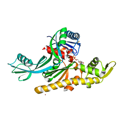
1Y8J
 | | Crystal Structure of human NEP complexed with an imidazo[4,5-c]pyridine inhibitor | | 分子名称: | 2-[(1S)-1-BENZYL-2-SULFANYLETHYL]-1H-IMIDAZO[4,5-C]PYRIDIN-5-IUM, 2-acetamido-2-deoxy-beta-D-glucopyranose, ACETATE ION, ... | | 著者 | Sahli, S, Frank, B, Schweizer, W.B, Diederich, F, Blum-Kaelin, D, Aebi, J.D, Bohm, H.J, Oefner, C, Dale, G.E. | | 登録日 | 2004-12-13 | | 公開日 | 2005-06-07 | | 最終更新日 | 2023-08-23 | | 実験手法 | X-RAY DIFFRACTION (2.25 Å) | | 主引用文献 | Second-Generation Inhibitors for the Metalloprotease Neprilysin Based on Bicyclic Heteroaromatic Scaffolds: Synthesis, Biological Activity, and X-ray Crystal Structure Analysis
HELV.CHIM.ACTA, 88, 2005
|
|
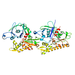
1YF0
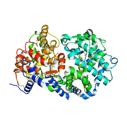
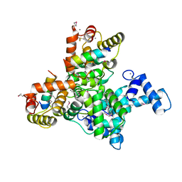
 | | Structural and biochemical analysis of the link between enzymatic activity and oligomerization in AhpC, a bacterial peroxiredoxin. | | 分子名称: | Alkyl hydroperoxide reductase subunit C, SULFATE ION | | 著者 | Parsonage, D, Youngblood, D.S, Sarma, G.N, Wood, Z.A, Karplus, P.A, Poole, L.B. | | 登録日 | 2004-12-29 | | 公開日 | 2005-08-16 | | 最終更新日 | 2021-10-20 | | 実験手法 | X-RAY DIFFRACTION (2.5 Å) | | 主引用文献 | Analysis of the Link between Enzymatic Activity and Oligomeric State in AhpC, a Bacterial Peroxiredoxin.
Biochemistry, 44, 2005
|
|
5K48
 | | VIM-2 Metallo Beta Lactamase in complex with 3-(mercaptomethyl)-[1,1'-biphenyl]-4-carboxylic acid | | 分子名称: | 4-phenyl-2-(sulfanylmethyl)benzoic acid, Beta-lactamase VIM-2, FORMIC ACID, ... | | 著者 | Zollman, D, McDonough, M, Brem, J, Schofield, C. | | 登録日 | 2016-05-20 | | 公開日 | 2017-06-07 | | 最終更新日 | 2024-01-10 | | 実験手法 | X-RAY DIFFRACTION (1.744 Å) | | 主引用文献 | In Silico Fragment-Based Design Identifies Subfamily B1 Metallo-beta-lactamase Inhibitors.
J. Med. Chem., 61, 2018
|
|
1K51
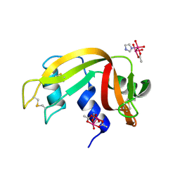
 | | A G55A Mutation Induces 3D Domain Swapping in the B1 Domain of Protein L from Peptostreptococcus magnus | | 分子名称: | Protein L, ZINC ION | | 著者 | O'Neill, J.W, Kim, D.E, Johnsen, K, Baker, D, Zhang, K.Y.J. | | 登録日 | 2001-10-09 | | 公開日 | 2001-12-05 | | 最終更新日 | 2023-08-16 | | 実験手法 | X-RAY DIFFRACTION (1.8 Å) | | 主引用文献 | Single-site mutations induce 3D domain swapping in the B1 domain of protein L from Peptostreptococcus magnus.
Structure, 9, 2001
|
|
1K9D
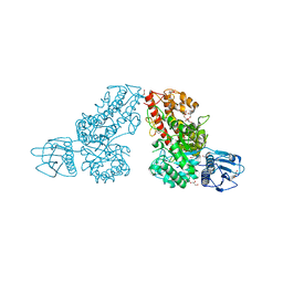 | | The 1.7 A crystal structure of alpha-D-glucuronidase, a family-67 glycoside hydrolase from Bacillus stearothermophilus T-1 | | 分子名称: | GLYCEROL, alpha-D-glucuronidase | | 著者 | Golan, G, Shallom, D, Teplitsky, A, Zaide, G, Shulami, S, Baasov, T, Stojanoff, V, Thompson, A, Shoham, Y, Shoham, G. | | 登録日 | 2001-10-29 | | 公開日 | 2002-10-29 | | 最終更新日 | 2024-04-03 | | 実験手法 | X-RAY DIFFRACTION (1.7 Å) | | 主引用文献 | Crystal Structures of Geobacillus stearothermophilus {alpha}-Glucuronidase Complexed with Its Substrate and Products: MECHANISTIC IMPLICATIONS.
J.Biol.Chem., 279, 2004
|
|
5J0J
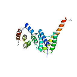
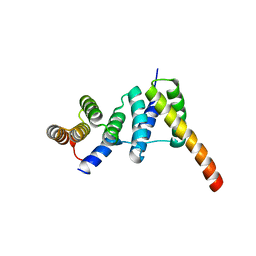
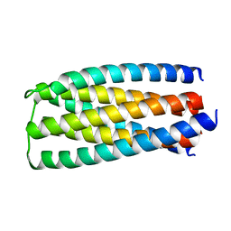 | | De novo design of protein homo-oligomers with modular hydrogen bond network-mediated specificity | | 分子名称: | designed protein 2L6HC3_6 | | 著者 | Sankaran, B, Zwart, P.H, Pereira, J.H, Baker, D, Boyken, S, Chen, Z, Groves, B, Langan, R.A, Oberdorfer, G, Ford, A, Gilmore, J, Xu, C, DiMaio, F, Seelig, G. | | 登録日 | 2016-03-28 | | 公開日 | 2016-05-25 | | 最終更新日 | 2024-03-06 | | 実験手法 | X-RAY DIFFRACTION (2.256 Å) | | 主引用文献 | De novo design of protein homo-oligomers with modular hydrogen-bond network-mediated specificity.
Science, 352, 2016
|
|
6J0O
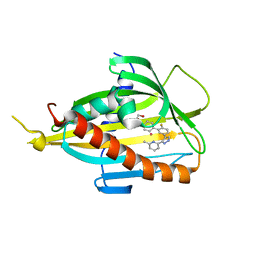 | | Crystal structure of CERT START domain in complex with compound SC1 | | 分子名称: | 2-[4-[2-fluoranyl-5-[3-(6-methylpyridin-2-yl)-1~{H}-pyrazol-4-yl]phenyl]phenyl]sulfonylethanol, LIPID-TRANSFER PROTEIN CERT, UNKNOWN ATOM OR ION | | 著者 | Suzuki, M, Nakao, N, Ueno, M, Sakai, S, Egawa, D, Hanzawa, H, Kawasaki, S, Kumagai, K, Kobayashi, S, Hanada, K. | | 登録日 | 2018-12-25 | | 公開日 | 2019-02-27 | | 最終更新日 | 2024-03-27 | | 実験手法 | X-RAY DIFFRACTION (1.8 Å) | | 主引用文献 | Natural ligand-nonmimetic inhibitors of the lipid-transfer protein CERT
Commun Chem, 2019
|
|
1K3R
 | | Crystal Structure of the Methyltransferase with a Knot from Methanobacterium thermoautotrophicum | | 分子名称: | conserved protein MT0001 | | 著者 | Zarembinski, T.I, Kim, Y, Peterson, K, Christendat, D, Dharamsi, A, Arrowsmith, C.H, Edwards, A.M, Joachimiak, A, Midwest Center for Structural Genomics (MCSG) | | 登録日 | 2001-10-03 | | 公開日 | 2002-05-15 | | 最終更新日 | 2024-02-07 | | 実験手法 | X-RAY DIFFRACTION (2.3 Å) | | 主引用文献 | Deep trefoil knot implicated in RNA binding found in an archaebacterial protein.
Proteins, 50, 2003
|
|
7QWZ
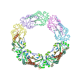
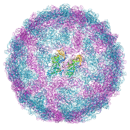
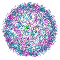 | | Full capsid of Saccharomyces cerevisiae virus L-BCLa | | 分子名称: | Major capsid protein | | 著者 | Grybchuk, D, Prochazkova, M, Fuzik, T, Konovalovas, A, Serva, S, Yurchenko, V, Plevka, P. | | 登録日 | 2022-01-26 | | 公開日 | 2022-09-07 | | 最終更新日 | 2024-07-17 | | 実験手法 | ELECTRON MICROSCOPY (3.7 Å) | | 主引用文献 | Structures of L-BC virus and its open particle provide insight into Totivirus capsid assembly.
Commun Biol, 5, 2022
|
|
5J2U
 | | Tubulin-MMAF complex | | 分子名称: | CALCIUM ION, GUANOSINE-5'-DIPHOSPHATE, GUANOSINE-5'-TRIPHOSPHATE, ... | | 著者 | Waight, A.B, Bargsten, K, Doronina, S, Steinmetz, M.O, Sussman, D, Prota, A.E. | | 登録日 | 2016-03-30 | | 公開日 | 2016-08-17 | | 最終更新日 | 2024-01-10 | | 実験手法 | X-RAY DIFFRACTION (2.5 Å) | | 主引用文献 | Structural Basis of Microtubule Destabilization by Potent Auristatin Anti-Mitotics.
Plos One, 11, 2016
|
|
1K52
 | | Monomeric Protein L B1 Domain with a K54G mutation | | 分子名称: | Protein L, ZINC ION | | 著者 | O'Neill, J.W, Kim, D.E, Johnsen, K, Baker, D, Zhang, K.Y.J. | | 登録日 | 2001-10-09 | | 公開日 | 2001-12-05 | | 最終更新日 | 2023-08-16 | | 実験手法 | X-RAY DIFFRACTION (1.8 Å) | | 主引用文献 | Single-site mutations induce 3D domain swapping in the B1 domain of protein L from Peptostreptococcus magnus.
Structure, 9, 2001
|
|
6JLH
 | | Structure of SCGN in complex with a Snap25 peptide | | 分子名称: | CALCIUM ION, CHLORIDE ION, Secretagogin, ... | | 著者 | Qin, J, Sun, Q, Jia, D. | | 登録日 | 2019-03-05 | | 公開日 | 2020-03-11 | | 最終更新日 | 2020-04-08 | | 実験手法 | X-RAY DIFFRACTION (2.37 Å) | | 主引用文献 | Structural and mechanistic insights into secretagogin-mediated exocytosis.
Proc.Natl.Acad.Sci.USA, 117, 2020
|
|
7QWX
 | | Empty capsid of Saccharomyces cerevisiae virus L-BCLa | | 分子名称: | Major capsid protein | | 著者 | Grybchuk, D, Prochazkova, M, Fuzik, T, Konovalovas, A, Serva, S, Yurchenko, V, Plevka, P. | | 登録日 | 2022-01-26 | | 公開日 | 2022-09-07 | | 最終更新日 | 2024-07-17 | | 実験手法 | ELECTRON MICROSCOPY (2.9 Å) | | 主引用文献 | Structures of L-BC virus and its open particle provide insight into Totivirus capsid assembly.
Commun Biol, 5, 2022
|
|
1K6G
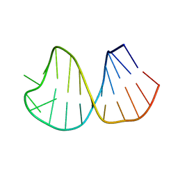 | | Solution Structure of Conserved AGNN Tetraloops: Insights into Rnt1p RNA Processing | | 分子名称: | RNA (5'-R(P*GP*GP*CP*GP*UP*CP*AP*UP*GP*AP*GP*UP*CP*CP*AP*UP*GP*GP*CP*GP*CP*C)-3') | | 著者 | Lebars, I, Lamontagne, B, Yoshizawa, S, Abou Elela, S, Fourmy, D. | | 登録日 | 2001-10-16 | | 公開日 | 2001-12-19 | | 最終更新日 | 2024-05-22 | | 実験手法 | SOLUTION NMR | | 主引用文献 | Solution structure of conserved AGNN tetraloops: insights into Rnt1p RNA processing.
EMBO J., 20, 2001
|
|
1K72
 | | The X-ray Crystal Structure Of Cel9G Complexed With cellotriose | | 分子名称: | CALCIUM ION, Endoglucanase 9G, GLYCEROL, ... | | 著者 | Mandelman, D, Belaich, A, Belaich, J.P, Aghajari, N, Driguez, H, Haser, R. | | 登録日 | 2001-10-18 | | 公開日 | 2003-07-15 | | 最終更新日 | 2023-08-16 | | 実験手法 | X-RAY DIFFRACTION (1.8 Å) | | 主引用文献 | X-Ray Crystal Structure of the Multidomain Endoglucanase Cel9G from Clostridium
cellulolyticum Complexed with Natural and Synthetic Cello-Oligosaccharides
J.BACTERIOL., 185, 2003
|
|
5J4N
 | |
1YC0
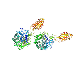
 | | short form HGFA with first Kunitz domain from HAI-1 | | 分子名称: | Hepatocyte growth factor activator, Kunitz-type protease inhibitor 1, PHOSPHATE ION | | 著者 | Shia, S, Stamos, J, Kirchhofer, D, Fan, B, Wu, J, Corpuz, R.T, Santell, L, Lazarus, R.A, Eigenbrot, C. | | 登録日 | 2004-12-21 | | 公開日 | 2005-02-15 | | 最終更新日 | 2023-08-23 | | 実験手法 | X-RAY DIFFRACTION (2.6 Å) | | 主引用文献 | Conformational lability in serine protease active sites: structures of hepatocyte growth factor activator (HGFA) alone and with the inhibitory domain from HGFA inhibitor-1B.
J.Mol.Biol., 346, 2005
|
|
1YCL
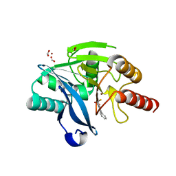
 | | Crystal Structure of B. subtilis LuxS in Complex with a Catalytic 2-Ketone Intermediate | | 分子名称: | (S)-2-AMINO-4-[(2S,3R)-2,3,5-TRIHYDROXY-4-OXO-PENTYL]MERCAPTO-BUTYRIC ACID, COBALT (II) ION, S-ribosylhomocysteinase, ... | | 著者 | Rajan, R, Zhu, J, Hu, X, Pei, D, Bell, C.E. | | 登録日 | 2004-12-22 | | 公開日 | 2005-03-15 | | 最終更新日 | 2024-03-13 | | 実験手法 | X-RAY DIFFRACTION (1.8 Å) | | 主引用文献 | Crystal Structure of S-Ribosylhomocysteinase (LuxS) in Complex with a Catalytic 2-Ketone Intermediate.
Biochemistry, 44, 2005
|
|
7Q6D
 | | E. coli FtsA 1-405 ATP 3 Ni | | 分子名称: | ADENOSINE-5'-TRIPHOSPHATE, Cell division protein FtsA, MAGNESIUM ION, ... | | 著者 | Nierhaus, T, Kureisaite-Ciziene, D, Lowe, J. | | 登録日 | 2021-11-06 | | 公開日 | 2022-09-21 | | 最終更新日 | 2024-01-31 | | 実験手法 | X-RAY DIFFRACTION (2.8 Å) | | 主引用文献 | Bacterial divisome protein FtsA forms curved antiparallel double filaments when binding to FtsN.
Nat Microbiol, 7, 2022
|
|
6IZV
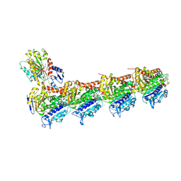
 | | Averaged strand structure of a 15-stranded ParM filament from Clostridium botulinum | | 分子名称: | ADENOSINE-5'-DIPHOSPHATE, MAGNESIUM ION, Putative plasmid segregation protein ParM | | 著者 | Koh, F, Narita, A, Lee, L.J, Tan, Y.Z, Dandey, V.P, Tanaka, K, Popp, D, Robinson, R.C. | | 登録日 | 2018-12-20 | | 公開日 | 2019-06-19 | | 最終更新日 | 2024-05-29 | | 実験手法 | ELECTRON MICROSCOPY (4.2 Å) | | 主引用文献 | The structure of a 15-stranded actin-like filament from Clostridium botulinum.
Nat Commun, 10, 2019
|
|


