2OVF
 
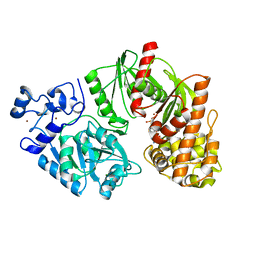
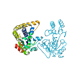 | | Crystal Structure of StaL-PAP complex | | 分子名称: | ADENOSINE-3'-5'-DIPHOSPHATE, StaL | | 著者 | Shi, R, Matte, A, Cygler, M, Montreal-Kingston Bacterial Structural Genomics Initiative (BSGI) | | 登録日 | 2007-02-13 | | 公開日 | 2007-02-27 | | 最終更新日 | 2023-08-30 | | 実験手法 | X-RAY DIFFRACTION (2.95 Å) | | 主引用文献 | Crystal structure of StaL, a glycopeptide antibiotic sulfotransferase from Streptomyces toyocaensis.
J.Biol.Chem., 282, 2007
|
|
2Q1F
 | |
5UAS
 | |
5UAM
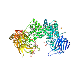
 | | Structure of a new family of Polysaccharide lyase PL25-Ulvanlyase. | | 分子名称: | 1,2-ETHANEDIOL, CHLORIDE ION, GLYCEROL, ... | | 著者 | Ulaganathan, T.S, Boniecki, M.T, Cygler, M. | | 登録日 | 2016-12-19 | | 公開日 | 2017-03-29 | | 最終更新日 | 2024-03-06 | | 実験手法 | X-RAY DIFFRACTION (1.45 Å) | | 主引用文献 | New Ulvan-Degrading Polysaccharide Lyase Family: Structure and Catalytic Mechanism Suggests Convergent Evolution of Active Site Architecture.
ACS Chem. Biol., 12, 2017
|
|
5UFK
 | | Structure of the effector protein SidK (lpg0968) from Legionella pneumophila | | 分子名称: | GLYCEROL, effector protein SidK | | 著者 | Beyrakhova, K, Xu, C, Boniecki, M.T, Cygler, M. | | 登録日 | 2017-01-04 | | 公開日 | 2017-05-10 | | 最終更新日 | 2020-01-08 | | 実験手法 | X-RAY DIFFRACTION (2.3 Å) | | 主引用文献 | Molecular basis for the binding and modulation of V-ATPase by a bacterial effector protein.
PLoS Pathog., 13, 2017
|
|
2RB9
 | | Crystal structure of E.coli HypE | | 分子名称: | HypE protein | | 著者 | Asinas, A.E, Rangarajan, E.S, Min, T, Matte, A, Proteau, A, Munger, C, Cygler, M, Montreal-Kingston Bacterial Structural Genomics Initiative (BSGI) | | 登録日 | 2007-09-18 | | 公開日 | 2007-10-23 | | 最終更新日 | 2023-08-30 | | 実験手法 | X-RAY DIFFRACTION (2 Å) | | 主引用文献 | Structure of [NiFe] hydrogenase maturation protein HypE from Escherichia coli and its interaction with HypF.
J.Bacteriol., 190, 2008
|
|
5UF5
 | | Structure of the effector protein SidK (lpg0968) from Legionella pneumophila (domain-swapped dimer) | | 分子名称: | GLYCEROL, effector protein SidK | | 著者 | Beyrakhova, K, Xu, C, Boniecki, M.T, Cygler, M. | | 登録日 | 2017-01-03 | | 公開日 | 2017-05-10 | | 最終更新日 | 2024-03-06 | | 実験手法 | X-RAY DIFFRACTION (2.4 Å) | | 主引用文献 | Molecular basis for the binding and modulation of V-ATPase by a bacterial effector protein.
PLoS Pathog., 13, 2017
|
|
2VHE
 | | PglD-CoA complex: An acetyl transferase from Campylobacter jejuni | | 分子名称: | ACETYLTRANSFERASE, COENZYME A, SULFATE ION | | 著者 | Rangarajan, E.S, Ruane, K.M, Sulea, T, Watson, D.C, Proteau, A, Leclerc, S, Cygler, M, Matte, A, Young, N.M. | | 登録日 | 2007-11-21 | | 公開日 | 2008-01-29 | | 最終更新日 | 2024-01-31 | | 実験手法 | X-RAY DIFFRACTION (1.8 Å) | | 主引用文献 | Structure and Active Site Residues of Pgld, an N-Acetyltransferase from the Bacillosamine Synthetic Pathway Required for N-Glycan Synthesis in Campylobacter Jejuni
Biochemistry, 47, 2008
|
|
2LIP
 | |
5WKP
 | |
5WLW
 | |
5WGB
 | |
1AC5
 | | CRYSTAL STRUCTURE OF KEX1(DELTA)P, A PROHORMONE-PROCESSING CARBOXYPEPTIDASE FROM SACCHAROMYCES CEREVISIAE | | 分子名称: | 2-acetamido-2-deoxy-beta-D-glucopyranose, 2-acetamido-2-deoxy-beta-D-glucopyranose-(1-4)-2-acetamido-2-deoxy-beta-D-glucopyranose, KEX1(DELTA)P | | 著者 | Shilton, B.H, Thomas, D.Y, Cygler, M. | | 登録日 | 1997-02-13 | | 公開日 | 1997-05-15 | | 最終更新日 | 2023-08-02 | | 実験手法 | X-RAY DIFFRACTION (2.4 Å) | | 主引用文献 | Crystal structure of Kex1deltap, a prohormone-processing carboxypeptidase from Saccharomyces cerevisiae.
Biochemistry, 36, 1997
|
|
3PT5
 | | Crystal structure of NanS | | 分子名称: | NANS (YJHS), A 9-O-acetyl N-acetylneuraminic acid esterase | | 著者 | Ruane, K.M, Rangarajan, E.S, Proteau, A, Schrag, J.D, Cygler, M, Montreal-Kingston Bacterial Structural Genomics Initiative (BSGI) | | 登録日 | 2010-12-02 | | 公開日 | 2011-05-18 | | 最終更新日 | 2024-02-21 | | 実験手法 | X-RAY DIFFRACTION (1.6 Å) | | 主引用文献 | Structural and enzymatic characterization of NanS (YjhS), a 9-O-Acetyl N-acetylneuraminic acid esterase from Escherichia coli O157:H7.
Protein Sci., 20, 2011
|
|
1XHN
 | |
1CRL
 | |
1DEU
 | | CRYSTAL STRUCTURE OF HUMAN PROCATHEPSIN X: A CYSTEINE PROTEASE WITH THE PROREGION COVALENTLY LINKED TO THE ACTIVE SITE CYSTEINE | | 分子名称: | PROCATHEPSIN X | | 著者 | Sivaraman, J, Nagler, D.K, Zhang, R, Menard, R, Cygler, M. | | 登録日 | 1999-11-15 | | 公開日 | 2000-02-18 | | 最終更新日 | 2023-08-09 | | 実験手法 | X-RAY DIFFRACTION (1.7 Å) | | 主引用文献 | Crystal structure of human procathepsin X: a cysteine protease with the proregion covalently linked to the active site cysteine.
J.Mol.Biol., 295, 2000
|
|
1DBO
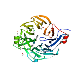
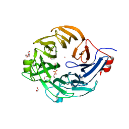
 | | CRYSTAL STRUCTURE OF CHONDROITINASE B | | 分子名称: | 4-deoxy-alpha-D-glucopyranose-(1-3)-[beta-D-glucopyranose-(1-4)]2-O-methyl-beta-L-fucopyranose-(1-4)-beta-D-xylopyranose-(1-4)-alpha-D-glucopyranuronic acid-(1-2)-[alpha-L-rhamnopyranose-(1-4)]alpha-D-mannopyranose, 4-deoxy-beta-D-glucopyranuronic acid-(1-3)-2-acetamido-2-deoxy-4-O-sulfo-beta-D-galactopyranose, CHONDROITINASE B | | 著者 | Huang, W, Matte, A, Li, Y, Kim, Y.S, Linhardt, R.J, Su, H, Cygler, M. | | 登録日 | 1999-11-03 | | 公開日 | 2000-01-12 | | 最終更新日 | 2020-07-29 | | 実験手法 | X-RAY DIFFRACTION (1.7 Å) | | 主引用文献 | Crystal structure of chondroitinase B from Flavobacterium heparinum and its complex with a disaccharide product at 1.7 A resolution.
J.Mol.Biol., 294, 1999
|
|
3BE5
 | | Crystal structure of FitE (crystal form 1), a group III periplasmic siderophore binding protein | | 分子名称: | CHLORIDE ION, Putative iron compound-binding protein of ABC transporter family | | 著者 | Shi, R, Matte, A, Cygler, M, Montreal-Kingston Bacterial Structural Genomics Initiative (BSGI) | | 登録日 | 2007-11-16 | | 公開日 | 2008-10-28 | | 最終更新日 | 2011-07-13 | | 実験手法 | X-RAY DIFFRACTION (2.2 Å) | | 主引用文献 | Trapping open and closed forms of FitE-A group III periplasmic binding protein.
Proteins, 75, 2008
|
|
4LRY
 | | Crystal Structure of the E.coli DhaR(N)-DhaK(T79L) complex | | 分子名称: | GLYCEROL, PTS-dependent dihydroxyacetone kinase operon regulatory protein, PTS-dependent dihydroxyacetone kinase, ... | | 著者 | Shi, R, McDonald, L, Cygler, M, Ekiel, I. | | 登録日 | 2013-07-21 | | 公開日 | 2014-01-29 | | 最終更新日 | 2024-02-28 | | 実験手法 | X-RAY DIFFRACTION (2.83 Å) | | 主引用文献 | Coiled-Coil Helix Rotation Selects Repressing or Activating State of Transcriptional Regulator DhaR.
Structure, 22, 2014
|
|
3BFP
 | | Crystal Structure of apo-PglD from Campylobacter jejuni | | 分子名称: | Acetyltransferase, CITRATE ANION | | 著者 | Rangarajan, E.S, Watson, D.C, Leclerc, S, Proteau, A, Cygler, M, Matte, A, Young, N.M, Montreal-Kingston Bacterial Structural Genomics Initiative (BSGI) | | 登録日 | 2007-11-22 | | 公開日 | 2008-01-22 | | 最終更新日 | 2024-02-21 | | 実験手法 | X-RAY DIFFRACTION (1.75 Å) | | 主引用文献 | Structure and Active Site Residues of PglD, an N-Acetyltransferase from the Bacillosamine Synthetic Pathway Required for N-Glycan Synthesis in Campylobacter jejuni.
Biochemistry, 47, 2008
|
|
3BE6
 | | Crystal structure of FitE (crystal form 2), a group III periplasmic siderophore binding protein | | 分子名称: | CHLORIDE ION, GLYCEROL, MAGNESIUM ION, ... | | 著者 | Shi, R, Matte, A, Cygler, M, Montreal-Kingston Bacterial Structural Genomics Initiative (BSGI) | | 登録日 | 2007-11-16 | | 公開日 | 2008-10-28 | | 最終更新日 | 2023-11-15 | | 実験手法 | X-RAY DIFFRACTION (1.82 Å) | | 主引用文献 | Trapping open and closed forms of FitE-A group III periplasmic binding protein.
Proteins, 75, 2008
|
|
4LRX
 | | Crystal Structure of the E.coli DhaR(N)-DhaK complex | | 分子名称: | GLYCEROL, PTS-dependent dihydroxyacetone kinase operon regulatory protein, PTS-dependent dihydroxyacetone kinase, ... | | 著者 | Shi, R, McDonald, L, Cygler, M, Ekiel, I. | | 登録日 | 2013-07-21 | | 公開日 | 2014-01-29 | | 最終更新日 | 2024-02-28 | | 実験手法 | X-RAY DIFFRACTION (3.25 Å) | | 主引用文献 | Coiled-Coil Helix Rotation Selects Repressing or Activating State of Transcriptional Regulator DhaR.
Structure, 22, 2014
|
|
4LRZ
 | | Crystal Structure of the E.coli DhaR(N)-DhaL complex | | 分子名称: | ADENOSINE-5'-DIPHOSPHATE, MAGNESIUM ION, PTS-dependent dihydroxyacetone kinase operon regulatory protein, ... | | 著者 | Shi, R, McDonald, L, Cygler, M, Ekiel, I. | | 登録日 | 2013-07-21 | | 公開日 | 2014-01-29 | | 最終更新日 | 2024-02-28 | | 実験手法 | X-RAY DIFFRACTION (2.32 Å) | | 主引用文献 | Coiled-Coil Helix Rotation Selects Repressing or Activating State of Transcriptional Regulator DhaR.
Structure, 22, 2014
|
|
3TTF
 | | Crystal structure of E. coli HypF with AMP and carbamoyl phosphate | | 分子名称: | ADENOSINE MONOPHOSPHATE, MAGNESIUM ION, Transcriptional regulatory protein, ... | | 著者 | Petkun, S, Shi, R, Li, Y, Cygler, M. | | 登録日 | 2011-09-14 | | 公開日 | 2011-12-28 | | 最終更新日 | 2024-02-28 | | 実験手法 | X-RAY DIFFRACTION (1.92 Å) | | 主引用文献 | Structure of Hydrogenase Maturation Protein HypF with Reaction Intermediates Shows Two Active Sites.
Structure, 19, 2011
|
|


