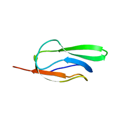
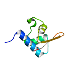
2B8F
 
 | |
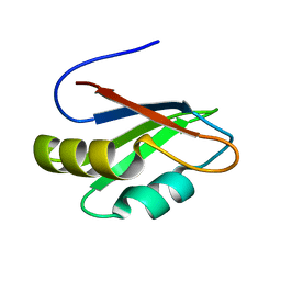
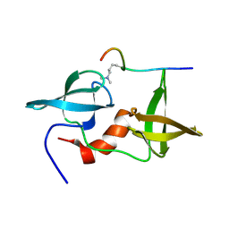
2B0G
 | |
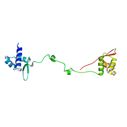
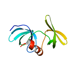
2B8G
 | |
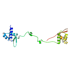
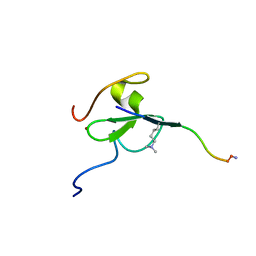
6VE5
 | |
6MXZ
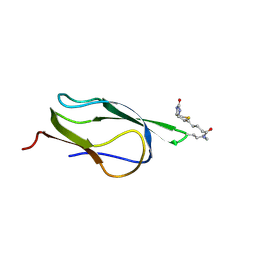
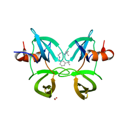 | | Structure of 53BP1 Tudor domains in complex with small molecule UNC3474 | | 分子名称: | FORMIC ACID, N-[3-(tert-butylamino)propyl]-3-(propan-2-yl)benzamide, TP53-binding protein 1 | | 著者 | Cui, G, Botuyan, M.V, Schuller, D.J, Mer, G. | | 登録日 | 2018-10-31 | | 公開日 | 2019-11-27 | | 最終更新日 | 2023-10-11 | | 実験手法 | X-RAY DIFFRACTION (2.5 Å) | | 主引用文献 | An autoinhibited state of 53BP1 revealed by small molecule antagonists and protein engineering.
Nat Commun, 14, 2023
|
|
6MXX
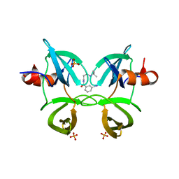 | | Structure of 53BP1 tandem Tudor domains in complex with small molecule UNC2991 | | 分子名称: | FORMIC ACID, N-[3-(tert-butylamino)propyl]-3-iodobenzamide, PHOSPHATE ION, ... | | 著者 | Cui, G, Botuyan, M.V, Mer, G. | | 登録日 | 2018-10-31 | | 公開日 | 2019-11-27 | | 最終更新日 | 2023-10-11 | | 実験手法 | X-RAY DIFFRACTION (2.298 Å) | | 主引用文献 | An autoinhibited state of 53BP1 revealed by small molecule antagonists and protein engineering.
Nat Commun, 14, 2023
|
|
2KTF
 | |
2L0F
 | |
2MWO
 | |
2MWP
 | |
6MXY
 | |
1Z6H
 | |
1Z7T
 | |
2AYM
 | |
2LY1
 | |
2LY2
 | |
6CO1
 | |
2L0G
 | |
5VZM
 | |
2LDM
 | |
2LH9
 | |
2LVM
 | |
6MY0
 | |
5TBN
 | |
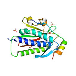
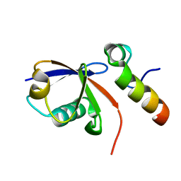
6D0L
 | | Structure of human TIRR | | 分子名称: | Tudor-interacting repair regulator protein | | 著者 | Cui, G, Botuyan, M.V, Mer, G. | | 登録日 | 2018-04-10 | | 公開日 | 2018-06-06 | | 最終更新日 | 2023-10-04 | | 実験手法 | X-RAY DIFFRACTION (1.97 Å) | | 主引用文献 | Mechanism of 53BP1 activity regulation by RNA-binding TIRR and a designer protein.
Nat. Struct. Mol. Biol., 25, 2018
|
|


