3CS4
 
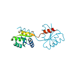
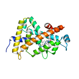 | | Structure-based design of a superagonist ligand for the vitamin D nuclear receptor | | 分子名称: | (1S,3R,5Z,7E,14beta,17alpha)-17-[(2S,4S)-4-(2-hydroxy-2-methylpropyl)-2-methyltetrahydrofuran-2-yl]-9,10-secoandrosta-5,7,10-triene-1,3-diol, Vitamin D3 receptor | | 著者 | Hourai, S, Rodriguez, L.C, Antony, P, Reina-San-Martin, B, Ciesielski, F, Magnier, B.C, Schoonjans, K, Mourino, A, Rochel, N, Moras, D. | | 登録日 | 2008-04-09 | | 公開日 | 2008-05-27 | | 最終更新日 | 2024-02-21 | | 実験手法 | X-RAY DIFFRACTION (2 Å) | | 主引用文献 | Structure-based design of a superagonist ligand for the vitamin d nuclear receptor.
Chem.Biol., 15, 2008
|
|
1K41
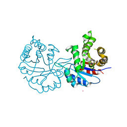
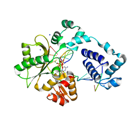
 | | Crystal structure of KSI Y57S mutant | | 分子名称: | Ketosteroid Isomerase | | 著者 | Cha, S.S, Oh, B.H, Nam, G.H, Jang, D.S, Lee, T.H, Choi, K.Y. | | 登録日 | 2001-10-05 | | 公開日 | 2002-10-16 | | 最終更新日 | 2024-05-29 | | 実験手法 | X-RAY DIFFRACTION (2.2 Å) | | 主引用文献 | Maintenance of alpha-helical structures by phenyl rings in the active-site tyrosine triad contributes to catalysis and stability of ketosteroid isomerase from Pseudomonas putida biotype B
Biochemistry, 40, 2001
|
|
8J7C
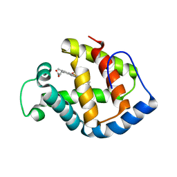
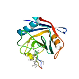
 | | Crystal structure of triosephosphate isomerase from Leishmania orientalis at 1.88A with an arsenic ion bound at Cys57 | | 分子名称: | ARSENIC, Triosephosphate isomerase | | 著者 | Kuaprasert, B, Attarataya, J, Riangrungroj, P, Pornthanakasem, W, Suginta, W, Mungthin, M, Leelayoova, S, Choowongkomon, K, Leartsakulpanich, U. | | 登録日 | 2023-04-27 | | 公開日 | 2024-05-01 | | 実験手法 | X-RAY DIFFRACTION (1.88 Å) | | 主引用文献 | Leishmania orientalis triosephosphate isomerase crystal structure at 1.45 angstroms resolution and its potential specific inhibitors
To be published
|
|
1IT2
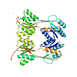
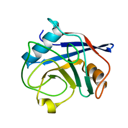
 | | Hagfish deoxy hemoglobin | | 分子名称: | PROTOPORPHYRIN IX CONTAINING FE, hemoglobin | | 著者 | Mito, M, Chong, K.T, Park, S.-Y, Tame, J.R. | | 登録日 | 2002-01-05 | | 公開日 | 2002-01-23 | | 最終更新日 | 2023-10-25 | | 実験手法 | X-RAY DIFFRACTION (1.6 Å) | | 主引用文献 | Crystal structures of deoxy- and carbonmonoxyhemoglobin F1 from the hagfish Eptatretus burgeri
J.Biol.Chem., 277, 2002
|
|
1DBQ
 | | DNA-BINDING REGULATORY PROTEIN | | 分子名称: | MAGNESIUM ION, PURINE REPRESSOR | | 著者 | Schumacher, M.A, Choi, K.Y, Lu, F, Zalkin, H, Brennan, R.G. | | 登録日 | 1996-02-13 | | 公開日 | 1996-12-07 | | 最終更新日 | 2024-02-07 | | 実験手法 | X-RAY DIFFRACTION (2.2 Å) | | 主引用文献 | Mechanism of corepressor-mediated specific DNA binding by the purine repressor.
Cell(Cambridge,Mass.), 83, 1995
|
|
1DZ3
 | | DOMAIN-SWAPPING IN THE SPORULATION RESPONSE REGULATOR SPO0A | | 分子名称: | SULFATE ION, Stage 0 sporulation protein A | | 著者 | Lewis, R.J, Brannigan, J.A, Muchova, K, Leonard, G, Barak, I, Wilkinson, A.J. | | 登録日 | 2000-02-15 | | 公開日 | 2000-04-10 | | 最終更新日 | 2024-05-08 | | 実験手法 | X-RAY DIFFRACTION (1.65 Å) | | 主引用文献 | Domain swapping in the sporulation response regulator Spo0A.
J. Mol. Biol., 297, 2000
|
|
3MDC
 | | DNA polymerase lambda in complex with dFdCTP | | 分子名称: | 2'-deoxy-2',2'-difluorocytidine 5'-(tetrahydrogen triphosphate), DNA (5'-D(*CP*AP*GP*TP*AP*C)-3'), DNA (5'-D(*CP*GP*GP*CP*GP*GP*TP*AP*CP*TP*G)-3'), ... | | 著者 | Garcia-Diaz, M, Murray, M, Kunkel, T, Chou, K.M. | | 登録日 | 2010-03-30 | | 公開日 | 2010-04-28 | | 最終更新日 | 2024-02-21 | | 実験手法 | X-RAY DIFFRACTION (1.999 Å) | | 主引用文献 | Interaction between DNA Polymerase lambda and anticancer nucleoside analogs.
J.Biol.Chem., 285, 2010
|
|
3PMP
 | | Crystal Structure of Cyclophilin A from Moniliophthora perniciosa in complex with Cyclosporin A | | 分子名称: | CYCLOSPORIN A, Cyclophilin A | | 著者 | Monzani, P, Pereira, H.M, Gramacho, K.P, Meirelles, F.V, Oliva, G, Cascardo, J.C.C. | | 登録日 | 2010-11-17 | | 公開日 | 2011-11-23 | | 最終更新日 | 2023-05-31 | | 実験手法 | X-RAY DIFFRACTION (1.47 Å) | | 主引用文献 | Crystal Structure of Cyclophilin A from Moniliophthora perniciosa
To be Published
|
|
3O7T
 | | Crystal Structure of Cyclophilin A from Moniliophthora perniciosa | | 分子名称: | Cyclophilin A | | 著者 | Monzani, P.S, Pereira, H.M, Gramacho, K.P, Meirelles, F.V, Oliva, G, Cascardo, J.C.M. | | 登録日 | 2010-07-31 | | 公開日 | 2011-08-10 | | 最終更新日 | 2024-02-21 | | 実験手法 | X-RAY DIFFRACTION (1.85 Å) | | 主引用文献 | Crystal Structures of apo-cyclophilin and bounded cyclosporine A from Moniliophthora perniciosa
To be Published
|
|
2JS2
 | |
2JS0
 | |
2V5Y
 | | Crystal structure of the receptor protein tyrosine phosphatase mu ectodomain | | 分子名称: | 2-acetamido-2-deoxy-beta-D-glucopyranose, RECEPTOR-TYPE TYROSINE-PROTEIN PHOSPHATASE MU, SODIUM ION | | 著者 | Aricescu, A.R, Siebold, C, Choudhuri, K, Chang, V.T, Lu, W, Davis, S.J, van der Merwe, P.A, Jones, E.Y. | | 登録日 | 2007-07-11 | | 公開日 | 2007-09-11 | | 最終更新日 | 2023-12-13 | | 実験手法 | X-RAY DIFFRACTION (3.1 Å) | | 主引用文献 | Structure of a Tyrosine Phosphatase Adhesive Interaction Reveals a Spacer-Clamp Mechanism.
Science, 317, 2007
|
|


