7L33
 
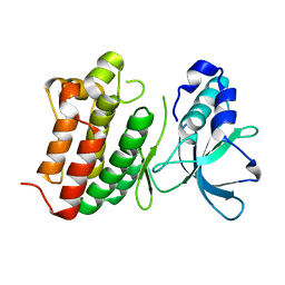
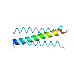 | | X-ray Structure of a Cu-Bound De Novo Designed Peptide Trimer | | 分子名称: | COPPER (II) ION, Cu-3SCC | | 著者 | Chakraborty, S, Wawrzak, Z, Prasad, P, Mitra, S, Prakash, D. | | 登録日 | 2020-12-17 | | 公開日 | 2021-08-11 | | 最終更新日 | 2023-10-18 | | 実験手法 | X-RAY DIFFRACTION (1.45 Å) | | 主引用文献 | De Novo Design of a Self-Assembled Artificial Copper Peptide that Activates and Reduces Peroxide
Acs Catalysis, 11, 2021
|
|
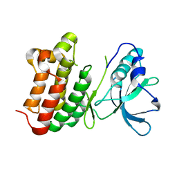
3LJM
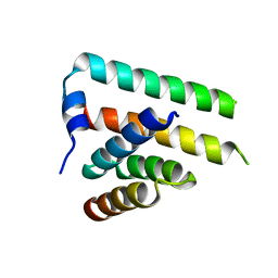
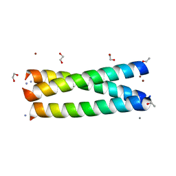 | | Structure of de novo designed apo peptide coil SER L9C | | 分子名称: | 1,2-ETHANEDIOL, CALCIUM ION, COIL SER L9C, ... | | 著者 | Chakraborty, S. | | 登録日 | 2010-01-26 | | 公開日 | 2010-09-15 | | 最終更新日 | 2023-09-06 | | 実験手法 | X-RAY DIFFRACTION (1.36 Å) | | 主引用文献 | Structural comparisons of apo- and metalated three-stranded coiled coils clarify metal binding determinants in thiolate containing designed peptides.
J.Am.Chem.Soc., 132, 2010
|
|
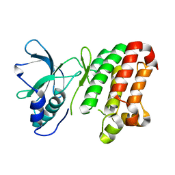
2X6P
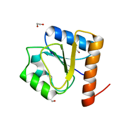
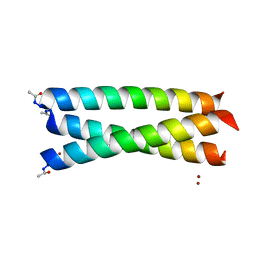 | | Crystal Structure of Coil Ser L19C | | 分子名称: | COIL SER L19C, ZINC ION | | 著者 | Chakraborty, S, Touw, D.S, Peacock, A.F.A, Stuckey, J.A, Pecoraro, V.L. | | 登録日 | 2010-02-18 | | 公開日 | 2010-09-22 | | 最終更新日 | 2023-12-20 | | 実験手法 | X-RAY DIFFRACTION (2.15 Å) | | 主引用文献 | Structural Comparisons of Apo- and Metalated Three-Stranded Coiled Coils Clarify Metal Binding Determinants in Thiolate Containing Designed Peptides.
J.Am.Chem.Soc., 132, 2010
|
|
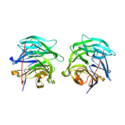
8GT0
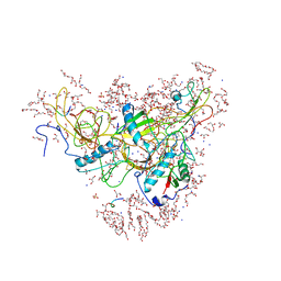
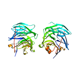
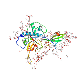 | | Structure of falcipain and human Stefin A complex | | 分子名称: | 1,2-ETHANEDIOL, 3,6,9,12,15,18,21-HEPTAOXATRICOSANE-1,23-DIOL, CHLORIDE ION, ... | | 著者 | Chakraborty, S, Biswas, S. | | 登録日 | 2022-09-07 | | 公開日 | 2023-09-13 | | 実験手法 | X-RAY DIFFRACTION (3.28 Å) | | 主引用文献 | Structure of falcipain and human Stefin A complex
To Be Published
|
|
8GT7
 | | Structure of falcipain and human Stefin A mutant complex | | 分子名称: | 1,2-ETHANEDIOL, 3,6,9,12,15,18,21-HEPTAOXATRICOSANE-1,23-DIOL, Cystatin-A, ... | | 著者 | Chakraborty, S, Biswas, S. | | 登録日 | 2022-09-07 | | 公開日 | 2023-09-13 | | 実験手法 | X-RAY DIFFRACTION (3.28 Å) | | 主引用文献 | Structure of falcipain and human Stefin A complex
To Be Published
|
|
4MXL
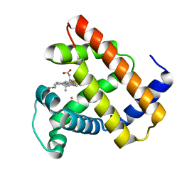
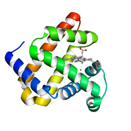
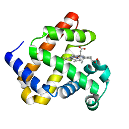 | | X-ray structure of ZnPFeBMb1 | | 分子名称: | Myoglobin, PROTOPORPHYRIN IX CONTAINING ZN | | 著者 | Chakraborty, S, Lu, Y, Petrik, I. | | 登録日 | 2013-09-26 | | 公開日 | 2014-02-12 | | 最終更新日 | 2024-02-28 | | 実験手法 | X-RAY DIFFRACTION (1.5 Å) | | 主引用文献 | Spectroscopic and computational study of a nonheme iron nitrosyl center in a biosynthetic model of nitric oxide reductase.
Angew.Chem.Int.Ed.Engl., 53, 2014
|
|
4MXK
 | | X-ray structure of Fe(II)-ZnPIXFeBMb1 | | 分子名称: | FE (II) ION, Myoglobin, PROTOPORPHYRIN IX CONTAINING ZN | | 著者 | Chakraborty, S, Lu, Y, Petrik, I. | | 登録日 | 2013-09-26 | | 公開日 | 2014-02-12 | | 最終更新日 | 2024-02-28 | | 実験手法 | X-RAY DIFFRACTION (1.52 Å) | | 主引用文献 | Spectroscopic and computational study of a nonheme iron nitrosyl center in a biosynthetic model of nitric oxide reductase.
Angew.Chem.Int.Ed.Engl., 53, 2014
|
|
7EI0
 | |
6JW9
 | |
7EEF
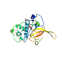
 | | Crystal structure of EphA7 mutant G656E | | 分子名称: | Ephrin type-A receptor 7 | | 著者 | Chakraborty, S, Varma, A.K. | | 登録日 | 2021-03-18 | | 公開日 | 2021-07-07 | | 最終更新日 | 2023-11-29 | | 実験手法 | X-RAY DIFFRACTION (2.6 Å) | | 主引用文献 | Crystal structure of clinically reported mutations Gly656Arg, Gly656Glu and Asp751His identified in the kinase domain of EphA7.
Biochem.Biophys.Res.Commun., 568, 2021
|
|
7EED
 | | Crystal structure of EphA7 mutant D751H | | 分子名称: | Ephrin type-A receptor 7 | | 著者 | Chakraborty, S, Varma, A.K. | | 登録日 | 2021-03-18 | | 公開日 | 2021-07-07 | | 最終更新日 | 2023-11-29 | | 実験手法 | X-RAY DIFFRACTION (3.05 Å) | | 主引用文献 | Crystal structure of clinically reported mutations Gly656Arg, Gly656Glu and Asp751His identified in the kinase domain of EphA7.
Biochem.Biophys.Res.Commun., 568, 2021
|
|
7EEC
 | | Crystal structure of EphA7 mutant G656R | | 分子名称: | Ephrin type-A receptor 7 | | 著者 | Chakraborty, S, Varma, A.K. | | 登録日 | 2021-03-18 | | 公開日 | 2021-07-07 | | 最終更新日 | 2023-11-29 | | 実験手法 | X-RAY DIFFRACTION (3.1 Å) | | 主引用文献 | Crystal structure of clinically reported mutations Gly656Arg, Gly656Glu and Asp751His identified in the kinase domain of EphA7.
Biochem.Biophys.Res.Commun., 568, 2021
|
|
3G4H
 | |
3G4E
 | |
3PH0
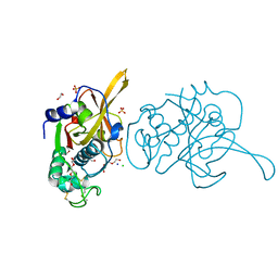
 | | Crystal structure of the heteromolecular chaperone, AscE-AscG, from the type III secretion system in Aeromonas hydrophila | | 分子名称: | AscE, AscG | | 著者 | Chatterjee, C, Kumar, S, Chakraborty, S, Tan, Y.W, Leung, K.Y, Sivaraman, J, Mok, Y.K. | | 登録日 | 2010-11-03 | | 公開日 | 2011-07-20 | | 最終更新日 | 2024-03-20 | | 実験手法 | X-RAY DIFFRACTION (2.4 Å) | | 主引用文献 | Crystal structure of the heteromolecular chaperone, AscE-AscG, from the type III secretion system in Aeromonas hydrophila
Plos One, 6, 2011
|
|
8H5V
 | |
5VRT
 | | Nonheme Iron Replacement in a Biosynthetic Nitric Oxide Reductase Model Performing O2 Reduction to Water: Co-bound FeBMb | | 分子名称: | COBALT (II) ION, Myoglobin, PROTOPORPHYRIN IX CONTAINING FE | | 著者 | Reed, J, Shi, Y, Zhu, Q, Chakraborty, S, Mirs, E.N, Petrik, I.D, Bhagi-Damodaran, A, Ross, M, Moenne-Loccoz, P, Zhang, Y, Lu, Y. | | 登録日 | 2017-05-11 | | 公開日 | 2017-08-16 | | 最終更新日 | 2023-10-04 | | 実験手法 | X-RAY DIFFRACTION (1.995 Å) | | 主引用文献 | Manganese and Cobalt in the Nonheme-Metal-Binding Site of a Biosynthetic Model of Heme-Copper Oxidase Superfamily Confer Oxidase Activity through Redox-Inactive Mechanism.
J. Am. Chem. Soc., 139, 2017
|
|
5VNU
 | | Nonheme Iron Replacement in a Biosynthetic Nitric Oxide Reductase Model Performing O2 Reduction to Water: Mn-bound FeBMb | | 分子名称: | MANGANESE (II) ION, Myoglobin, PROTOPORPHYRIN IX CONTAINING FE | | 著者 | Reed, J, Shi, Y, Zhu, Q, Chakraborty, S, Mirs, E.N, Petrik, I.D, Bhagi-Damodaran, A, Ross, M, Moenne-Loccoz, P, Zhang, Y, Lu, Y. | | 登録日 | 2017-05-01 | | 公開日 | 2017-08-16 | | 最終更新日 | 2024-03-13 | | 実験手法 | X-RAY DIFFRACTION (1.584 Å) | | 主引用文献 | Manganese and Cobalt in the Nonheme-Metal-Binding Site of a Biosynthetic Model of Heme-Copper Oxidase Superfamily Confer Oxidase Activity through Redox-Inactive Mechanism.
J. Am. Chem. Soc., 139, 2017
|
|
5WLS
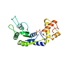
 | | Crystal Structure of a Pollen Receptor Kinase 3 | | 分子名称: | 2-acetamido-2-deoxy-beta-D-glucopyranose, 2-acetamido-2-deoxy-beta-D-glucopyranose-(1-4)-2-acetamido-2-deoxy-beta-D-glucopyranose, Pollen receptor-like kinase 3 | | 著者 | Xu, G, Chakraborty, S, Pan, H. | | 登録日 | 2017-07-27 | | 公開日 | 2018-06-06 | | 最終更新日 | 2023-10-04 | | 実験手法 | X-RAY DIFFRACTION (2.496 Å) | | 主引用文献 | The Extracellular Domain of Pollen Receptor Kinase 3 is structurally similar to the SERK family of co-receptors.
Sci Rep, 8, 2018
|
|
6LUA
 | |
6LUF
 | |
6CDX
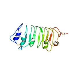
 | | High-resolution crystal structure of fluoropropylated cystine knot, binding to alpha-5 beta-6 integrin | | 分子名称: | cystine knot (fluoropropylated) | | 著者 | Kimura, R, Nix, J, Bongura, C, Chakraborti, S, Gambhir, S, Filipp, F.V. | | 登録日 | 2018-02-09 | | 公開日 | 2019-08-14 | | 最終更新日 | 2023-10-04 | | 実験手法 | X-RAY DIFFRACTION (1 Å) | | 主引用文献 | Evaluation of integrin alpha v beta6cystine knot PET tracers to detect cancer and idiopathic pulmonary fibrosis.
Nat Commun, 10, 2019
|
|
6TXN
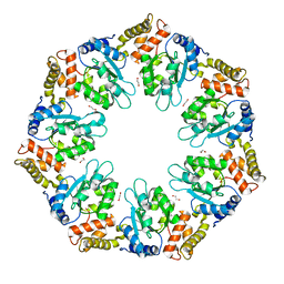
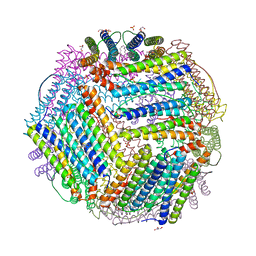 | | Crystal structure of thermotoga maritima Ferritin in apo form | | 分子名称: | EICOSANE, Ferritin, GLYCEROL, ... | | 著者 | Wilk, P, Grudnik, P, Kumar, M, Heddle, J, Chakraborti, S. | | 登録日 | 2020-01-14 | | 公開日 | 2021-07-28 | | 最終更新日 | 2024-01-24 | | 実験手法 | X-RAY DIFFRACTION (2.01 Å) | | 主引用文献 | A single residue can modulate nanocage assembly in salt dependent ferritin.
Nanoscale, 13, 2021
|
|
6TXM
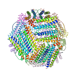 | | Crystal structure of thermotoga maritima E65R Ferritin | | 分子名称: | EICOSANE, Ferritin, GLYCEROL, ... | | 著者 | Wilk, P, Grudnik, P, Kumar, M, Heddle, J, Chakraborti, S. | | 登録日 | 2020-01-14 | | 公開日 | 2021-07-28 | | 最終更新日 | 2024-01-24 | | 実験手法 | X-RAY DIFFRACTION (2.198 Å) | | 主引用文献 | A single residue can modulate nanocage assembly in salt dependent ferritin.
Nanoscale, 13, 2021
|
|
6TXH
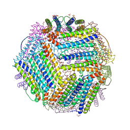 | | Crystal structure of thermotoga maritima Ferritin in apo form | | 分子名称: | EICOSANE, Ferritin, GLYCEROL, ... | | 著者 | Wilk, P, Grudnik, P, Kumar, M, Heddle, J, Chakraborti, S. | | 登録日 | 2020-01-14 | | 公開日 | 2021-07-28 | | 最終更新日 | 2024-01-24 | | 実験手法 | X-RAY DIFFRACTION (2.198 Å) | | 主引用文献 | A single residue can modulate nanocage assembly in salt dependent ferritin.
Nanoscale, 13, 2021
|
|


