Loading
PDBj
MenuPDBj@FacebookPDBj@TwitterPDBj@YouTubewwPDB FoundationwwPDB
RCSB PDBPDBeBMRBAdv. SearchSearch help
PDB: 307 results

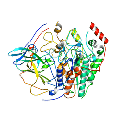
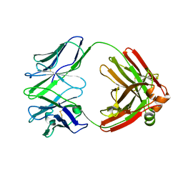
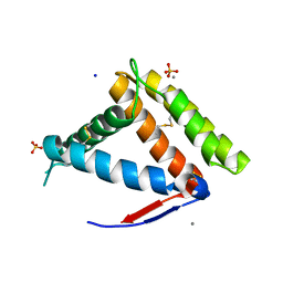
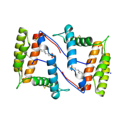
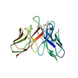
2A9N
DownloadVisualize
BU of 2a9n by Molmil
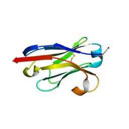
A Mutation Designed to Alter Crystal Packing Permits Structural Analysis of a Tight-binding Fluorescein-scFv complex
Descriptor: 4-(2,7-DIFLUORO-6-HYDROXY-3-OXO-3H-XANTHEN-9-YL)ISOPHTHALIC ACID, fluorescein-scfv
Authors:Cambillau, C, Spinelli, S, Honegger, A, Pluckthun, A.
Deposit date:2005-07-12
Release date:2005-10-25
Last modified:2024-10-30
Method:X-RAY DIFFRACTION (3 Å)
Cite:A mutation designed to alter crystal packing permits structural analysis of a tight-binding fluorescein-scFv complex.
Protein Sci., 14, 2005
8BOZ
DownloadVisualize
BU of 8boz by Molmil
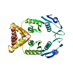
structure of the Adherent-Invasive Escherichia coli Tle3/Tli3 T6SS effector/immunity complex
Descriptor: CALCIUM ION, Lipoprotein, Transmembrane protein
Authors:Cambillau, C, Roussel, A.
Deposit date:2022-11-15
Release date:2023-02-08
Last modified:2024-05-01
Method:X-RAY DIFFRACTION (3.6 Å)
Cite:Activity and Crystal Structure of the Adherent-Invasive Escherichia coli Tle3/Tli3 T6SS Effector/Immunity Complex Determined Using an AlphaFold2 Predicted Model.
Int J Mol Sci, 24, 2023
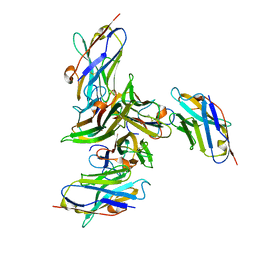
7PHS
DownloadVisualize
BU of 7phs by Molmil
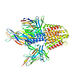
highly potent IL6 antagonistic antibody selected from a camelid immune phage display repertoire
Descriptor: Heavy chain of Fab129D3, Light chain of Fab129D3
Authors:Cambillau, C, Pannecoucke, E, De Haard, H.
Deposit date:2021-08-18
Release date:2022-09-07
Last modified:2024-01-31
Method:X-RAY DIFFRACTION (2.77 Å)
Cite:Non-promiscuous pairing in a highly potent IL6 antagonistic antibody selected from a camelid immune phage display repertoire
To Be Published
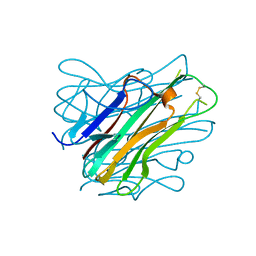
7NYJ
DownloadVisualize
BU of 7nyj by Molmil
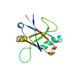
Structure of OBP1 from Varroa destructor, form P3<2>21
Descriptor: CALCIUM ION, Odorant Binding Protein 1 from Varoa destructor, form P3<2>21, ...
Authors:Cambillau, C, Amigues, B, Roussel, A, Leone, P, Gaubert, A, Pelosi, P.
Deposit date:2021-03-22
Release date:2021-07-07
Last modified:2024-10-09
Method:X-RAY DIFFRACTION (1.81 Å)
Cite:A new non-classical fold of varroa odorant-binding proteins reveals a wide open internal cavity.
Sci Rep, 11, 2021
7NZA
DownloadVisualize
BU of 7nza by Molmil
Structure of OBP1 from Varroa destructor, form P2<1>
Descriptor: 2-[N-CYCLOHEXYLAMINO]ETHANE SULFONIC ACID, Odorant Binding Protein from Varroa destructor, form P2<1>, ...
Authors:Cambillau, C, Amigues, B, Roussel, A, Leone, P, Gaubert, A, Pelosi, P.
Deposit date:2021-03-23
Release date:2021-07-07
Last modified:2024-01-31
Method:X-RAY DIFFRACTION (1.199 Å)
Cite:A new non-classical fold of varroa odorant-binding proteins reveals a wide open internal cavity.
Sci Rep, 11, 2021
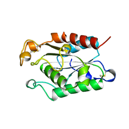
1FFD
DownloadVisualize
BU of 1ffd by Molmil
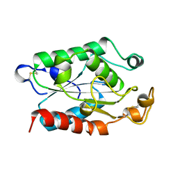
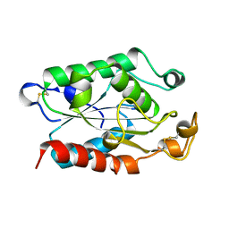
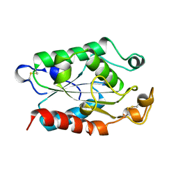
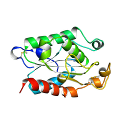
CONTRIBUTION OF CUTINASE SERINE 42 SIDE CHAIN TO THE STABILIZATION OF THE OXYANION TRANSITION STATE
Descriptor: CUTINASE
Authors:Cambillau, C, Martinez, C, Nicolas, A.
Deposit date:1995-10-07
Release date:1996-03-08
Last modified:2021-11-03
Method:X-RAY DIFFRACTION (1.69 Å)
Cite:Contribution of cutinase serine 42 side chain to the stabilization of the oxyanion transition state.
Biochemistry, 35, 1996
1FFA
DownloadVisualize
BU of 1ffa by Molmil
CONTRIBUTION OF CUTINASE SERINE 42 SIDE CHAIN TO THE STABILIZATION OF THE OXYANION TRANSITION STATE
Descriptor: CUTINASE
Authors:Cambillau, C, Martinez, C, Nicolas, A.
Deposit date:1995-10-07
Release date:1996-03-08
Last modified:2024-10-16
Method:X-RAY DIFFRACTION (1.69 Å)
Cite:Contribution of cutinase serine 42 side chain to the stabilization of the oxyanion transition state.
Biochemistry, 35, 1996
1FFE
DownloadVisualize
BU of 1ffe by Molmil
CONTRIBUTION OF CUTINASE SERINE 42 SIDE CHAIN TO THE STABILIZATION OF THE OXYANION TRANSITION STATE
Descriptor: CUTINASE
Authors:Cambillau, C, Martinez, C, Nicolas, A.
Deposit date:1995-10-07
Release date:1996-03-08
Last modified:2024-10-30
Method:X-RAY DIFFRACTION (1.69 Å)
Cite:Contribution of cutinase serine 42 side chain to the stabilization of the oxyanion transition state.
Biochemistry, 35, 1996
1FFC
DownloadVisualize
BU of 1ffc by Molmil
CONTRIBUTION OF CUTINASE SERINE 42 SIDE CHAIN TO THE STABILIZATION OF THE OXYANION TRANSITION STATE
Descriptor: CUTINASE
Authors:Cambillau, C, Martinez, C, Nicolas, A.
Deposit date:1995-10-07
Release date:1996-03-08
Last modified:2024-10-23
Method:X-RAY DIFFRACTION (1.75 Å)
Cite:Contribution of cutinase serine 42 side chain to the stabilization of the oxyanion transition state.
Biochemistry, 35, 1996
1FFB
DownloadVisualize
BU of 1ffb by Molmil
CONTRIBUTION OF CUTINASE SERINE 42 SIDE CHAIN TO THE STABILIZATION OF THE OXYANION TRANSITION STATE
Descriptor: CUTINASE
Authors:Cambillau, C, Martinez, C, Nicolas, A.
Deposit date:1995-10-07
Release date:1996-03-08
Last modified:2024-10-16
Method:X-RAY DIFFRACTION (1.75 Å)
Cite:Contribution of cutinase serine 42 side chain to the stabilization of the oxyanion transition state.
Biochemistry, 35, 1996
5M2J
DownloadVisualize
BU of 5m2j by Molmil
Complex between human TNF alpha and Llama VHH2
Descriptor: Anti-(ED-B) scFV, Tumor necrosis factor
Authors:Cambillau, C, Spinelli, S, Desmyter, A, de Haard, H.
Deposit date:2016-10-13
Release date:2017-08-30
Last modified:2024-01-17
Method:X-RAY DIFFRACTION (1.9 Å)
Cite:Bivalent Llama Single-Domain Antibody Fragments against Tumor Necrosis Factor Have Picomolar Potencies due to Intramolecular Interactions.
Front Immunol, 8, 2017
5M2M
DownloadVisualize
BU of 5m2m by Molmil
Complex between human TNF alpha and Llama VHH3
Descriptor: Llama nanobody VHH3, Tumor necrosis factor
Authors:Cambillau, C, Spinelli, S, Desmyter, A, de Haard, H.
Deposit date:2016-10-13
Release date:2017-08-30
Last modified:2024-01-17
Method:X-RAY DIFFRACTION (2.3 Å)
Cite:Bivalent Llama Single-Domain Antibody Fragments against Tumor Necrosis Factor Have Picomolar Potencies due to Intramolecular Interactions.
Front Immunol, 8, 2017
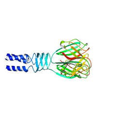
2F0C
DownloadVisualize
BU of 2f0c by Molmil
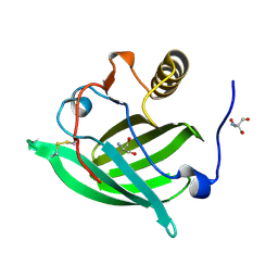
Structure of the Receptor Binding Protein (ORF49, bbp) from lactophage tp901-1
Descriptor: GLYCEROL, Phage tp901-1 ORF49 (BPP)
Authors:Cambillau, C, Spinelli, S.
Deposit date:2005-11-13
Release date:2006-03-28
Last modified:2023-08-23
Method:X-RAY DIFFRACTION (1.65 Å)
Cite:Modular structure of the receptor binding proteins of Lactococcus lactis phages. The RBP structure of the temperate phage TP901-1.
J.Biol.Chem., 281, 2006
5NGH
DownloadVisualize
BU of 5ngh by Molmil
Structure of Odorant Binding Protein 3 from Giant Panda (Ailuropoda melanoleuca)
Descriptor: 2-AMINO-2-HYDROXYMETHYL-PROPANE-1,3-DIOL, Odorant Binding Protein 3
Authors:Cambillau, C, Spinelli, S, Pelosi, P.
Deposit date:2017-03-17
Release date:2017-11-01
Last modified:2024-01-17
Method:X-RAY DIFFRACTION (2.8 Å)
Cite:Reverse chemical ecology: Olfactory proteins from the giant panda and their interactions with putative pheromones and bamboo volatiles.
Proc. Natl. Acad. Sci. U.S.A., 114, 2017
5LY8
DownloadVisualize
BU of 5ly8 by Molmil
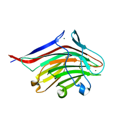
Structure of the CBM2 module of Lactobacillus casei BL23 phage J-1 evolved Dit.
Descriptor: MAGNESIUM ION, Tail component
Authors:Cambillau, C, Spinelli, S, Dieterle, M.-E, Piuri, M.
Deposit date:2016-09-26
Release date:2017-03-22
Last modified:2024-05-01
Method:X-RAY DIFFRACTION (1.28 Å)
Cite:Evolved distal tail carbohydrate binding modules of Lactobacillus phage J-1: a novel type of anti-receptor widespread among lactic acid bacteria phages.
Mol. Microbiol., 104, 2017
6EYY
DownloadVisualize
BU of 6eyy by Molmil
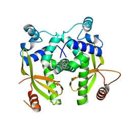
Anti-CRISPR AcrIIa6 cubic form
Descriptor: AcrIIa6
Authors:Cambillau, C, Amigues, B, Moineau, S.
Deposit date:2017-11-13
Release date:2018-06-06
Last modified:2024-05-08
Method:X-RAY DIFFRACTION (2.5 Å)
Cite:Widespread anti-CRISPR proteins in virulent bacteriophages inhibit a range of Cas9 proteins.
Nat Commun, 9, 2018
6EYX
DownloadVisualize
BU of 6eyx by Molmil
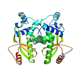
Anti-CRISPR AcrIIa6 tetragonal form
Descriptor: AcrIIa6
Authors:Cambillau, C, Amigues, B, Moineau, S.
Deposit date:2017-11-13
Release date:2018-06-06
Last modified:2024-05-08
Method:X-RAY DIFFRACTION (1.96 Å)
Cite:Widespread anti-CRISPR proteins in virulent bacteriophages inhibit a range of Cas9 proteins.
Nat Commun, 9, 2018
5M38
DownloadVisualize
BU of 5m38 by Molmil
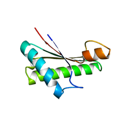
Structure of the TagL peptidoglycan binding domain from EAEC T6SS
Descriptor: OmpA family protein
Authors:Cambillau, C, Nguyen, V.S, Spinelli, S, Cascales, E.
Deposit date:2016-10-14
Release date:2018-02-21
Last modified:2024-01-17
Method:X-RAY DIFFRACTION (2.6 Å)
Cite:Structure of the TagL peptidoglycan binding domain from EAEC T6SS
To Be Published
1U0Q
DownloadVisualize
BU of 1u0q by Molmil
Structure of a Llama VHH domain raised against a carbazole molecule
Descriptor: immunoglobulin heavy chain variable domain
Authors:Cambillau, C, Spinelli, S, Desmyter, A, Verrips, T.
Deposit date:2004-07-14
Release date:2005-06-28
Last modified:2019-12-25
Method:X-RAY DIFFRACTION (1.6 Å)
Cite:Crystal Structure of a Llama VHH Domain Raised Against a Carbazole Hapten
TO BE PUBLISHED
5NGI
DownloadVisualize
BU of 5ngi by Molmil
Structure of XcpQN012
Descriptor: Type II secretion system protein D
Authors:Cambillau, C, Roussel, A, Trinh, T.N.
Deposit date:2017-03-17
Release date:2018-01-24
Last modified:2024-01-17
Method:X-RAY DIFFRACTION (2.98 Å)
Cite:Unraveling the Self-Assembly of the Pseudomonas aeruginosa XcpQ Secretin Periplasmic Domain Provides New Molecular Insights into Type II Secretion System Secreton Architecture and Dynamics.
MBio, 8, 2017
6FW5
DownloadVisualize
BU of 6fw5 by Molmil
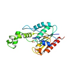
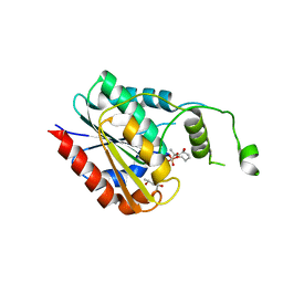
TesA a major thioesterase from Mycobacterium tuberculosis
Descriptor: PHOSPHATE ION, Probable thioesterase TesA
Authors:Cambillau, C, Nguyen, V.S, Canaan, S.
Deposit date:2018-03-05
Release date:2018-10-24
Last modified:2019-01-23
Method:X-RAY DIFFRACTION (2.75 Å)
Cite:Biochemical and Structural Characterization of TesA, a Major Thioesterase Required for Outer-Envelope Lipid Biosynthesis in Mycobacterium tuberculosis.
J. Mol. Biol., 430, 2018
6FVJ
DownloadVisualize
BU of 6fvj by Molmil
TesA a major thioesterase from Mycobacterium tuberculosis
Descriptor: (4S)-2-METHYL-2,4-PENTANEDIOL, CALCIUM ION, Thioesterase, ...
Authors:Cambillau, C, Nguyen, V.S, Canaan, S.
Deposit date:2018-03-03
Release date:2018-10-24
Last modified:2018-12-19
Method:X-RAY DIFFRACTION (2.6 Å)
Cite:Biochemical and Structural Characterization of TesA, a Major Thioesterase Required for Outer-Envelope Lipid Biosynthesis in Mycobacterium tuberculosis.
J. Mol. Biol., 430, 2018
5M2W
DownloadVisualize
BU of 5m2w by Molmil
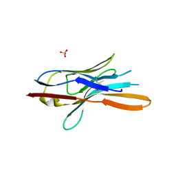
Structure of nanobody nb18 raised against TssK from E. coli T6SS
Descriptor: Llama nanobody nb8 against TssK from T6SS, SULFATE ION
Authors:Cambillau, C, Nguyen, V.S, Spinelli, S, Desmyter, A.
Deposit date:2016-10-13
Release date:2017-06-28
Last modified:2017-08-30
Method:X-RAY DIFFRACTION (1.5 Å)
Cite:Type VI secretion TssK baseplate protein exhibits structural similarity with phage receptor-binding proteins and evolved to bind the membrane complex.
Nat Microbiol, 2, 2017
5MWN
DownloadVisualize
BU of 5mwn by Molmil
Structure of the EAEC T6SS component TssK N-terminal domain in complex with llama nanobodies nbK18 and nbK27
Descriptor: PHOSPHATE ION, Type VI secretion protein, llama nanobody raised against TssK, ...
Authors:Cambillau, C, Nguyen, V.S, Spinelli, S, Legrand, P.
Deposit date:2017-01-18
Release date:2017-06-28
Last modified:2024-10-16
Method:X-RAY DIFFRACTION (2.2 Å)
Cite:Type VI secretion TssK baseplate protein exhibits structural similarity with phage receptor-binding proteins and evolved to bind the membrane complex.
Nat Microbiol, 2, 2017
5M2Y
DownloadVisualize
BU of 5m2y by Molmil
Structure of TssK C-terminal domain from E. coli T6SS
Descriptor: TssK C
Authors:Cambillau, C, Nguyen, V.S, Spinelli, S, Desmyter, A, Legrand, P.
Deposit date:2016-10-13
Release date:2017-06-28
Last modified:2024-01-17
Method:X-RAY DIFFRACTION (1.61 Å)
Cite:Type VI secretion TssK baseplate protein exhibits structural similarity with phage receptor-binding proteins and evolved to bind the membrane complex.
Nat Microbiol, 2, 2017

226707

건을2024-10-30부터공개중

PDB statisticsPDBj update infoContact PDBjnumon