8AH8
 
 | |
8AH4
 | |
8AH7
 | |
4J9E
 | |
4J9B
 | |
4J9D
 | |
4JJB
 | |
4J9H
 | |
4J9C
 | |
4J9G
 | |
4JJD
 | |
3EG2
 | |
3EG0
 | |
3EG1
 | |
3EGU
 | |
3EG3
 | |
6S7N
 | |
6SYC
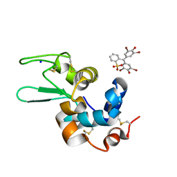
 | | Crystal structure of the lysozyme in presence of bromophenol blue at pH 6.5 | | 分子名称: | CHLORIDE ION, IMIDAZOLE, Lysozyme, ... | | 著者 | Camara-Artigas, A, Plaza-Garrido, M, Salinas-Garcia, M.C. | | 登録日 | 2019-09-27 | | 公開日 | 2020-09-09 | | 最終更新日 | 2024-01-24 | | 実験手法 | X-RAY DIFFRACTION (1.38 Å) | | 主引用文献 | Lysozyme crystals dyed with bromophenol blue: where has the dye gone?
Acta Crystallogr D Struct Biol, 76, 2020
|
|
6SYD
 | |
6SYE
 | |
1M3X
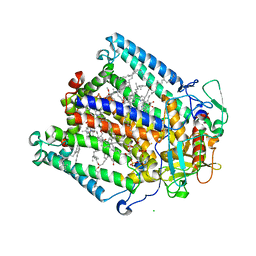
 | | Photosynthetic Reaction Center From Rhodobacter Sphaeroides | | 分子名称: | 1,2-DIACYL-SN-GLYCERO-3-PHOSPHOCHOLINE, BACTERIOCHLOROPHYLL A, BACTERIOPHEOPHYTIN A, ... | | 著者 | Camara-Artigas, A, Brune, D, Allen, J.P. | | 登録日 | 2002-07-01 | | 公開日 | 2002-08-28 | | 最終更新日 | 2024-02-14 | | 実験手法 | X-RAY DIFFRACTION (2.55 Å) | | 主引用文献 | Interactions between lipids and bacterial reaction centers determined by protein crystallography.
Proc.Natl.Acad.Sci.USA, 99, 2002
|
|
1KBY
 | | Structure of Photosynthetic Reaction Center with bacteriochlorophyll-bacteriopheophytin heterodimer | | 分子名称: | BACTERIOCHLOROPHYLL A, BACTERIOPHEOPHYTIN A, CARDIOLIPIN, ... | | 著者 | Camara-Artigas, A, Magee, C, Goetsch, A, Allen, J.P. | | 登録日 | 2001-11-07 | | 公開日 | 2002-11-13 | | 最終更新日 | 2024-02-07 | | 実験手法 | X-RAY DIFFRACTION (2.5 Å) | | 主引用文献 | The structure of the heterodimer reaction center from Rhodobacter sphaeroides at 2.55 a resolution.
Photosynth.Res., 74, 2002
|
|
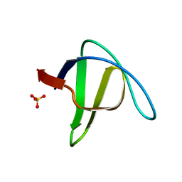
2F2V
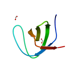 | | alpha-spectrin SH3 domain A56G mutant | | 分子名称: | FORMIC ACID, Spectrin alpha chain, brain | | 著者 | Camara-Artigas, A, Conejero-Lara, F, Casares, S, Lopez-Mayorga, O, Vega, C. | | 登録日 | 2005-11-18 | | 公開日 | 2006-10-31 | | 最終更新日 | 2023-08-23 | | 実験手法 | X-RAY DIFFRACTION (1.85 Å) | | 主引用文献 | Cooperative propagation of local stability changes from low-stability and high-stability regions in a SH3 domain
Proteins, 67, 2007
|
|
2F2W
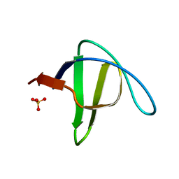 | | alpha-spectrin SH3 domain R21A mutant | | 分子名称: | SULFATE ION, Spectrin alpha chain, brain | | 著者 | Camara-Artigas, A, Conejero-Lara, F, Casares, S, Lopez-Mayorga, O, Vega, C. | | 登録日 | 2005-11-18 | | 公開日 | 2006-10-31 | | 最終更新日 | 2023-08-23 | | 実験手法 | X-RAY DIFFRACTION (1.7 Å) | | 主引用文献 | Cooperative propagation of local stability changes from low-stability and high-stability regions in a SH3 domain
Proteins, 67, 2007
|
|
2F2X
 | | alpha-spectrin SH3 domain R21G mutant | | 分子名称: | SULFATE ION, Spectrin alpha chain, brain | | 著者 | Camara-Artigas, A, Conejero-Lara, F, Casares, S, Lopez-Mayorga, O, Vega, C. | | 登録日 | 2005-11-18 | | 公開日 | 2006-10-31 | | 最終更新日 | 2023-08-23 | | 実験手法 | X-RAY DIFFRACTION (1.6 Å) | | 主引用文献 | Cooperative propagation of local stability changes from low-stability and high-stability regions in a SH3 domain
Proteins, 67, 2007
|
|


