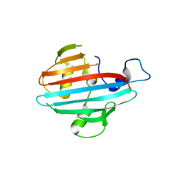
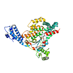
5AIP
 
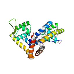 | | Crystal structure of NadR in complex with 4-hydroxyphenylacetate | | 分子名称: | 4-HYDROXYPHENYLACETATE, TRANSCRIPTIONAL REGULATOR, MARR FAMILY | | 著者 | Liguori, A, Malito, E, Bottomley, M.J. | | 登録日 | 2015-02-16 | | 公開日 | 2016-03-02 | | 最終更新日 | 2024-01-10 | | 実験手法 | X-RAY DIFFRACTION (2.3 Å) | | 主引用文献 | Molecular Basis of Ligand-Dependent Regulation of Nadr, the Transcriptional Repressor of Meningococcal Virulence Factor Nada.
Plos Pathog., 12, 2016
|
|
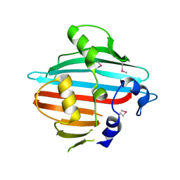
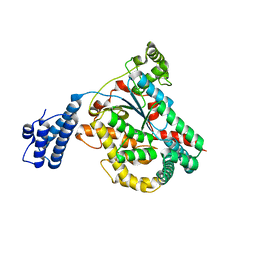
1OQJ
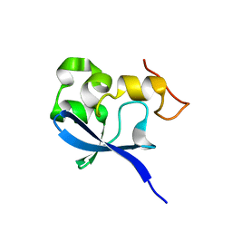 | | Crystal structure of the SAND domain from glucocorticoid modulatory element binding protein-1 (GMEB1) | | 分子名称: | Glucocorticoid Modulatory Element Binding protein-1, ZINC ION | | 著者 | Surdo, P.L, Bottomley, M.J, Sattler, M, Scheffzek, K. | | 登録日 | 2003-03-10 | | 公開日 | 2003-11-11 | | 最終更新日 | 2024-02-14 | | 実験手法 | X-RAY DIFFRACTION (1.55 Å) | | 主引用文献 | Crystal structure and nuclear magnetic resonance analyses of the SAND domain from glucocorticoid modulatory element binding protein-1 reveals deoxyribonucleic acid and zinc binding regions
MOL.ENDOCRINOL., 17, 2003
|
|
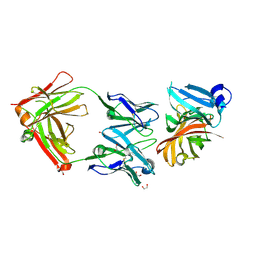
3P5B
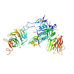 | | The structure of the LDLR/PCSK9 complex reveals the receptor in an extended conformation | | 分子名称: | CALCIUM ION, Low density lipoprotein receptor variant, Proprotein convertase subtilisin/kexin type 9 | | 著者 | Lo Surdo, P, Bottomley, M.J, Calzetta, A, Settembre, E.C, Cirillo, A, Pandit, S, Ni, Y, Hubbard, B, Sitlani, A, Carfi, A. | | 登録日 | 2010-10-08 | | 公開日 | 2011-10-26 | | 最終更新日 | 2017-11-08 | | 実験手法 | X-RAY DIFFRACTION (3.3 Å) | | 主引用文献 | Mechanistic implications for LDL receptor degradation from the PCSK9/LDLR structure at neutral pH.
Embo Rep., 12, 2011
|
|
3P5C
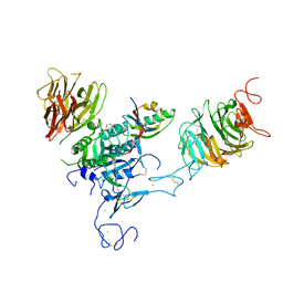 | | The structure of the LDLR/PCSK9 complex reveals the receptor in an extended conformation | | 分子名称: | CALCIUM ION, Low density lipoprotein receptor variant, Proprotein convertase subtilisin/kexin type 9 | | 著者 | Lo Surdo, P, Bottomley, M.J, Calzetta, A, Settembre, E.C, Cirillo, A, Pandit, S, Ni, Y, Hubbard, B, Sitlani, A, Carfi, A. | | 登録日 | 2010-10-08 | | 公開日 | 2011-10-26 | | 最終更新日 | 2024-01-17 | | 実験手法 | X-RAY DIFFRACTION (4.2 Å) | | 主引用文献 | Mechanistic implications for LDL receptor degradation from the PCSK9/LDLR structure at neutral pH.
Embo Rep., 12, 2011
|
|
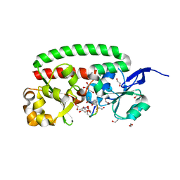
4BIH
 | | Crystal structure of the conserved staphylococcal antigen 1A, Csa1A | | 分子名称: | CALCIUM ION, UNCHARACTERIZED LIPOPROTEIN SAOUHSC_00053 | | 著者 | Malito, E, Bottomley, M.J, Spraggon, G, Schluepen, C, Liberatori, S. | | 登録日 | 2013-04-10 | | 公開日 | 2013-08-07 | | 最終更新日 | 2023-12-20 | | 実験手法 | X-RAY DIFFRACTION (2.459 Å) | | 主引用文献 | Mining the Bacterial Unknown Proteome: Identification and Characterization of a Novel Family of Highly Conserved Protective Antigens in Staphylococcus Aureus
Biochem.J., 455, 2013
|
|
4BIG
 | | Crystal structure of the conserved staphylococcal antigen 1B, Csa1B | | 分子名称: | UNCHARACTERIZED LIPOPROTEIN SAOUHSC_00053 | | 著者 | Malito, E, Bottomley, M.J, Schluepen, C, Liberatori, S. | | 登録日 | 2013-04-10 | | 公開日 | 2013-08-07 | | 最終更新日 | 2013-10-30 | | 実験手法 | X-RAY DIFFRACTION (2.274 Å) | | 主引用文献 | Mining the Bacterial Unknown Proteome: Identification and Characterization of a Novel Family of Highly Conserved Protective Antigens in Staphylococcus Aureus
Biochem.J., 455, 2013
|
|
4B8Y
 | | Ferrichrome-bound FhuD2 | | 分子名称: | 1,2-ETHANEDIOL, CHLORIDE ION, FE (III) ION, ... | | 著者 | Malito, E, Bottomley, M.J, Spraggon, G. | | 登録日 | 2012-08-31 | | 公開日 | 2012-11-21 | | 最終更新日 | 2023-12-20 | | 実験手法 | X-RAY DIFFRACTION (1.9 Å) | | 主引用文献 | Structural and Functional Characterization of the Staphylococcus Aureus Virulence Factor and Vaccine Candidate Fhud2.
Biochem.J., 449, 2013
|
|
4CJD
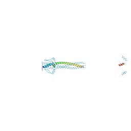 | | Crystal structure of Neisseria meningitidis trimeric autotransporter and vaccine antigen NadA | | 分子名称: | IODIDE ION, NADA | | 著者 | Malito, E, Biancucci, M, Spraggon, G, Bottomley, M.J. | | 登録日 | 2013-12-19 | | 公開日 | 2014-11-26 | | 最終更新日 | 2024-05-08 | | 実験手法 | X-RAY DIFFRACTION (2.056 Å) | | 主引用文献 | Structure of the Meningococcal Vaccine Antigen Nada and Epitope Mapping of a Bactericidal Antibody.
Proc.Natl.Acad.Sci.USA, 111, 2014
|
|
4DMW
 | | Crystal structure of the GT domain of Clostridium difficile toxin A (TcdA) in complex with UDP and Manganese | | 分子名称: | MANGANESE (II) ION, Toxin A, URIDINE-5'-DIPHOSPHATE | | 著者 | Malito, E, D'Urzo, N, Bottomley, M.J, Biancucci, M, Scarselli, M, Maione, D, Martinelli, M. | | 登録日 | 2012-02-08 | | 公開日 | 2012-07-18 | | 最終更新日 | 2023-09-13 | | 実験手法 | X-RAY DIFFRACTION (2.5 Å) | | 主引用文献 | The structure of Clostridium difficile toxin A glucosyltransferase domain bound to Mn2+ and UDP provides insights into glucosyltransferase activity and product release.
Febs J., 279, 2012
|
|
4DMV
 | | Crystal structure of the GT domain of Clostridium difficile Toxin A | | 分子名称: | Toxin A | | 著者 | Malito, E, D'Urzo, N, Bottomley, M.J, Biancucci, M, Maione, D, Scarselli, M, Martinelli, M. | | 登録日 | 2012-02-08 | | 公開日 | 2012-07-18 | | 最終更新日 | 2023-09-13 | | 実験手法 | X-RAY DIFFRACTION (1.5 Å) | | 主引用文献 | The structure of Clostridium difficile toxin A glucosyltransferase domain bound to Mn2+ and UDP provides insights into glucosyltransferase activity and product release.
Febs J., 279, 2012
|
|
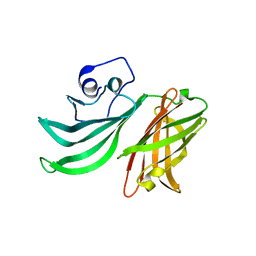
2YPV
 | | Crystal structure of the Meningococcal vaccine antigen factor H binding protein in complex with a bactericidal antibody | | 分子名称: | 1,2-ETHANEDIOL, FAB 12C1, LIPOPROTEIN | | 著者 | Malito, E, Veggi, D, Bottomley, M.J. | | 登録日 | 2012-11-01 | | 公開日 | 2013-02-20 | | 最終更新日 | 2023-12-20 | | 実験手法 | X-RAY DIFFRACTION (1.8 Å) | | 主引用文献 | Defining a Protective Epitope on Factor H Binding Protein, a Key Meningococcal Virulence Factor and Vaccine Antigen.
Proc.Natl.Acad.Sci.USA, 110, 2013
|
|
2Y7S
 | |
4D7W
 | | Crystal structure of sortase C1 (SrtC1) from Streptococcus agalactiae | | 分子名称: | 1,2-ETHANEDIOL, SORTASE FAMILY PROTEIN | | 著者 | Malito, E, Lazzarin, M, Cozzi, R, Bottomley, M.J. | | 登録日 | 2014-11-28 | | 公開日 | 2015-08-19 | | 最終更新日 | 2023-12-20 | | 実験手法 | X-RAY DIFFRACTION (1.95 Å) | | 主引用文献 | Noncanonical Sortase-Mediated Assembly of Pilus Type 2B in Group B Streptococcus.
Faseb J., 29, 2015
|
|


