8AYU
 
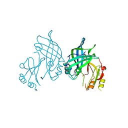 | | Crystal structure of SUDV VP40 L117A mutant | | 分子名称: | Matrix protein VP40 | | 著者 | Werner, A.-D, Steinchen, W, Werel, L, Kowalski, K, Essen, L.-O, Becker, S. | | 登録日 | 2022-09-03 | | 公開日 | 2023-09-13 | | 最終更新日 | 2024-04-24 | | 実験手法 | X-RAY DIFFRACTION (2 Å) | | 主引用文献 | Crystal structure of SUDV VP40 L117A mutant
To Be Published
|
|
5ABS
 
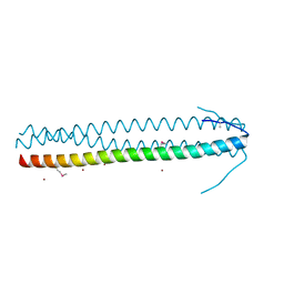 | | CRYSTAL STRUCTURE OF THE C-TERMINAL COILED-COIL DOMAIN OF CIN85 IN SPACE GROUP P321 | | 分子名称: | SH3 DOMAIN-CONTAINING KINASE-BINDING PROTEIN 1, ZINC ION | | 著者 | Wong, L, Habeck, M, Griesinger, C, Becker, S. | | 登録日 | 2015-08-07 | | 公開日 | 2016-07-13 | | 最終更新日 | 2019-02-06 | | 実験手法 | X-RAY DIFFRACTION (1.74 Å) | | 主引用文献 | The Adaptor Protein Cin85 Assembles Intracellular Signaling Clusters for B Cell Activation.
Sci.Signal., 9, 2016
|
|
4UN2
 
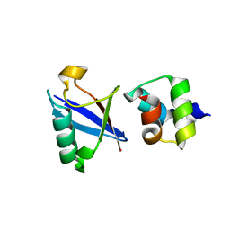 | | Crystal structure of the UBA domain of Dsk2 in complex with Ubiquitin | | 分子名称: | UBIQUITIN, UBIQUITIN DOMAIN-CONTAINING PROTEIN DSK2 | | 著者 | Michielssens, S, Peters, J.H, Ban, D, Pratihar, S, Seeliger, D, Sharma, M, Giller, K, Sabo, T.M, Becker, S, Lee, D, Griesinger, C, de Groot, B.L. | | 登録日 | 2014-05-23 | | 公開日 | 2014-08-27 | | 最終更新日 | 2024-01-10 | | 実験手法 | X-RAY DIFFRACTION (1.51 Å) | | 主引用文献 | A Designed Conformational Shift to Control Protein Binding Specificity.
Angew.Chem.Int.Ed.Engl., 53, 2014
|
|
3UFE
 
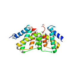 | | Structure of transcriptional antiterminator (BGLG-family) at 1.5 A resolution | | 分子名称: | CHLORIDE ION, GLYCEROL, PHOSPHATE ION, ... | | 著者 | Grosse, C, Himmel, S, Becker, S, Sheldrick, G.M, Uson, I. | | 登録日 | 2011-11-01 | | 公開日 | 2012-02-01 | | 最終更新日 | 2023-09-13 | | 実験手法 | X-RAY DIFFRACTION (1.5 Å) | | 主引用文献 | Structure of transcriptional antiterminator (BGLG-family) at 1.5 A resolution
To be Published
|
|
2W0N
 
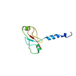 | | Plasticity of PAS domain and potential role for signal transduction in the histidine-kinase DcuS | | 分子名称: | SENSOR PROTEIN DCUS | | 著者 | Etzkorn, M, Kneuper, H, Duennwald, P, Vijayan, V, Kraemer, J, Griesinger, C, Becker, S, Unden, G, Baldus, M. | | 登録日 | 2008-08-19 | | 公開日 | 2008-09-30 | | 最終更新日 | 2024-05-15 | | 実験手法 | SOLID-STATE NMR | | 主引用文献 | Plasticity of the Pas Domain and a Potential Role for Signal Transduction in the Histidine Kinase Dcus.
Nat.Struct.Mol.Biol., 15, 2008
|
|
4MHX
 
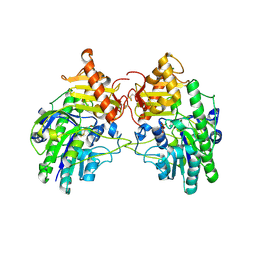 | | Crystal Structure of Sulfamidase | | 分子名称: | 2-acetamido-2-deoxy-beta-D-glucopyranose, 2-acetamido-2-deoxy-beta-D-glucopyranose-(1-4)-2-acetamido-2-deoxy-beta-D-glucopyranose, CALCIUM ION, ... | | 著者 | Sidhu, N.S, Uson, I, Schreiber, K, Proepper, K, Becker, S, Gaertner, J, Kraetzner, R, Steinfeld, R, Sheldrick, G.M. | | 登録日 | 2013-08-30 | | 公開日 | 2014-05-14 | | 最終更新日 | 2021-06-02 | | 実験手法 | X-RAY DIFFRACTION (2 Å) | | 主引用文献 | Structure of sulfamidase provides insight into the molecular pathology of mucopolysaccharidosis IIIA.
Acta Crystallogr.,Sect.D, 70, 2014
|
|
8B1S
 
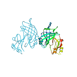 | | co-crystal of SUDV VP40 with salicylic acid | | 分子名称: | 2-HYDROXYBENZOIC ACID, Matrix protein VP40 | | 著者 | Werner, A.-D, Krapoth, N, Norris, M.J, Heine, A, Klebe, G, Ollmann Saphire, E, Becker, S. | | 登録日 | 2022-09-12 | | 公開日 | 2023-09-27 | | 最終更新日 | 2024-08-14 | | 実験手法 | X-RAY DIFFRACTION (1.6 Å) | | 主引用文献 | Development of a Crystallographic Screening to Identify Sudan Virus VP40 Ligands.
Acs Omega, 9, 2024
|
|
8B2U
 
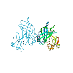 | | Crystal structure of SUDV VP40 in complex with salicylic acid | | 分子名称: | 2-HYDROXYBENZOIC ACID, Matrix protein VP40 | | 著者 | Werner, A.-D, Krapoth, N, Norris, M.J, Heine, A, Klebe, G, Ollmann Saphire, E, Becker, S. | | 登録日 | 2022-09-14 | | 公開日 | 2023-09-27 | | 最終更新日 | 2024-08-14 | | 実験手法 | X-RAY DIFFRACTION (1.8 Å) | | 主引用文献 | Development of a Crystallographic Screening to Identify Sudan Virus VP40 Ligands.
Acs Omega, 9, 2024
|
|
8OWJ
 
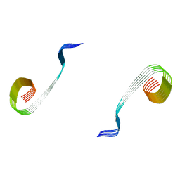 | | Lipidic amyloid-beta(1-40) fibril - polymorph L2-L2 | | 分子名称: | Amyloid-beta A4 protein | | 著者 | Frieg, B, Han, M, Giller, K, Dienemann, C, Riedel, D, Becker, S, Andreas, L.B, Griesinger, C, Schroeder, G.F. | | 登録日 | 2023-04-28 | | 公開日 | 2024-03-06 | | 実験手法 | ELECTRON MICROSCOPY (3.75 Å) | | 主引用文献 | Cryo-EM structures of lipidic fibrils of amyloid-beta (1-40).
Nat Commun, 15, 2024
|
|
8OVM
 
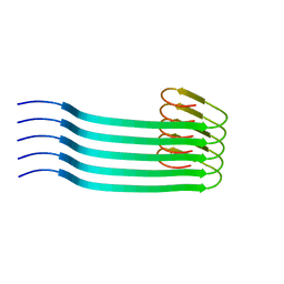 | | Lipidic amyloid-beta(1-40) fibril - polymorph L2 | | 分子名称: | Amyloid-beta A4 protein | | 著者 | Frieg, B, Han, M, Giller, K, Dienemann, C, Riedel, D, Becker, S, Andreas, L.B, Griesinger, C, Schroeder, G.F. | | 登録日 | 2023-04-26 | | 公開日 | 2024-03-06 | | 実験手法 | ELECTRON MICROSCOPY (3.24 Å) | | 主引用文献 | Cryo-EM structures of lipidic fibrils of amyloid-beta (1-40).
Nat Commun, 15, 2024
|
|
8OWE
 
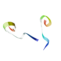 | | Lipidic amyloid-beta(1-40) fibril - polymorph L2-L3 | | 分子名称: | Amyloid-beta A4 protein | | 著者 | Frieg, B, Han, M, Giller, K, Dienemann, C, Riedel, D, Becker, S, Andreas, L.B, Griesinger, C, Schroeder, G.F. | | 登録日 | 2023-04-27 | | 公開日 | 2024-03-06 | | 実験手法 | ELECTRON MICROSCOPY (3.75 Å) | | 主引用文献 | Cryo-EM structures of lipidic fibrils of amyloid-beta (1-40).
Nat Commun, 15, 2024
|
|
8OVK
 
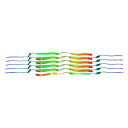 | | Lipidic amyloid-beta(1-40) fibril - polymorph L1 | | 分子名称: | Amyloid-beta A4 protein | | 著者 | Frieg, B, Han, M, Giller, K, Dienemann, C, Riedel, D, Becker, S, Andreas, L.B, Griesinger, C, Schroeder, G.F. | | 登録日 | 2023-04-26 | | 公開日 | 2024-03-06 | | 実験手法 | ELECTRON MICROSCOPY (2.88 Å) | | 主引用文献 | Cryo-EM structures of lipidic fibrils of amyloid-beta (1-40).
Nat Commun, 15, 2024
|
|
8OWK
 
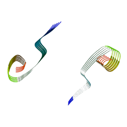 | | Lipidic amyloid-beta(1-40) fibril - polymorph L3-L3 | | 分子名称: | Amyloid-beta A4 protein | | 著者 | Frieg, B, Han, M, Giller, K, Dienemann, C, Riedel, D, Becker, S, Andreas, L.B, Griesinger, C, Schroeder, G.F. | | 登録日 | 2023-04-28 | | 公開日 | 2024-03-06 | | 実験手法 | ELECTRON MICROSCOPY (3.86 Å) | | 主引用文献 | Cryo-EM structures of lipidic fibrils of amyloid-beta (1-40).
Nat Commun, 15, 2024
|
|
8OWD
 
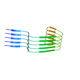 | | Lipidic amyloid-beta(1-40) fibril - polymorph L3 | | 分子名称: | Amyloid-beta A4 protein | | 著者 | Frieg, B, Han, M, Giller, K, Dienemann, C, Riedel, D, Becker, S, Andreas, L.B, Griesinger, C, Schroeder, G.F. | | 登録日 | 2023-04-27 | | 公開日 | 2024-03-06 | | 実験手法 | ELECTRON MICROSCOPY (3.28 Å) | | 主引用文献 | Cryo-EM structures of lipidic fibrils of amyloid-beta (1-40).
Nat Commun, 15, 2024
|
|
6GEZ
 
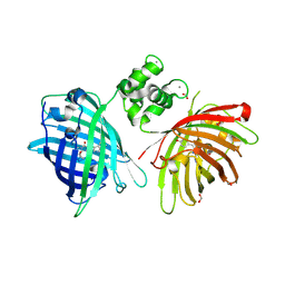 | | THE STRUCTURE OF TWITCH-2B N532F | | 分子名称: | CALCIUM ION, FORMIC ACID, Green fluorescent protein,Optimized Ratiometric Calcium Sensor,Green fluorescent protein,Green fluorescent protein | | 著者 | Trigo Mourino, P, Paulat, M, Thestrup, T, Griesbeck, O, Griesinger, C, Becker, S. | | 登録日 | 2018-04-27 | | 公開日 | 2019-08-21 | | 最終更新日 | 2024-10-16 | | 実験手法 | X-RAY DIFFRACTION (2.47 Å) | | 主引用文献 | Dynamic tuning of FRET in a green fluorescent protein biosensor.
Sci Adv, 5, 2019
|
|
6GEL
 
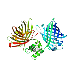 | | The structure of TWITCH-2B | | 分子名称: | CALCIUM ION, FORMIC ACID, GLYCEROL, ... | | 著者 | Trigo Mourino, P, Paulat, M, Thestrup, T, Griesbeck, O, Griesinger, C, Becker, S. | | 登録日 | 2018-04-26 | | 公開日 | 2019-08-21 | | 最終更新日 | 2019-09-11 | | 実験手法 | X-RAY DIFFRACTION (2.51 Å) | | 主引用文献 | Dynamic tuning of FRET in a green fluorescent protein biosensor.
Sci Adv, 5, 2019
|
|
3GWH
 
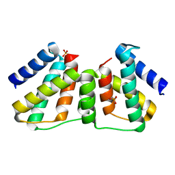 | | Crystallographic Ab Initio protein solution far below atomic resolution | | 分子名称: | PHOSPHATE ION, Transcriptional antiterminator (BglG family) | | 著者 | Rodriguez, D.D, Grosse, C, Himmel, S, Gonzalez, C, Becker, S, Sheldrick, G.M, Uson, I. | | 登録日 | 2009-04-01 | | 公開日 | 2010-04-07 | | 最終更新日 | 2024-03-20 | | 実験手法 | X-RAY DIFFRACTION (1.95 Å) | | 主引用文献 | Crystallographic ab initio protein structure solution below atomic resolution
Nat.Methods, 6, 2009
|
|
2UVS
 
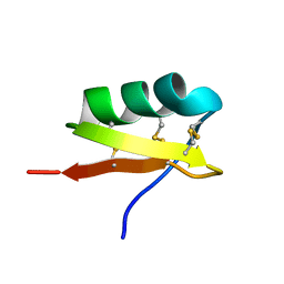 | | High Resolution Solid-state NMR structure of Kaliotoxin | | 分子名称: | POTASSIUM CHANNEL TOXIN ALPHA-KTX 3.1 | | 著者 | Korukottu, J, Lange, A, Vijayan, V, Schneider, R, Pongs, O, Becker, S, Baldus, M, Zweckstetter, M. | | 登録日 | 2007-03-14 | | 公開日 | 2008-05-27 | | 最終更新日 | 2020-01-15 | | 実験手法 | SOLID-STATE NMR | | 主引用文献 | Conformational Plasticity in Ion Channel Recognition of a Peptide Toxin
To be Published
|
|
7ABT
 
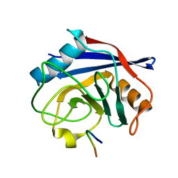 | |
1XSW
 
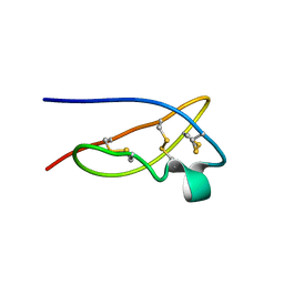 | | The solid-state NMR structure of Kaliotoxin | | 分子名称: | Kaliotoxin 1 | | 著者 | Lange, A, Becker, S, Seidel, K, Giller, K, Pongs, O, Baldus, M. | | 登録日 | 2004-10-20 | | 公開日 | 2005-04-05 | | 最終更新日 | 2022-03-02 | | 実験手法 | SOLID-STATE NMR | | 主引用文献 | A Concept for Rapid Protein-Structure Determination by Solid-State NMR Spectroscopy
Angew.Chem.Int.Ed.Engl., 44, 2005
|
|
2V9A
 
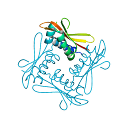 | | Structure of Citrate-free Periplasmic Domain of Sensor Histidine Kinase CitA | | 分子名称: | SENSOR KINASE CITA | | 著者 | Sevvana, M, Vijayan, V, Zweckstetter, M, Reinelt, S, Madden, D.R, Sheldrick, G.M, Bott, M, Griesinger, C, Becker, S. | | 登録日 | 2007-08-23 | | 公開日 | 2008-03-25 | | 最終更新日 | 2023-12-13 | | 実験手法 | X-RAY DIFFRACTION (2 Å) | | 主引用文献 | A Ligand-Induced Switch in the Periplasmic Domain of Sensor Histidine Kinase Cita.
J.Mol.Biol., 377, 2008
|
|
4C4Z
 
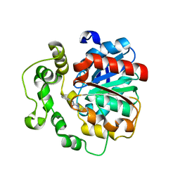 | | Crystal structure of human bifunctional epoxide hydroxylase 2 complexed with A8 | | 分子名称: | 1-ethyl-3-naphthalen-1-ylurea, BIFUNCTIONAL EPOXIDE HYDROLASE 2 | | 著者 | Pilger, J, Mazur, A, Monecke, P, Schreuder, H, Elshorst, B, Langer, T, Schiffer, A, Krimm, I, Wegstroth, M, Lee, D, Hessler, G, Wendt, K.-U, Becker, S, Griesinger, C. | | 登録日 | 2013-09-09 | | 公開日 | 2014-10-01 | | 最終更新日 | 2024-05-08 | | 実験手法 | X-RAY DIFFRACTION (2.06 Å) | | 主引用文献 | A Combination of Spin Diffusion Methods for the Determination of Protein-Ligand Complex Structural Ensembles.
Angew.Chem.Int.Ed.Engl., 54, 2015
|
|
4C4Y
 
 | | Crystal structure of human bifunctional epoxide hydroxylase 2 complexed with A4 | | 分子名称: | 1-(3-chlorophenyl)-3-(2-methoxyethyl)urea, BIFUNCTIONAL EPOXIDE HYDROLASE 2 | | 著者 | Pilger, J, Mazur, A, Monecke, P, Schreuder, H, Elshorst, B, Langer, T, Schiffer, A, Krimm, I, Wegstroth, M, Lee, D, Hessler, G, Wendt, K.-U, Becker, S, Griesinger, C. | | 登録日 | 2013-09-09 | | 公開日 | 2014-10-01 | | 最終更新日 | 2023-12-20 | | 実験手法 | X-RAY DIFFRACTION (2.41 Å) | | 主引用文献 | A Combination of Spin Diffusion Methods for the Determination of Protein-Ligand Complex Structural Ensembles.
Angew.Chem.Int.Ed.Engl., 54, 2015
|
|
4C4X
 
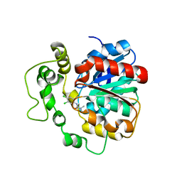 | | Crystal structure of human bifunctional epoxide hydroxylase 2 complexed with C9 | | 分子名称: | 3-(3,4-dichlorophenyl)-1,1-dimethyl-urea, BIFUNCTIONAL EPOXIDE HYDROLASE 2 | | 著者 | Pilger, J, Mazur, A, Monecke, P, Schreuder, H, Elshorst, B, Langer, T, Schiffer, A, Krimm, I, Wegstroth, M, Lee, D, Hessler, G, Wendt, K.-U, Becker, S, Griesinger, C. | | 登録日 | 2013-09-09 | | 公開日 | 2014-10-01 | | 最終更新日 | 2023-12-20 | | 実験手法 | X-RAY DIFFRACTION (2.17 Å) | | 主引用文献 | A Combination of Spin Diffusion Methods for the Determination of Protein-Ligand Complex Structural Ensembles.
Angew.Chem.Int.Ed.Engl., 54, 2015
|
|
1GL2
 
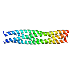 | | Crystal structure of an endosomal SNARE core complex | | 分子名称: | ENDOBREVIN, SYNTAXIN 7, SYNTAXIN 8, ... | | 著者 | Antonin, W, Becker, S, Jahn, R, Schneider, T.R. | | 登録日 | 2001-08-22 | | 公開日 | 2002-01-15 | | 最終更新日 | 2023-12-13 | | 実験手法 | X-RAY DIFFRACTION (1.9 Å) | | 主引用文献 | Crystal Structure of the Endosomal Snare Complex Reveals Common Structural Principles of All Snares.
Nat.Struct.Biol., 9, 2001
|
|
