2FKF
 
 | |
2H5A
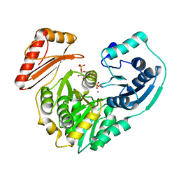
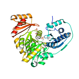
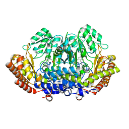
 | | Complex of the enzyme PMM/PGM with xylose 1-phosphate | | 分子名称: | 1-O-phosphono-alpha-D-xylopyranose, Phosphomannomutase/phosphoglucomutase, ZINC ION | | 著者 | Regni, C, Shackelford, G.S, Beamer, L.J. | | 登録日 | 2006-05-25 | | 公開日 | 2006-08-08 | | 最終更新日 | 2020-07-29 | | 実験手法 | X-RAY DIFFRACTION (1.72 Å) | | 主引用文献 | Complexes of the enzyme phosphomannomutase/phosphoglucomutase with a slow substrate and an inhibitor.
Acta Crystallogr.,Sect.F, 62, 2006
|
|
6UIQ
 | |
6UXJ
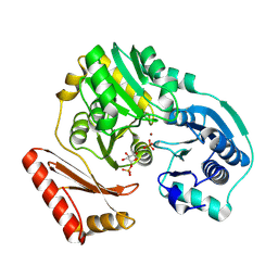
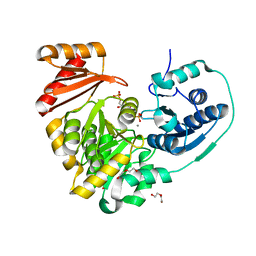
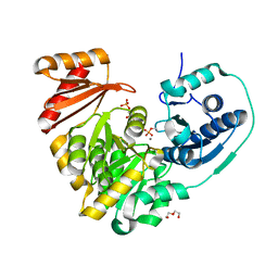
 | | Structure of serine hydroxymethyltransferase 8 from Glycine max cultivar Essex complexed with PLP-glycine and 5-formyltetrahydrofolate | | 分子名称: | 1,2-ETHANEDIOL, N-GLYCINE-[3-HYDROXY-2-METHYL-5-PHOSPHONOOXYMETHYL-PYRIDIN-4-YL-METHANE], N-[4-({[(6S)-2-amino-5-formyl-4-oxo-3,4,5,6,7,8-hexahydropteridin-6-yl]methyl}amino)benzoyl]-L-glutamic acid, ... | | 著者 | Korasick, D.A, Tanner, J.J, Beamer, L.J. | | 登録日 | 2019-11-07 | | 公開日 | 2020-02-12 | | 最終更新日 | 2023-10-11 | | 実験手法 | X-RAY DIFFRACTION (1.4 Å) | | 主引用文献 | Impaired folate binding of serine hydroxymethyltransferase 8 from soybean underlies resistance to the soybean cyst nematode.
J.Biol.Chem., 295, 2020
|
|
6UXH
 | |
6UXL
 | |
6UXI
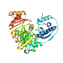
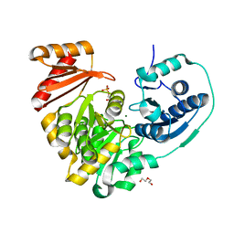
 | | Structure of serine hydroxymethyltransferase 8 from Glycine max cultivar Essex complexed with PLP-Glycine | | 分子名称: | 1,2-ETHANEDIOL, N-GLYCINE-[3-HYDROXY-2-METHYL-5-PHOSPHONOOXYMETHYL-PYRIDIN-4-YL-METHANE], Serine hydroxymethyltransferase | | 著者 | Korasick, D.A, Tanner, J.J, Beamer, L.J. | | 登録日 | 2019-11-07 | | 公開日 | 2020-02-12 | | 最終更新日 | 2023-10-11 | | 実験手法 | X-RAY DIFFRACTION (2.1 Å) | | 主引用文献 | Impaired folate binding of serine hydroxymethyltransferase 8 from soybean underlies resistance to the soybean cyst nematode.
J.Biol.Chem., 295, 2020
|
|
6UXK
 | |
6NN2
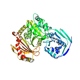
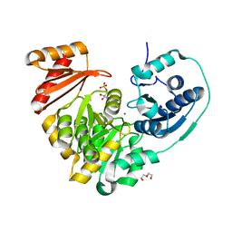
 | | Xanthomonas citri PGM Apo-Phospho | | 分子名称: | CALCIUM ION, HEXAETHYLENE GLYCOL, Phosphoglucomutase | | 著者 | Stiers, K.M, Beamer, L.J. | | 登録日 | 2019-01-14 | | 公開日 | 2019-04-10 | | 最終更新日 | 2023-10-11 | | 実験手法 | X-RAY DIFFRACTION (1.44 Å) | | 主引用文献 | Structural and dynamical description of the enzymatic reaction of a phosphohexomutase.
Struct Dyn., 6, 2019
|
|
6NNT
 | |
6NN1
 | | Xanthomonas citri PGM Apo-Dephospho | | 分子名称: | DI(HYDROXYETHYL)ETHER, MAGNESIUM ION, PHOSPHATE ION, ... | | 著者 | Stiers, K.M, Beamer, L.J. | | 登録日 | 2019-01-14 | | 公開日 | 2019-04-10 | | 最終更新日 | 2023-10-11 | | 実験手法 | X-RAY DIFFRACTION (1.5 Å) | | 主引用文献 | Structural and dynamical description of the enzymatic reaction of a phosphohexomutase.
Struct Dyn., 6, 2019
|
|
6NQF
 | |
6NQE
 | |
6NOQ
 | |
6NP8
 | |
6NNP
 | |
6NNN
 | |
6NOL
 | |
8DOM
 | |
8DSK
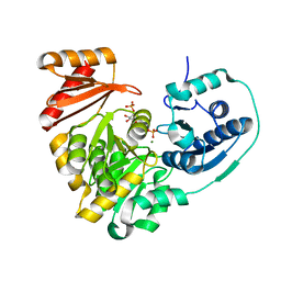
 | | Structure of the N358Y variant of serine hydroxymethyltransferase 8 in complex with PLP, glycine, and formyl tetrahydrofolate | | 分子名称: | 1,2-ETHANEDIOL, N-GLYCINE-[3-HYDROXY-2-METHYL-5-PHOSPHONOOXYMETHYL-PYRIDIN-4-YL-METHANE], N-[4-({[(6S)-2-amino-5-formyl-4-oxo-3,4,5,6,7,8-hexahydropteridin-6-yl]methyl}amino)benzoyl]-L-glutamic acid, ... | | 著者 | Korasick, D.A, Beamer, L.J. | | 登録日 | 2022-07-22 | | 公開日 | 2023-10-18 | | 最終更新日 | 2024-01-31 | | 実験手法 | X-RAY DIFFRACTION (1.63 Å) | | 主引用文献 | Structural and functional analysis of two SHMT8 variants associated with soybean cyst nematode resistance.
Febs J., 291, 2024
|
|
6NNS
 | |
6NNU
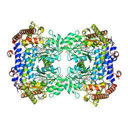
 | | Xanthomonas citri Phospho-PGM in complex with glucose-1,6-phosphate | | 分子名称: | 1,6-di-O-phosphono-alpha-D-glucopyranose, CALCIUM ION, DI(HYDROXYETHYL)ETHER, ... | | 著者 | Stiers, K.M, Beamer, L.J. | | 登録日 | 2019-01-15 | | 公開日 | 2019-04-10 | | 最終更新日 | 2023-10-11 | | 実験手法 | X-RAY DIFFRACTION (1.46 Å) | | 主引用文献 | Structural and dynamical description of the enzymatic reaction of a phosphohexomutase.
Struct Dyn., 6, 2019
|
|
6NPX
 | |
6NQG
 | |
6NQH
 | |


