Loading
PDBj
MenuPDBj@FacebookPDBj@TwitterPDBj@YouTubewwPDB FoundationwwPDB
RCSB PDBPDBeBMRBAdv. SearchSearch help
PDB: 67 results

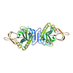
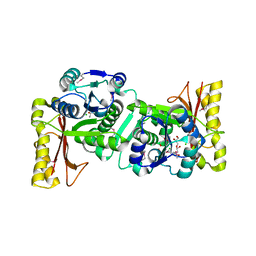
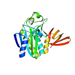
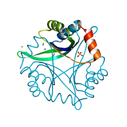
1NZA
DownloadVisualize
BU of 1nza by Molmil
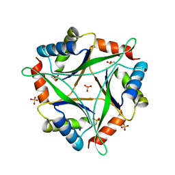
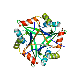
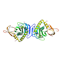
Divalent cation tolerance protein (Cut A1) from thermus thermophilus HB8
Descriptor: CHLORIDE ION, Divalent cation tolerance protein, GLYCEROL, ...
Authors:Bagautdinov, B, Miyano, M, Tahirov, T.H, RIKEN Structural Genomics/Proteomics Initiative (RSGI)
Deposit date:2003-02-17
Release date:2003-03-11
Last modified:2023-10-25
Method:X-RAY DIFFRACTION (1.7 Å)
Cite:The structures of the CutA1 proteins from Thermus thermophilus and Pyrococcus horikoshii: characterization of metal-binding sites and metal-induced assembly.
Acta Crystallogr.,Sect.F, 70, 2014
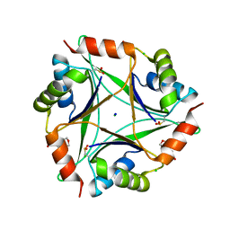
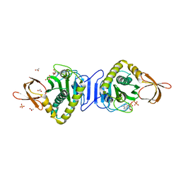
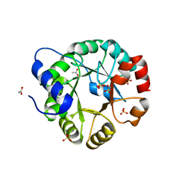
1J3N
DownloadVisualize
BU of 1j3n by Molmil
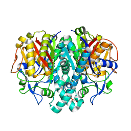
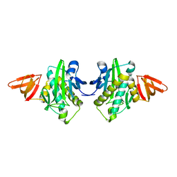
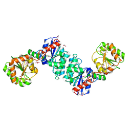
Crystal Structure of 3-oxoacyl-(acyl-carrier protein) Synthase II from Thermus thermophilus HB8
Descriptor: 3-oxoacyl-(acyl-carrier protein) synthase II, CITRIC ACID, MAGNESIUM ION
Authors:Bagautdinov, B, Miyano, M, Tahirov, T.H, RIKEN Structural Genomics/Proteomics Initiative (RSGI)
Deposit date:2003-02-10
Release date:2003-03-11
Last modified:2023-10-25
Method:X-RAY DIFFRACTION (2 Å)
Cite:Structure of 3-oxoacyl-(acyl-carrier protein) synthase II from Thermus thermophilus HB8.
Acta Crystallogr.,Sect.F, 64, 2008
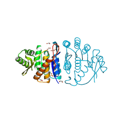
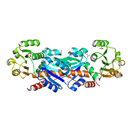
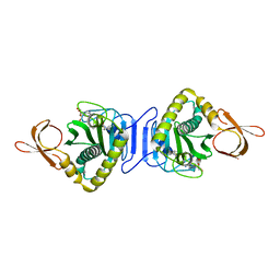
2ZGW
DownloadVisualize
BU of 2zgw by Molmil
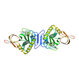
Crystal Structure Of Biotin Protein Ligase From Pyrococcus Horikoshii Complexed with Adenosine and Biotin, Mutations R48A and K111A
Descriptor: ADENOSINE, BIOTIN, biotin--[acetyl-CoA-carboxylase] ligase
Authors:Bagautdinov, B, Matsuura, Y, Bagautdinova, S, Kunishima, N, RIKEN Structural Genomics/Proteomics Initiative (RSGI)
Deposit date:2008-01-28
Release date:2008-02-05
Last modified:2023-11-01
Method:X-RAY DIFFRACTION (1.5 Å)
Cite:Protein biotinylation visualized by a complex structure of biotin protein ligase with a substrate
J.Biol.Chem., 283, 2008
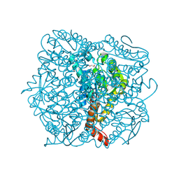
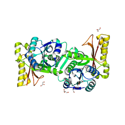
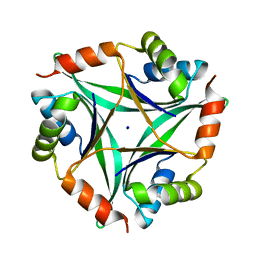
4NYP
DownloadVisualize
BU of 4nyp by Molmil
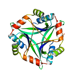
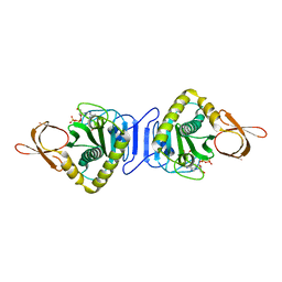
The 2.0 Angstrom Crystal Structure of Pyrococcus Horikoshii Cuta1 Complexed With NA+
Descriptor: Divalent-cation tolerance protein CutA, SODIUM ION, SULFATE ION
Authors:Bagautdinov, B, Tahirov, T.H, RIKEN Structural Genomics/Proteomics Initiative (RSGI)
Deposit date:2013-12-11
Release date:2014-01-01
Last modified:2024-10-30
Method:X-RAY DIFFRACTION (2 Å)
Cite:The 2.0 Angstrom Crystal Structure of Pyrococcus Horikoshii Complexed with Na+
To be Published
2HNI
DownloadVisualize
BU of 2hni by Molmil
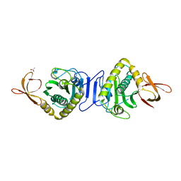
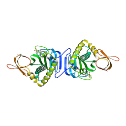
Crystal Structure Of Biotin Protein Ligase From Pyrococcus Horikoshii OT3, K111A mutation
Descriptor: 235aa long hypothetical biotin-[acetyl-CoA-carboxylase] ligase, ACETIC ACID
Authors:Bagautdinov, B, Kunishima, N, RIKEN Structural Genomics/Proteomics Initiative (RSGI)
Deposit date:2006-07-13
Release date:2007-01-13
Last modified:2023-10-25
Method:X-RAY DIFFRACTION (1.9 Å)
Cite:Ligand Structures Of Biotin Protein Ligase From Pyrococcus Horikoshii OT3
To be Published
2FYK
DownloadVisualize
BU of 2fyk by Molmil
Crystal Structure Of Biotin Protein Ligase From Pyrococcus Horikoshii OT3 in complex with ADP and Biotin
Descriptor: ADENOSINE-5'-DIPHOSPHATE, BIOTIN, biotin--protein ligase
Authors:Bagautdinov, B, Kunishima, N, RIKEN Structural Genomics/Proteomics Initiative (RSGI)
Deposit date:2006-02-08
Release date:2006-08-08
Last modified:2023-11-15
Method:X-RAY DIFFRACTION (1.6 Å)
Cite:Ligand Structures Of Biotin Protein Ligase From Pyrococcus Horikoshii Ot3
To be Published
4NYO
DownloadVisualize
BU of 4nyo by Molmil
The 1.8 Angstrom Crystal Structure of the Periplasmic Divalent Cation Tolerance Protein Cuta from Pyrococcus Horikoshii OT3
Descriptor: 2-(N-MORPHOLINO)-ETHANESULFONIC ACID, CHLORIDE ION, Divalent-cation tolerance protein CutA, ...
Authors:Bagautdinov, B, Tahirov, T.H, RIKEN Structural Genomics/Proteomics Initiative (RSGI)
Deposit date:2013-12-11
Release date:2014-01-01
Last modified:2023-11-08
Method:X-RAY DIFFRACTION (1.8 Å)
Cite:The structures of the CutA1 proteins from Thermus thermophilus and Pyrococcus horikoshii: characterization of metal-binding sites and metal-induced assembly
ACTA CRYSTALLOGR.,SECT.F, 70, 2014
1UB0
DownloadVisualize
BU of 1ub0 by Molmil
Crystal Structure Analysis of Phosphomethylpyrimidine Kinase (ThiD) from Thermus Thermophilus Hb8
Descriptor: 1,4-DIETHYLENE DIOXIDE, GLYCEROL, Phosphomethylpyrimidine Kinase, ...
Authors:Bagautdinov, B, Kuramitsu, S, Yokoyama, S, Miyano, M, Tahirov, T.H, RIKEN Structural Genomics/Proteomics Initiative (RSGI)
Deposit date:2003-03-26
Release date:2003-04-08
Last modified:2023-10-25
Method:X-RAY DIFFRACTION (2.05 Å)
Cite:Crystal Structure Analysis of Phosphomethylpyrimidine Kinase (ThiD) from Thermus Thermophilus Hb8
To be Published
1UIY
DownloadVisualize
BU of 1uiy by Molmil
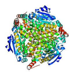
Crystal Structure of Enoyl-CoA Hydratase from Thermus Thermophilus HB8
Descriptor: 1,4-DIETHYLENE DIOXIDE, Enoyl-CoA Hydratase, GLYCEROL
Authors:Bagautdinov, B, Kuramitsu, S, Yokoyama, S, Miyano, M, Tahirov, T.H, RIKEN Structural Genomics/Proteomics Initiative (RSGI)
Deposit date:2003-07-24
Release date:2003-08-05
Last modified:2023-11-15
Method:X-RAY DIFFRACTION (2.85 Å)
Cite:Crystal structure of enoyl-CoA hydratase from Thermus thermophilus HB8.
Acta Crystallogr.,Sect.F, 77, 2021
1WQ7
DownloadVisualize
BU of 1wq7 by Molmil
Crystal Structure Of Biotin-(Acetyl-CoA-Carboxylase) ligase From Pyrococcus Horikoshii Ot3
Descriptor: biotin--[acetyl-CoA-carboxylase] ligase
Authors:Bagautdinov, B, Kunishima, N, RIKEN Structural Genomics/Proteomics Initiative (RSGI)
Deposit date:2004-09-23
Release date:2005-10-04
Last modified:2024-03-13
Method:X-RAY DIFFRACTION (1.6 Å)
Cite:Crystal Structures of Biotin Protein Ligase from Pyrococcus horikoshii OT3 and its Complexes: Structural Basis of Biotin Activation
J.Mol.Biol., 353, 2005
2ZFH
DownloadVisualize
BU of 2zfh by Molmil
Crystal structure of putative CutA1 from Homo sapiens at 2.05A resolution
Descriptor: CutA
Authors:Bagautdinov, B, Yutani, K, RIKEN Structural Genomics/Proteomics Initiative (RSGI)
Deposit date:2008-01-07
Release date:2008-01-22
Last modified:2023-11-01
Method:X-RAY DIFFRACTION (2.05 Å)
Cite:Structure of putative CutA1 from Homo sapiens determined at 2.05 A resolution.
Acta Crystallogr.,Sect.F, 64, 2008
1V6H
DownloadVisualize
BU of 1v6h by Molmil
The Trimeric Structure Of Divalent Cation Tolerance Protein CutA1 From Thermus Thermophilus HB8
Descriptor: Divalent Cation Tolerance Protein CutA1, SULFATE ION
Authors:Bagautdinov, B, Tahirov, T.H, RIKEN Structural Genomics/Proteomics Initiative (RSGI)
Deposit date:2003-11-29
Release date:2003-12-23
Last modified:2023-10-25
Method:X-RAY DIFFRACTION (1.9 Å)
Cite:The structures of the CutA1 proteins from Thermus thermophilus and Pyrococcus horikoshii: characterization of metal-binding sites and metal-induced assembly.
Acta Crystallogr.,Sect.F, 70, 2014
1WQW
DownloadVisualize
BU of 1wqw by Molmil
Crystal Structure Of Biotin Protein Ligase From Pyrococcus Horikoshii Ot3 in complex with Biotinyl-5-AMP
Descriptor: BIOTINYL-5-AMP, biotin--[acetyl-CoA-carboxylase] ligase
Authors:Bagautdinov, B, Kunishima, N, RIKEN Structural Genomics/Proteomics Initiative (RSGI)
Deposit date:2004-10-04
Release date:2005-10-04
Last modified:2023-10-25
Method:X-RAY DIFFRACTION (1.45 Å)
Cite:Crystal Structures of Biotin Protein Ligase from Pyrococcus horikoshii OT3 and its Complexes: Structural Basis of Biotin Activation
J.Mol.Biol., 353, 2005
1WPY
DownloadVisualize
BU of 1wpy by Molmil
Crystal Structure Of Biotin-(Acetyl-CoA-Carboxylase) ligase From Pyrococcus Horikoshii Ot3 in complex with biotin
Descriptor: BIOTIN, biotin--[acetyl-CoA-carboxylase] ligase
Authors:Bagautdinov, B, Kunishima, N, RIKEN Structural Genomics/Proteomics Initiative (RSGI)
Deposit date:2004-09-17
Release date:2005-10-04
Last modified:2024-10-30
Method:X-RAY DIFFRACTION (1.6 Å)
Cite:Crystal Structures of Biotin Protein Ligase from Pyrococcus horikoshii OT3 and its Complexes: Structural Basis of Biotin Activation
J.Mol.Biol., 353, 2005
1V4V
DownloadVisualize
BU of 1v4v by Molmil
Crystal Structure Of UDP-N-Acetylglucosamine 2-Epimerase From Thermus Thermophilus HB8
Descriptor: ACETIC ACID, GLYCEROL, UDP-N-Acetylglucosamine 2-Epimerase
Authors:Bagautdinov, B, Tahirov, T.H, RIKEN Structural Genomics/Proteomics Initiative (RSGI)
Deposit date:2003-11-19
Release date:2003-12-02
Last modified:2023-11-15
Method:X-RAY DIFFRACTION (1.8 Å)
Cite:The Structure Of UDP-N-Acetylglucosamine 2-Epimerase From Thermus Thermophilus HB8
To be Published
1WNL
DownloadVisualize
BU of 1wnl by Molmil
Crystal Structure Of Biotin-(Acetyl-CoA-Carboxylase) ligase From Pyrococcus Horikoshii Ot3 in complex with ADP
Descriptor: ADENOSINE-5'-DIPHOSPHATE, GLYCEROL, biotin--[acetyl-CoA-carboxylase] ligase
Authors:Bagautdinov, B, Kunishima, N, RIKEN Structural Genomics/Proteomics Initiative (RSGI)
Deposit date:2004-08-05
Release date:2005-08-16
Last modified:2024-04-03
Method:X-RAY DIFFRACTION (1.6 Å)
Cite:Crystal Structures of Biotin Protein Ligase from Pyrococcus horikoshii OT3 and its Complexes: Structural Basis of Biotin Activation
J.Mol.Biol., 353, 2005
1WZ8
DownloadVisualize
BU of 1wz8 by Molmil
Crystal Structure of Probable Enoyl-CoA Dehydratase from Thermus Thermophilus HB8
Descriptor: (4S)-2-METHYL-2,4-PENTANEDIOL, enoyl-CoA hydratase
Authors:Bagautdinov, B, Kunishima, N, RIKEN Structural Genomics/Proteomics Initiative (RSGI)
Deposit date:2005-03-02
Release date:2005-03-15
Last modified:2023-10-25
Method:X-RAY DIFFRACTION (1.8 Å)
Cite:Crystal Structure of Probable Enoyl-CoA Dehydratase from Thermus Thermophilus HB8
To be Published
1UFV
DownloadVisualize
BU of 1ufv by Molmil
Crystal Structure Of Pantothenate Synthetase From Thermus Thermophilus HB8
Descriptor: CHLORIDE ION, GLYCEROL, Pantoate-beta-alanine ligase
Authors:Bagautdinov, B, Kuramitsu, S, Yokoyama, S, Miyano, M, Tahirov, T.H, RIKEN Structural Genomics/Proteomics Initiative (RSGI)
Deposit date:2003-06-10
Release date:2003-06-24
Last modified:2024-10-30
Method:X-RAY DIFFRACTION (2.05 Å)
Cite:Crystal Structure Of Pantothenate Synthetase From Thermus Thermophilus HB8
To be Published
1X01
DownloadVisualize
BU of 1x01 by Molmil
Crystal Structure Of Biotin Protein Ligase From Pyrococcus Horikoshii Ot3 in complex with ATP
Descriptor: ACETIC ACID, ADENOSINE-5'-TRIPHOSPHATE, PHOSPHATE ION, ...
Authors:Bagautdinov, B, Kunishima, N, RIKEN Structural Genomics/Proteomics Initiative (RSGI)
Deposit date:2005-03-11
Release date:2006-05-23
Last modified:2023-10-25
Method:X-RAY DIFFRACTION (2 Å)
Cite:Protein biotinylation visualized by a complex structure of biotin protein ligase with a substrate
J.Biol.Chem., 283, 2008
1WXD
DownloadVisualize
BU of 1wxd by Molmil
Crystal Structure of Shikimate 5-Dehydrogenase (AroE) from Thermus Thermophilus HB8
Descriptor: ACETATE ION, SULFATE ION, shikimate 5-dehydrogenase
Authors:Bagautdinov, B, Kunishima, N, RIKEN Structural Genomics/Proteomics Initiative (RSGI)
Deposit date:2005-01-21
Release date:2005-02-01
Last modified:2023-10-25
Method:X-RAY DIFFRACTION (2.1 Å)
Cite:Crystal Structures of Shikimate Dehydrogenase AroE from Thermus thermophilus HB8 and its Cofactor and Substrate Complexes: Insights into the Enzymatic Mechanism
J.Mol.Biol., 373, 2007
1V8F
DownloadVisualize
BU of 1v8f by Molmil
Crystal Structure of Pantoate-beta-Alanine (Pantothenate Synthetase) from Thermus Thermophilus HB8
Descriptor: CHLORIDE ION, GLYCEROL, HEXAETHYLENE GLYCOL, ...
Authors:Bagautdinov, B, Tahirov, T.H, RIKEN Structural Genomics/Proteomics Initiative (RSGI)
Deposit date:2004-01-08
Release date:2004-01-20
Last modified:2023-10-25
Method:X-RAY DIFFRACTION (1.9 Å)
Cite:Crystal Structure Of Pantothenate Synthetase From Thermus Thermophilus HB8
To be Published
2EJ9
DownloadVisualize
BU of 2ej9 by Molmil
Crystal Structure Of Biotin Protein Ligase From Methanococcus jannaschii
Descriptor: BIOTIN, Putative biotin ligase
Authors:Bagautdinov, B, Bagautdinova, S, Kunishima, N, RIKEN Structural Genomics/Proteomics Initiative (RSGI)
Deposit date:2007-03-16
Release date:2008-03-18
Last modified:2023-10-25
Method:X-RAY DIFFRACTION (2 Å)
Cite:Crystal Structure Of Biotin Protein Ligase From Methanococcus jannaschii
To be Published
1VC4
DownloadVisualize
BU of 1vc4 by Molmil
Crystal Structure of Indole-3-Glycerol Phosphate Synthase (TrpC) from Thermus Thermophilus At 1.8 A Resolution
Descriptor: ACETIC ACID, GLYCEROL, Indole-3-Glycerol Phosphate Synthase, ...
Authors:Bagautdinov, B, Tahirov, T.H, RIKEN Structural Genomics/Proteomics Initiative (RSGI)
Deposit date:2004-03-04
Release date:2004-03-23
Last modified:2023-10-25
Method:X-RAY DIFFRACTION (1.8 Å)
Cite:Structure of indole-3-glycerol phosphate synthase from Thermus thermophilus HB8: implications for thermal stability.
Acta Crystallogr.,Sect.D, 67, 2011
2E41
DownloadVisualize
BU of 2e41 by Molmil
Crystal Structure Of Biotin Protein Ligase From Pyrococcus Horikoshii Complexed with the Reaction Product Analog Biotinol-5'-AMP, Mutations R48A and K111A
Descriptor: ((2R,3S,4R,5R)-5-(6-AMINO-9H-PURIN-9-YL)-3,4-DIHYDROXY-TETRAHYDROFURAN-2-YL)METHYL 5-((3AS,4S,6AR)-2-OXO-HEXAHYDRO-1H-THIENO[3,4-D]IMIDAZOL-4-YL)PENTYL HYDROGEN PHOSPHATE, biotin--[acetyl-CoA-carboxylase] ligase
Authors:Bagautdinov, B, Matsuura, Y, Bagautdinova, S, Kunishima, N, RIKEN Structural Genomics/Proteomics Initiative (RSGI)
Deposit date:2006-12-01
Release date:2007-06-05
Last modified:2023-10-25
Method:X-RAY DIFFRACTION (1.75 Å)
Cite:Protein biotinylation visualized by a complex structure of biotin protein ligase with a substrate
J.Biol.Chem., 283, 2008
2E66
DownloadVisualize
BU of 2e66 by Molmil
Crystal Structure Of CutA1 From Pyrococcus Horikoshii OT3, Mutation D60A
Descriptor: CHLORIDE ION, Divalent-cation tolerance protein cutA, SODIUM ION
Authors:Bagautdinov, B, Sawano, M, Bagautdinova, S, Yutani, K, Kunishima, N, RIKEN Structural Genomics/Proteomics Initiative (RSGI)
Deposit date:2006-12-25
Release date:2007-06-26
Last modified:2023-10-25
Method:X-RAY DIFFRACTION (2 Å)
Cite:Structural basis of the hyper-thermostability of CutA1
To be Published


123>

226707

PDB entries from 2024-10-30

PDB statisticsPDBj update infoContact PDBjnumon