Loading
PDBj
MenuPDBj@FacebookPDBj@TwitterPDBj@YouTubewwPDB FoundationwwPDB
RCSB PDBPDBeBMRBAdv. SearchSearch help
PDB: 7 results

1QVE
DownloadVisualize
BU of 1qve by Molmil
Crystal structure of the truncated K122-4 pilin from Pseudomonas aeruginosa
Descriptor: Fimbrial protein
Authors:Audette, G.F, Irvin, R.T, Hazes, B.
Deposit date:2003-08-27
Release date:2004-09-07
Last modified:2023-08-16
Method:X-RAY DIFFRACTION (1.54 Å)
Cite:Crystallographic Analysis of the Pseudomonas aeruginosa Strain K122-4 Monomeric Pilin Reveals a Conserved Receptor-Binding Architecture
Biochemistry, 43, 2004
1RG0
DownloadVisualize
BU of 1rg0 by Molmil
Monoclinic crystal form of the truncated K122-4 pilin from Pseudomonas aeruginosa
Descriptor: Fimbrial protein
Authors:Audette, G.F, Irvin, R.T, Hazes, B.
Deposit date:2003-11-10
Release date:2004-09-07
Last modified:2023-12-06
Method:X-RAY DIFFRACTION (1.8 Å)
Cite:Crystallographic Analysis of the Pseudomonas aeruginosa Strain K122-4 Monomeric Pilin Reveals a Conserved Receptor-Binding Architecture
Biochemistry, 43, 2004
1JXN
DownloadVisualize
BU of 1jxn by Molmil
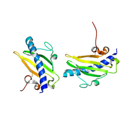
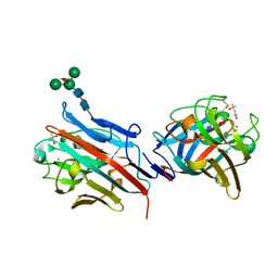
Crystal Structure of the Lectin I from Ulex europaeus in complex with the methyl glycoside of alpha-L-fucose
Descriptor: (4R)-2-METHYLPENTANE-2,4-DIOL, CALCIUM ION, MANGANESE (II) ION, ...
Authors:Audette, G.F, Olson, D.J.H, Ross, A.R.S, Quail, J.W, Delbaere, L.T.J.
Deposit date:2001-09-07
Release date:2002-12-06
Last modified:2023-08-16
Method:X-RAY DIFFRACTION (2.3 Å)
Cite:Examination of the Structural Basis for O(H) Blood Group Specificity by Ulex europaeus Lectin I
Can.J.Chem., 80, 2002
1FU0
DownloadVisualize
BU of 1fu0 by Molmil
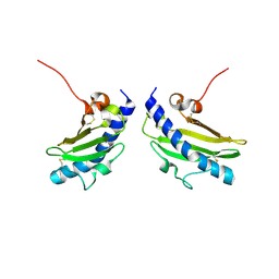
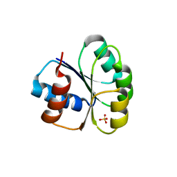
CRYSTAL STRUCTURE ANALYSIS OF THE PHOSPHO-SERINE 46 HPR FROM ENTEROCOCCUS FAECALIS
Descriptor: PHOSPHOCARRIER PROTEIN HPR
Authors:Audette, G.F, Engelmann, R, Hengstenberg, W, Deutscher, J, Hayakawa, K, Quail, J.W, Delbaere, L.T.J.
Deposit date:2000-09-13
Release date:2000-11-22
Last modified:2022-12-21
Method:X-RAY DIFFRACTION (1.9 Å)
Cite:The 1.9 A resolution structure of phospho-serine 46 HPr from Enterococcus faecalis.
J.Mol.Biol., 303, 2000
1FX5
DownloadVisualize
BU of 1fx5 by Molmil
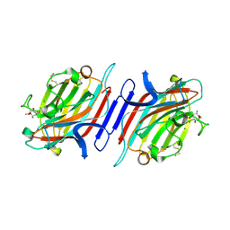
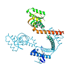
CRYSTAL STRUCTURE ANALYSIS OF ULEX EUROPAEUS LECTIN I
Descriptor: (4R)-2-METHYLPENTANE-2,4-DIOL, ANTI-H(O) LECTIN I, CALCIUM ION, ...
Authors:Audette, G.F, Vandonselaar, M, Delbaere, L.T.J.
Deposit date:2000-09-25
Release date:2001-01-17
Last modified:2023-08-09
Method:X-RAY DIFFRACTION (2.2 Å)
Cite:The 2.2 A resolution structure of the O(H) blood-group-specific lectin I from Ulex europaeus.
J.Mol.Biol., 304, 2000
6BHF
DownloadVisualize
BU of 6bhf by Molmil
Crystal structure of the petidylprolyl cis,trans-isomerase from Helicobacter pylori
Descriptor: Putative peptidyl-prolyl cis-trans isomerase HP_0175
Authors:Yaseen, A, Audette, G.F.
Deposit date:2017-10-30
Release date:2018-09-19
Last modified:2023-10-04
Method:X-RAY DIFFRACTION (2.09 Å)
Cite:Structural flexibility in the Helicobacter pylori peptidyl-prolyl cis,trans-isomerase HP0175 is achieved through an extension of the chaperone helices.
J. Struct. Biol., 204, 2018
6M8O
DownloadVisualize
BU of 6m8o by Molmil
Crystal structure of the receiver domain of LytR from Staphylococcus aureus
Descriptor: DNA-binding response regulator, SULFATE ION
Authors:Shala-Lawrence, A, Audette, G.F.
Deposit date:2018-08-22
Release date:2018-09-19
Last modified:2023-10-11
Method:X-RAY DIFFRACTION (2.5 Å)
Cite:Crystal structure of the receiver domain of LytR from Staphylococcus aureus
To Be Published

218853

PDB entries from 2024-04-24

PDB statisticsPDBj update infoContact PDBjnumon