7BU3
 
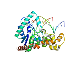
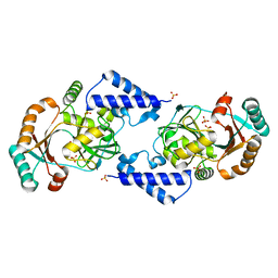
 | | Structure of alcohol dehydrogenase YjgB in complex with NADP from Escherichia coli | | 分子名称: | ASPARTIC ACID, Alcohol dehydrogenase, DI(HYDROXYETHYL)ETHER, ... | | 著者 | Nguyen, G.T, Kim, Y.-G, Ahn, J.-W, Chang, J.H. | | 登録日 | 2020-04-03 | | 公開日 | 2020-05-13 | | 最終更新日 | 2023-11-29 | | 実験手法 | X-RAY DIFFRACTION (2 Å) | | 主引用文献 | Structural Basis for Broad Substrate Selectivity of Alcohol Dehydrogenase YjgB from Escherichia coli .
Molecules, 25, 2020
|
|
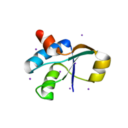
5GT2
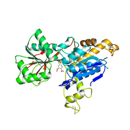
 | | Crystal Structure and Biochemical Features of dye-decolorizing peroxidase YfeX from Escherichia coli O157 | | 分子名称: | PROTOPORPHYRIN IX CONTAINING FE, Probable deferrochelatase/peroxidase YfeX | | 著者 | Ma, Y.L, Yuan, Z.G, Liu, S, Wang, J.X, Gu, L.C, Liu, X.H. | | 登録日 | 2016-08-18 | | 公開日 | 2017-02-08 | | 最終更新日 | 2024-03-20 | | 実験手法 | X-RAY DIFFRACTION (2.093 Å) | | 主引用文献 | Crystal structure and biochemical features of dye-decolorizing peroxidase YfeX from Escherichia coli O157 Asp(143) and Arg(232) play divergent roles toward different substrates
Biochem. Biophys. Res. Commun., 484, 2017
|
|
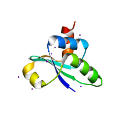
5AYY
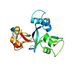
 | | CRYSTAL STRUCTURE OF HUMAN QUINOLINATE PHOSPHORIBOSYLTRANSFERASE IN COMPLEX WITH THE REACTANT QUINOLINATE | | 分子名称: | Nicotinate-nucleotide pyrophosphorylase [carboxylating], QUINOLINIC ACID | | 著者 | Youn, H.S, Kim, T.G, Kim, M.K, Kang, G.B, Kang, J.Y, Seo, Y.J, Lee, J.G, An, J.Y, Park, K.R, Lee, Y, Im, Y.J, Lee, J.H, Fukuoka, S.I, Eom, S.H. | | 登録日 | 2015-09-14 | | 公開日 | 2016-02-03 | | 最終更新日 | 2023-11-08 | | 実験手法 | X-RAY DIFFRACTION (3.09 Å) | | 主引用文献 | Structural Insights into the Quaternary Catalytic Mechanism of Hexameric Human Quinolinate Phosphoribosyltransferase, a Key Enzyme in de novo NAD Biosynthesis
Sci Rep, 6, 2016
|
|
5YR2
 | |
8IR0
 | |
6JG0
 | | Crystal structure of the N-terminal domain single mutant (S92E) of the human mitochondrial calcium uniporter fused with T4 lysozyme | | 分子名称: | Endolysin,Calcium uniporter protein, SULFATE ION | | 著者 | Lee, Y, Park, J, Min, C.K, Kang, J.Y, Kim, T.G, Yamamoto, T, Kim, D.H, Eom, S.H. | | 登録日 | 2019-02-13 | | 公開日 | 2020-02-19 | | 最終更新日 | 2023-11-22 | | 実験手法 | X-RAY DIFFRACTION (2.5 Å) | | 主引用文献 | Crystal structure of calcium channel domain
To Be Published
|
|
5MGH
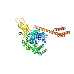
 | | Crystal structure of pathogenic mutants of human mitochodnrial PheRS | | 分子名称: | PHENYLALANINE, Phenylalanine--tRNA ligase, mitochondrial | | 著者 | Kartvelishvili, E, Tworowski, D, Vernon, H, Chrzanowska-Lightowlers, Z, Moor, N, Wang, J, Wong, L.-J, Safro, M. | | 登録日 | 2016-11-21 | | 公開日 | 2017-05-03 | | 最終更新日 | 2024-05-08 | | 実験手法 | X-RAY DIFFRACTION (1.87 Å) | | 主引用文献 | Kinetic and structural changes in HsmtPheRS, induced by pathogenic mutations in human FARS2.
Protein Sci., 26, 2017
|
|
5HC4
 | | Structure of esterase Est22 | | 分子名称: | 2-AMINO-2-HYDROXYMETHYL-PROPANE-1,3-DIOL, GLYCEROL, Lipolytic enzyme | | 著者 | Li, J, Huang, J. | | 登録日 | 2016-01-04 | | 公開日 | 2017-01-18 | | 最終更新日 | 2024-03-20 | | 実験手法 | X-RAY DIFFRACTION (2 Å) | | 主引用文献 | Structural insights of a hormone sensitive lipase homologue Est22.
Sci Rep, 6, 2016
|
|
3KD7
 | |
3KHF
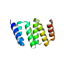
 | | The crystal structure of the PDZ domain of human Microtubule Associated Serine/Threonine Kinase 3 (MAST3) | | 分子名称: | 1,2-ETHANEDIOL, CHLORIDE ION, Microtubule-associated serine/threonine-protein kinase 3 | | 著者 | Roos, A, Elkins, J, Savitsky, P, Wang, J, Ugochukwu, E, Murray, J, Bountra, C, Arrowsmith, C.H, Weigelt, J, Edwards, A, von Delft, F, Knapp, S, Structural Genomics Consortium (SGC) | | 登録日 | 2009-10-30 | | 公開日 | 2009-12-08 | | 最終更新日 | 2023-09-06 | | 実験手法 | X-RAY DIFFRACTION (1.2 Å) | | 主引用文献 | The crystal structure of the PDZ domain of human Microtubule Associated Serine/Threonine Kinase 3 (MAST3)
To be Published
|
|
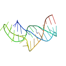
5HNK
 | | Crystal structure of T5Fen in complex intact substrate and metal ions. | | 分子名称: | DNA (5'-D(*AP*AP*AP*AP*GP*CP*GP*TP*AP*CP*GP*C)-3'), Exodeoxyribonuclease, GLYCEROL, ... | | 著者 | Almalki, F.A, Feng, M, Zhang, J, Sedelnikova, S.E, Rafferty, J.B, Sayers, J.R, Artymiuk, P.J. | | 登録日 | 2016-01-18 | | 公開日 | 2016-06-01 | | 最終更新日 | 2024-01-10 | | 実験手法 | X-RAY DIFFRACTION (2.22 Å) | | 主引用文献 | Direct observation of DNA threading in flap endonuclease complexes.
Nat.Struct.Mol.Biol., 23, 2016
|
|
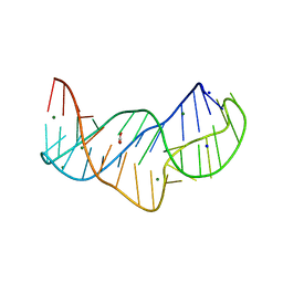
5NZ3
 | | The structure of the thermobifida fusca guanidine III riboswitch with methylguanidine | | 分子名称: | 1-METHYLGUANIDINE, MAGNESIUM ION, RNA (41-MER), ... | | 著者 | Huang, L, Wang, J, Lilley, D.M.J. | | 登録日 | 2017-05-12 | | 公開日 | 2017-10-18 | | 最終更新日 | 2024-05-08 | | 実験手法 | X-RAY DIFFRACTION (2.059 Å) | | 主引用文献 | Structure of the Guanidine III Riboswitch.
Cell Chem Biol, 24, 2017
|
|
6E5V
 | | human mGlu8 receptor amino terminal domain in complex with (S)-3,4-Dicarboxyphenylglycine (DCPG) | | 分子名称: | 4-[(S)-amino(carboxy)methyl]benzene-1,2-dicarboxylic acid, CHLORIDE ION, Metabotropic glutamate receptor 8 | | 著者 | Chen, Q, Ho, J.D, Ashok, S, Vargas, M.C, Wang, J, Atwell, S, Bures, M, Schkeryantz, J.M, Monn, J.A, Hao, J. | | 登録日 | 2018-07-23 | | 公開日 | 2018-11-07 | | 最終更新日 | 2023-10-11 | | 実験手法 | X-RAY DIFFRACTION (2.95 Å) | | 主引用文献 | Structural Basis for ( S)-3,4-Dicarboxyphenylglycine (DCPG) As a Potent and Subtype Selective Agonist of the mGlu8Receptor.
J. Med. Chem., 61, 2018
|
|
5JCD
 | | Crystal structure of OsCEBiP | | 分子名称: | 2-acetamido-2-deoxy-beta-D-glucopyranose, Chitin elicitor-binding protein | | 著者 | Chai, J.J, Liu, S.M, Wang, J.Z. | | 登録日 | 2016-04-15 | | 公開日 | 2017-02-22 | | 最終更新日 | 2020-07-29 | | 実験手法 | X-RAY DIFFRACTION (2.4 Å) | | 主引用文献 | Molecular Mechanism for Fungal Cell Wall Recognition by Rice Chitin Receptor OsCEBiP
Structure, 24, 2016
|
|
3JSL
 | |
3JTN
 | | Crystal Structure of the c-terminal domain of YpbH | | 分子名称: | Adapter protein mecA 2, IODIDE ION | | 著者 | Wang, F, Mei, Z, Qi, Y, Yan, C, Wang, J, Shi, Y. | | 登録日 | 2009-09-14 | | 公開日 | 2009-09-29 | | 最終更新日 | 2024-03-20 | | 実験手法 | X-RAY DIFFRACTION (2.09 Å) | | 主引用文献 | Crystal Structure of the MecA degradation tag
To be Published
|
|
3JTP
 | | crystal structure of the C-terminal domain of MecA | | 分子名称: | Adapter protein mecA 1, IODIDE ION | | 著者 | Wang, F, Mei, Z, Qi, Y, Yan, C, Wang, J, Shi, Y. | | 登録日 | 2009-09-14 | | 公開日 | 2009-09-29 | | 最終更新日 | 2024-05-29 | | 実験手法 | X-RAY DIFFRACTION (2.17 Å) | | 主引用文献 | crystal structure of the MecA degradation tag
To be Published
|
|
5NY8
 | | The structure of the thermobifida fusca guanidine III riboswitch with aminoguanidine | | 分子名称: | AMINOGUANIDINE, MAGNESIUM ION, RNA (41-MER), ... | | 著者 | Huang, L, Wang, J, Lilley, D.M.J. | | 登録日 | 2017-05-11 | | 公開日 | 2017-10-18 | | 最終更新日 | 2024-05-08 | | 実験手法 | X-RAY DIFFRACTION (2.04 Å) | | 主引用文献 | Structure of the Guanidine III Riboswitch.
Cell Chem Biol, 24, 2017
|
|
5NZ6
 | |
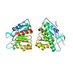
5JJA
 | | Crystal structure of a PP2A B56gamma/BubR1 complex | | 分子名称: | Mitotic checkpoint serine/threonine-protein kinase BUB1 beta, Serine/threonine-protein phosphatase 2A 56 kDa regulatory subunit gamma isoform | | 著者 | Wang, Z, Wang, J, Rao, Z, Xu, W. | | 登録日 | 2016-04-22 | | 公開日 | 2016-07-13 | | 最終更新日 | 2023-09-27 | | 実験手法 | X-RAY DIFFRACTION (2.35 Å) | | 主引用文献 | Crystal structure of a PP2A B56-BubR1 complex and its implications for PP2A substrate recruitment and localization.
Protein Cell, 7, 2016
|
|
5NWQ
 | | The structure of the thermobifida fusca guanidine III riboswitch with guanidine. | | 分子名称: | GUANIDINE, Guanidine III riboswitch, MAGNESIUM ION, ... | | 著者 | Huang, L, Wang, J, Lilley, D.M.J. | | 登録日 | 2017-05-08 | | 公開日 | 2017-10-18 | | 最終更新日 | 2024-05-08 | | 実験手法 | X-RAY DIFFRACTION (1.91 Å) | | 主引用文献 | Structure of the Guanidine III Riboswitch.
Cell Chem Biol, 24, 2017
|
|
5NZD
 | | The structure of the thermobifida fusca guanidine III riboswitch in space group P212121. | | 分子名称: | ACETATE ION, MAGNESIUM ION, SODIUM ION, ... | | 著者 | Huang, L, Wang, J, Lilley, D.M.J. | | 登録日 | 2017-05-13 | | 公開日 | 2017-10-18 | | 最終更新日 | 2024-05-08 | | 実験手法 | X-RAY DIFFRACTION (2.007 Å) | | 主引用文献 | Structure of the Guanidine III Riboswitch.
Cell Chem Biol, 24, 2017
|
|
5E84
 | | ATP-bound state of BiP | | 分子名称: | 78 kDa glucose-regulated protein, ADENOSINE-5'-TRIPHOSPHATE, MAGNESIUM ION, ... | | 著者 | Liu, Q, Yang, J, Nune, M, Zong, Y, Zhou, L. | | 登録日 | 2015-10-13 | | 公開日 | 2016-01-27 | | 最終更新日 | 2023-09-27 | | 実験手法 | X-RAY DIFFRACTION (2.99 Å) | | 主引用文献 | Close and Allosteric Opening of the Polypeptide-Binding Site in a Human Hsp70 Chaperone BiP.
Structure, 23, 2015
|
|
5DWD
 | | Crystal structure of esterase PE8 | | 分子名称: | 2-(2-{2-[2-(2-METHOXY-ETHOXY)-ETHOXY]-ETHOXY}-ETHOXY)-ETHANOL, Esterase, GLYCEROL | | 著者 | Li, J, Huang, J. | | 登録日 | 2015-09-22 | | 公開日 | 2016-10-26 | | 最終更新日 | 2024-03-20 | | 実験手法 | X-RAY DIFFRACTION (1.66 Å) | | 主引用文献 | Structure of esterase PE8
To Be Published
|
|
6MZL
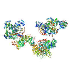 | | Human TFIID canonical state | | 分子名称: | TATA-box-binding protein, Transcription initiation factor TFIID subunit 1, Transcription initiation factor TFIID subunit 10, ... | | 著者 | Patel, A.B, Louder, R.K, Greber, B.J, Grunberg, S, Luo, J, Fang, J, Liu, Y, Ranish, J, Hahn, S, Nogales, E. | | 登録日 | 2018-11-05 | | 公開日 | 2018-11-28 | | 最終更新日 | 2019-11-20 | | 実験手法 | ELECTRON MICROSCOPY (23 Å) | | 主引用文献 | Structure of human TFIID and mechanism of TBP loading onto promoter DNA.
Science, 362, 2018
|
|


