Loading
PDBj
MenuPDBj@FacebookPDBj@TwitterPDBj@YouTubewwPDB FoundationwwPDB
RCSB PDBPDBeBMRBAdv. SearchSearch help
PDB: 263 results

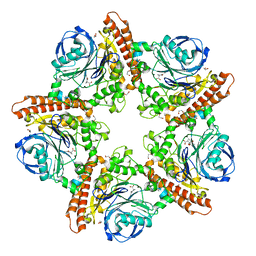
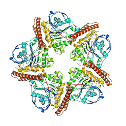
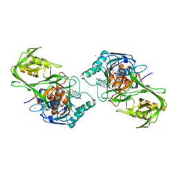
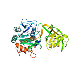
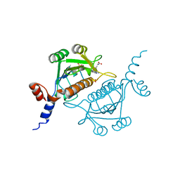
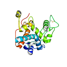
5IMH
DownloadVisualize
BU of 5imh by Molmil
D31P mutant of C69-family cysteine dipeptidase from Lactobacillus farciminis
Descriptor: 1,2-ETHANEDIOL, ACETATE ION, COBALT (II) ION, ...
Authors:Kono, R, Watanabe, K.
Deposit date:2016-03-06
Release date:2017-03-15
Last modified:2023-11-08
Method:X-RAY DIFFRACTION (2.471 Å)
Cite:A C69-family cysteine dipeptidase from Lactobacillus farciminis; substrate recognition mechanism and autoproteolytic mechanism in enzyme maturation
To Be Published
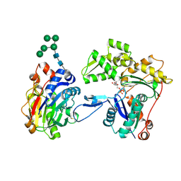
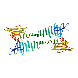
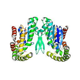
3W3E
DownloadVisualize
BU of 3w3e by Molmil
Structure of Vigna unguiculata chitinase with regulation activity of the plant cell wall
Descriptor: Cotyledoneous yieldin-like protein
Authors:Morohashi, K, Sasaki, K, Sakabe, N, Sakabe, K.
Deposit date:2012-12-20
Release date:2013-01-16
Last modified:2023-11-08
Method:X-RAY DIFFRACTION (1.5 Å)
Cite:Three-dimensional structure analysis of Vigna unguiculata chitinase with regulation activity of the yield threshold of cell wall
To be Published
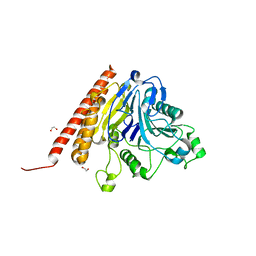
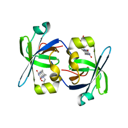
3W3D
DownloadVisualize
BU of 3w3d by Molmil
Crystal structure of smooth muscle G actin DNase I complex
Descriptor: ADENOSINE-5'-TRIPHOSPHATE, Actin, gamma-enteric smooth muscle, ...
Authors:Sakabe, N, Sakabe, K, Sasaki, K, Kondo, H, Shimomur, M.
Deposit date:2012-12-20
Release date:2013-01-30
Last modified:2020-07-29
Method:X-RAY DIFFRACTION (1.8 Å)
Cite:Refined structure and solvent network of chicken gizzard G-actin DNase 1 complex at 1.8A resolution
Acta Crystallogr.,Sect.A, 49, 1993
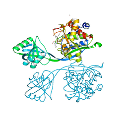
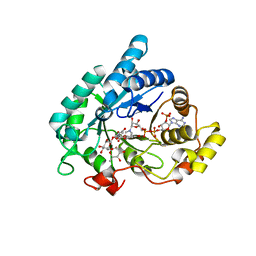
5INR
DownloadVisualize
BU of 5inr by Molmil
A C69-family cysteine dipeptidase in complex with Ala-Pro from Lactobacillus farciminis
Descriptor: 1,2-ETHANEDIOL, ACETATE ION, ALANINE, ...
Authors:Kono, R, Watanabe, K.
Deposit date:2016-03-07
Release date:2017-03-15
Last modified:2023-11-08
Method:X-RAY DIFFRACTION (1.652 Å)
Cite:A C69-family cysteine dipeptidase from Lactobacillus farciminis; substrate recognition mechanism and autoproteolytic mechanism in enzyme maturation
To Be Published
5IAU
DownloadVisualize
BU of 5iau by Molmil
A C69-family cysteine dipeptidase from Lactobacillus farciminis
Descriptor: 1,2-ETHANEDIOL, Dipeptidase
Authors:Kono, R, Watanabe, K.
Deposit date:2016-02-21
Release date:2017-03-01
Last modified:2024-03-20
Method:X-RAY DIFFRACTION (1.781 Å)
Cite:A C69-family cysteine dipeptidase from Lactobacillus farciminis; substrate recognition mechanism and autoproteolytic mechanism in enzyme maturation
To Be Published
5IB9
DownloadVisualize
BU of 5ib9 by Molmil
Crystal structure of aminopeptidase equipped with PAD from Aneurinibacillus sp. AM-1
Descriptor: 2-(3-AMINO-2-HYDROXY-4-PHENYL-BUTYRYLAMINO)-4-METHYL-PENTANOIC ACID, Aminopeptidase, ZINC ION
Authors:Tagawa, R, Nakano, H, Watanabe, K.
Deposit date:2016-02-22
Release date:2017-02-22
Last modified:2023-11-08
Method:X-RAY DIFFRACTION (1.4 Å)
Cite:Crystal structure of aminopeptidase
To Be Published
5IMG
DownloadVisualize
BU of 5img by Molmil
C9A mutant of C69-family cysteine dipeptidase from Lactobacillus farciminis
Descriptor: COBALT (II) ION, Dipeptidase, GLYCEROL
Authors:Kono, R, Watanabe, K.
Deposit date:2016-03-06
Release date:2017-03-15
Last modified:2023-11-08
Method:X-RAY DIFFRACTION (1.844 Å)
Cite:A C69-family cysteine dipeptidase from Lactobacillus farciminis; substrate recognition mechanism and autoproteolytic mechanism in enzyme maturation
To Be Published
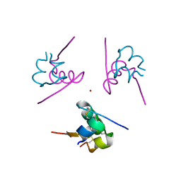
3W7Z
DownloadVisualize
BU of 3w7z by Molmil
1.15A structure of human 2Zn insulin at 293K
Descriptor: Insulin, ZINC ION
Authors:Hoshikawa, N, Sasaki, K, Sakabe, N, Sakabe, K.
Deposit date:2013-03-11
Release date:2013-07-03
Last modified:2023-11-08
Method:X-RAY DIFFRACTION (1.15 Å)
Cite:1.15A structure of human 2Zn insulin at 293K
To be Published
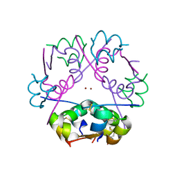
3W80
DownloadVisualize
BU of 3w80 by Molmil
Crystal structure of dodecamer human insulin with double C-axis length of the hexamer 2 Zn insulin cell
Descriptor: Insulin, ZINC ION
Authors:Murayoshi, M, Sasaki, K, Sakabe, N, Sakabe, K.
Deposit date:2013-03-11
Release date:2013-07-03
Last modified:2024-11-06
Method:X-RAY DIFFRACTION (1.4 Å)
Cite:Crystal structure of dodecamer insulin with double C-axis length of the hexamer 2 Zn insulin celll
To be Published
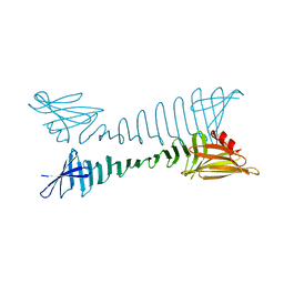
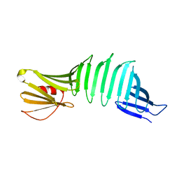
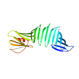
5YS7
DownloadVisualize
BU of 5ys7 by Molmil
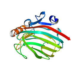
Crystal structure of an OspA mutant
Descriptor: Outer Surface Protein A
Authors:Takada, S, Makabe, K.
Deposit date:2017-11-13
Release date:2018-11-14
Last modified:2023-11-22
Method:X-RAY DIFFRACTION (2.1 Å)
Cite:Crystal structure of an OspA mutant
To Be Published
1WVM
DownloadVisualize
BU of 1wvm by Molmil
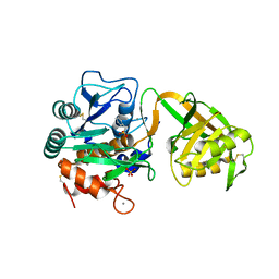
Crystal Structure of Psychrophilic Subtilisin-like Serine Protease APA1 from Antarctic Psychrotroph Pseudoaleromonas sp. AS-11, Complexed with Inhibitor Chymostatin
Descriptor: CALCIUM ION, CHYMOSTATIN, MAGNESIUM ION, ...
Authors:Dong, D, Watanabe, K.
Deposit date:2004-12-16
Release date:2006-03-21
Last modified:2024-10-30
Method:X-RAY DIFFRACTION (1.6 Å)
Cite:Crystal Structure of Psychrophilic Subtilisin-like Serine Protease from Antarctic Psychrotroph Pseudoalteromonas sp. AS-11 at 0.16 nm resolution
To be Published
5Z1O
DownloadVisualize
BU of 5z1o by Molmil
Crystal structure of an OspA mutant
Descriptor: Outer Surface Protein A
Authors:Takada, S, Makabe, K.
Deposit date:2017-12-27
Release date:2019-01-02
Last modified:2023-11-22
Method:X-RAY DIFFRACTION (2 Å)
Cite:Crystal structure of an OspA mutant.
To Be Published
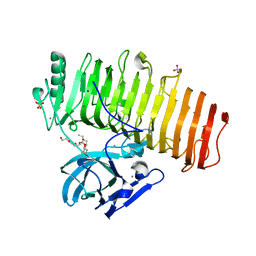
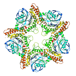
5ZRU
DownloadVisualize
BU of 5zru by Molmil
Crystal structure of Agl-KA catalytic domain
Descriptor: 3,6,9,12,15,18,21,24-OCTAOXAHEXACOSAN-1-OL, Alpha-1,3-glucanase, CALCIUM ION, ...
Authors:Yano, S, Makabe, K.
Deposit date:2018-04-25
Release date:2019-10-23
Last modified:2024-03-27
Method:X-RAY DIFFRACTION (1.833 Å)
Cite:Crystal structure of the catalytic unit of GH 87-type alpha-1,3-glucanase Agl-KA from Bacillus circulans.
Sci Rep, 9, 2019
6AIS
DownloadVisualize
BU of 6ais by Molmil
Loop deletion mutant (deleting two residues)
Descriptor: Outer surface protein A
Authors:Shiga, S, Makabe, K.
Deposit date:2018-08-24
Release date:2019-07-03
Last modified:2023-11-22
Method:X-RAY DIFFRACTION (1.3 Å)
Cite:Domain-Swapping Design by Polyproline Rod Insertion.
Chembiochem, 20, 2019
1Y9Z
DownloadVisualize
BU of 1y9z by Molmil
Crystal Structure of Psychrophilic Subtilisin-like Serine Protease from Antarctic Psychrotroph Pseudoalteromonas sp. AS-11 at 0.14 nm resolution
Descriptor: 2-AMINO-2-HYDROXYMETHYL-PROPANE-1,3-DIOL, CALCIUM ION, SODIUM ION, ...
Authors:Dong, D, Watanabe, K.
Deposit date:2004-12-16
Release date:2006-03-21
Last modified:2024-10-16
Method:X-RAY DIFFRACTION (1.4 Å)
Cite:Crystal Structure of Psychrophilic Subtilisin-like Serine Protease from Antarctic Psychrotroph Pseudoalteromonas sp. AS-11 at 0.14 nm resolution
To be Published
1Z9H
DownloadVisualize
BU of 1z9h by Molmil
Microsomal prostaglandin E synthase type-2
Descriptor: ACETATE ION, CHLORIDE ION, INDOMETHACIN, ...
Authors:Yamada, T, Komoto, J, Watanabe, K, Ohmiya, Y, Takusagawa, F.
Deposit date:2005-04-02
Release date:2005-05-17
Last modified:2024-02-14
Method:X-RAY DIFFRACTION (2.6 Å)
Cite:Crystal Structure and Possible Catalytic Mechanism of Microsomal Prostaglandin E Synthase Type 2 (mPGES-2).
J.Mol.Biol., 348, 2005
1HZE
DownloadVisualize
BU of 1hze by Molmil
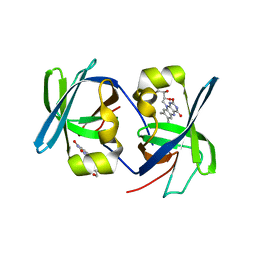
SOLUTION STRUCTURE OF THE N-TERMINAL DOMAIN OF RIBOFLAVIN SYNTHASE FROM E. COLI
Descriptor: RIBOFLAVIN, RIBOFLAVIN SYNTHASE ALPHA CHAIN
Authors:Truffault, V, Coles, M, Diercks, T, Abelmann, K, Eberhardt, S, Luettgen, H, Bacher, A, Kessler, H.
Deposit date:2001-01-24
Release date:2001-09-05
Last modified:2024-05-01
Method:SOLUTION NMR
Cite:The solution structure of the N-terminal domain of riboflavin synthase.
J.Mol.Biol., 309, 2001
1RY8
DownloadVisualize
BU of 1ry8 by Molmil
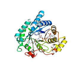
Prostaglandin F synthase complexed with NADPH and rutin
Descriptor: Aldo-keto reductase family 1 member C3, NADPH DIHYDRO-NICOTINAMIDE-ADENINE-DINUCLEOTIDE PHOSPHATE, RUTIN
Authors:Komoto, J, Yamada, T, Watanabe, K, Takusagawa, F.
Deposit date:2003-12-19
Release date:2004-11-02
Last modified:2023-08-23
Method:X-RAY DIFFRACTION (1.69 Å)
Cite:Crystal structure of human prostaglandin F synthase (AKR1C3).
Biochemistry, 43, 2004
1RY0
DownloadVisualize
BU of 1ry0 by Molmil
Structure of prostaglandin F synthase with prostaglandin D2
Descriptor: Aldo-keto reductase family 1 member C3, NADP NICOTINAMIDE-ADENINE-DINUCLEOTIDE PHOSPHATE, PROSTAGLANDIN D2
Authors:Komoto, J, Yamada, T, Watanabe, K, Takusagawa, F.
Deposit date:2003-12-19
Release date:2004-11-02
Last modified:2023-08-23
Method:X-RAY DIFFRACTION (1.69 Å)
Cite:Crystal structure of human prostaglandin F synthase (AKR1C3).
Biochemistry, 43, 2004
1I18
DownloadVisualize
BU of 1i18 by Molmil
SOLUTION STRUCTURE OF THE N-TERMINAL DOMAIN OF RIBOFLAVIN SYNTHASE FROM E. COLI
Descriptor: RIBOFLAVIN, RIBOFLAVIN SYNTHASE ALPHA CHAIN
Authors:Truffault, V, Coles, M, Diercks, T, Abelmann, K, Eberhardt, S, Luettgen, H, Bacher, A, Kessler, H.
Deposit date:2001-01-31
Release date:2001-09-05
Last modified:2024-05-01
Method:SOLUTION NMR
Cite:The solution structure of the N-terminal domain of riboflavin synthase.
J.Mol.Biol., 309, 2001
1J1T
DownloadVisualize
BU of 1j1t by Molmil
Alginate lyase from Alteromonas sp.272
Descriptor: Alginate Lyase, CALCIUM ION, SULFATE ION
Authors:Motoshima, H, Iwatomo, Y, Watanabe, K, Oda, T, Muramatsu, T.
Deposit date:2002-12-14
Release date:2004-02-03
Last modified:2024-10-16
Method:X-RAY DIFFRACTION (2 Å)
Cite:Crystal structure of Alginate Lyase from Alteromonas sp.272
To be published
1V6C
DownloadVisualize
BU of 1v6c by Molmil
Crystal Structure of Psychrophilic Subtilisin-like Protease Apa1 from Antarctic Psychrotroph Pseudoalteromonas sp. AS-11
Descriptor: CALCIUM ION, SULFATE ION, alkaline serine protease, ...
Authors:Dong, D, Ihara, T, Motoshima, H, Watanabe, K.
Deposit date:2003-11-28
Release date:2004-12-28
Last modified:2024-10-23
Method:X-RAY DIFFRACTION (1.8 Å)
Cite:Crystal Structure of Psychrophilic Subtilisin-like Protease Apa1 from Antarctic Psychrotroph Pseudoalteromonas sp. AS-11
To be Published
1V2X
DownloadVisualize
BU of 1v2x by Molmil
TrmH
Descriptor: PHOSPHATE ION, S-ADENOSYLMETHIONINE, tRNA (Gm18) methyltransferase
Authors:Nureki, O, Watanabe, K, Fukai, S, Ishii, R, Endo, Y, Hori, H, Yokoyama, S, RIKEN Structural Genomics/Proteomics Initiative (RSGI)
Deposit date:2003-10-17
Release date:2004-05-04
Last modified:2023-12-27
Method:X-RAY DIFFRACTION (1.5 Å)
Cite:Deep Knot Structure for Construction of Active Site and Cofactor Binding Site of tRNA Modification Enzyme
STRUCTURE, 12, 2004
1UMK
DownloadVisualize
BU of 1umk by Molmil
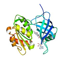
The Structure of Human Erythrocyte NADH-cytochrome b5 Reductase
Descriptor: FLAVIN-ADENINE DINUCLEOTIDE, NADH-cytochrome b5 reductase
Authors:Bando, S, Takano, T, Yubisui, T, Shirabe, K, Takeshita, M, Horii, C, Nakagawa, A.
Deposit date:2003-10-03
Release date:2004-11-02
Last modified:2023-12-27
Method:X-RAY DIFFRACTION (1.75 Å)
Cite:Structure of human erythrocyte NADH-cytochrome b5 reductase.
Acta Crystallogr.,Sect.D, 60, 2004
6J6E
DownloadVisualize
BU of 6j6e by Molmil
Borrelia burgdorferi OspA via surface entropy reduction (form5)
Descriptor: Outer surface protein A
Authors:Fujiwara, H, Makabe, K.
Deposit date:2019-01-14
Release date:2019-11-27
Last modified:2023-11-22
Method:X-RAY DIFFRACTION (1.5 Å)
Cite:Beta-sheet elasticity of peptide self-assembly mimic, PSAM, with a grafted sequence characterized by comprehensive analyses of isomorphous crystals
J Mol Liq, 290, 2019

227111

PDB entries from 2024-11-06

PDB statisticsPDBj update infoContact PDBjnumon