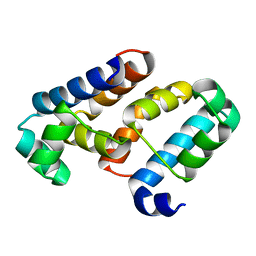
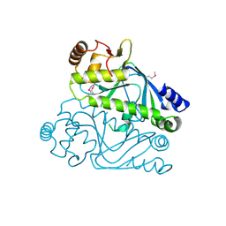
7T5Y
 
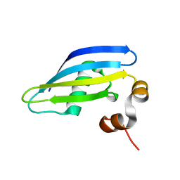
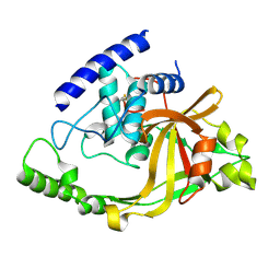
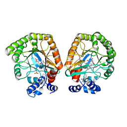 | | E. coli dihydroorotate dehydrogenase bound to the inhibitor HMNQ | | 分子名称: | 3-methyl-2-[(2E)-non-2-en-1-yl]quinolin-4(1H)-one, Dihydroorotate dehydrogenase (quinone), FLAVIN MONONUCLEOTIDE, ... | | 著者 | Horwitz, S.M, Ambarian, J.A, Davis, K.M. | | 登録日 | 2021-12-13 | | 公開日 | 2022-03-02 | | 最終更新日 | 2023-10-18 | | 実験手法 | X-RAY DIFFRACTION (2.62 Å) | | 主引用文献 | Structural insights into inhibition of the drug target dihydroorotate dehydrogenase by bacterial hydroxyalkylquinolines.
Rsc Chem Biol, 3, 2022
|
|
7ZL5
 | |
3EGO
 | | Crystal structure of Probable 2-dehydropantoate 2-reductase panE from Bacillus Subtilis | | 分子名称: | Probable 2-dehydropantoate 2-reductase | | 著者 | Ramagopal, U.A, Toro, R, Gilmore, M, Hu, S, Maletic, M, Gheyi, T, Burley, S.K, Almo, S.C, New York SGX Research Center for Structural Genomics (NYSGXRC) | | 登録日 | 2008-09-11 | | 公開日 | 2008-09-30 | | 最終更新日 | 2024-02-21 | | 実験手法 | X-RAY DIFFRACTION (1.9 Å) | | 主引用文献 | Crystal structure of Probable 2-dehydropantoate 2-reductase panE from Bacillus Subtilis
To be published
|
|
7ZU6
 | | Polyoxidovanadate interaction with proteins: crystal structure of lysozyme bound to tetra-vanadate ion (structure 1) | | 分子名称: | 4-(2-HYDROXYETHYL)-1-PIPERAZINE ETHANESULFONIC ACID, CYCLO-TETRAMETAVANADATE, Lysozyme, ... | | 著者 | Tito, G, Merlino, A, Ferraro, G. | | 登録日 | 2022-05-11 | | 公開日 | 2023-05-24 | | 最終更新日 | 2024-02-07 | | 実験手法 | X-RAY DIFFRACTION (1.183 Å) | | 主引用文献 | Stabilization and Binding of [V 4 O 12 ] 4- and Unprecedented [V 20 O 54 (NO 3 )] n- to Lysozyme upon Loss of Ligands and Oxidation of the Potential Drug V IV O(acetylacetonato) 2.
Angew.Chem.Int.Ed.Engl., 62, 2023
|
|
6IXF
 | |
3EEH
 | |
7ZRF
 | |
7ZRO
 | |
3EUS
 | |
8A5R
 | | Crystal structure of light-activated DNA-binding protein EL222 from Erythrobacter litoralis crystallized and measured in dark. | | 分子名称: | 2-(N-MORPHOLINO)-ETHANESULFONIC ACID, CHLORIDE ION, FLAVIN MONONUCLEOTIDE, ... | | 著者 | Koval, T, Chaudhari, A, Fuertes, G, Andersson, I, Dohnalek, J. | | 登録日 | 2022-06-15 | | 公開日 | 2023-07-05 | | 最終更新日 | 2024-02-07 | | 実験手法 | X-RAY DIFFRACTION (1.85 Å) | | 主引用文献 | EL222 from Erythrobacter litoralis.
To Be Published
|
|
8A9F
 | |
8A5S
 | | Crystal structure of light-activated DNA-binding protein EL222 from Erythrobacter litoralis crystallized in dark, measured illuminated. | | 分子名称: | 2-(N-MORPHOLINO)-ETHANESULFONIC ACID, CHLORIDE ION, FLAVIN MONONUCLEOTIDE, ... | | 著者 | Koval, T, Chaudhari, A, Fuertes, G, Andersson, I, Dohnalek, J. | | 登録日 | 2022-06-15 | | 公開日 | 2023-07-05 | | 最終更新日 | 2024-02-07 | | 実験手法 | X-RAY DIFFRACTION (1.85 Å) | | 主引用文献 | EL222 from Erythrobacter litoralis.
To Be Published
|
|
8A8S
 | |
6IYE
 | |
1T5J
 | |
6J12
 | |
2HY5
 | | Crystal structure of DsrEFH | | 分子名称: | DsrH, Intracellular sulfur oxidation protein dsrF, Putative sulfurtransferase dsrE | | 著者 | Shin, D.H, Schulte, A, Dahl, C, Kim, R, Kim, S.H, Berkeley Structural Genomics Center (BSGC) | | 登録日 | 2006-08-04 | | 公開日 | 2006-09-19 | | 最終更新日 | 2024-02-14 | | 実験手法 | X-RAY DIFFRACTION (1.72 Å) | | 主引用文献 | Crystal structure of DsrEFH
To be Published
|
|
4RFA
 | | Crystal structure of cyclic nucleotide-binding domain containing protein from Listeria monocytogenes EGD-e | | 分子名称: | Lmo0740 protein | | 著者 | Filippova, E.V, Minasov, G, Kiryukhina, O, Jedrzejczak, R, Joachimiak, A, Anderson, W.F, Midwest Center for Structural Genomics (MCSG) | | 登録日 | 2014-09-25 | | 公開日 | 2014-10-15 | | 最終更新日 | 2017-11-22 | | 実験手法 | X-RAY DIFFRACTION (2.21 Å) | | 主引用文献 | Crystal structure of cyclic nucleotide-binding domain containing protein from Listeria monocytogenes EGD-e
To be Published
|
|
2I5M
 | |
2I5L
 | |
7B6Z
 | |
2IM9
 | | Crystal structure of protein LPG0564 from Legionella pneumophila str. Philadelphia 1, Pfam DUF1460 | | 分子名称: | Hypothetical protein | | 著者 | Bonanno, J.B, Freeman, J, Bain, K.T, Powell, A, Ozyurt, S, Wasserman, S, Sauder, J.M, Burley, S.K, Almo, S.C, New York SGX Research Center for Structural Genomics (NYSGXRC) | | 登録日 | 2006-10-04 | | 公開日 | 2006-10-17 | | 最終更新日 | 2021-02-03 | | 実験手法 | X-RAY DIFFRACTION (1.67 Å) | | 主引用文献 | Crystal structure of hypothetical protein from Legionella pneumophila subsp. pneumophila str. Philadelphia 1
TO BE PUBLISHED
|
|
2IQY
 | | Rat Phosphatidylethanolamine-Binding Protein | | 分子名称: | CALCIUM ION, CHLORIDE ION, Phosphatidylethanolamine-binding protein 1 | | 著者 | Kim, Y, Joachimiak, G, Heil, G.L, Koide, S, Joachimiak, A. | | 登録日 | 2006-10-14 | | 公開日 | 2007-09-25 | | 最終更新日 | 2023-08-30 | | 実験手法 | X-RAY DIFFRACTION (1.4 Å) | | 主引用文献 | Crystal Structure of Rat Phosphatidylethanolamine-Binding Protein
To be Published
|
|
2IZS
 | | Structure of casein kinase gamma 3 in complex with inhibitor | | 分子名称: | CASEIN KINASE I ISOFORM GAMMA-3, CHLORIDE ION, MAGNESIUM ION, ... | | 著者 | Bunkoczi, G, Salah, E, Rellos, P, Das, S, Fedorov, O, Savitsky, P, Debreczeni, J.E, Gileadi, O, Sundstrom, M, Edwards, A, Arrowsmith, C, Weigelt, J, von Delft, F, Knapp, S. | | 登録日 | 2006-07-26 | | 公開日 | 2006-08-01 | | 最終更新日 | 2023-12-13 | | 実験手法 | X-RAY DIFFRACTION (1.95 Å) | | 主引用文献 | Inhibitor Binding by Casein Kinases
To be Published
|
|
6J9T
 | | Complex structure of Lactobacillus casei lactate dehydrogenase with fructose-1,6-bisphosphate | | 分子名称: | 1,6-di-O-phosphono-beta-D-fructofuranose, L-lactate dehydrogenase, SULFATE ION | | 著者 | Arai, K, Miyanaga, A, Uchikoba, H, Fushinobu, S, Taguchi, H. | | 登録日 | 2019-01-24 | | 公開日 | 2019-02-06 | | 最終更新日 | 2023-11-22 | | 実験手法 | X-RAY DIFFRACTION (2.7 Å) | | 主引用文献 | Crystal structure of penta mutant of L-lactate dehydrogenase from Lactobacillus casei
To Be Published
|
|


