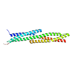
1Y19
 
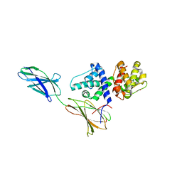
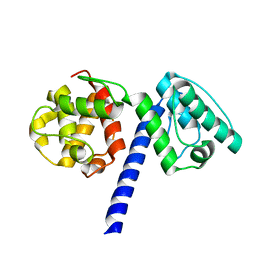
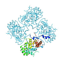
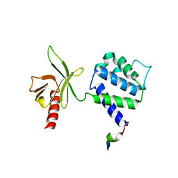 | | Structural basis for phosphatidylinositol phosphate kinase type I-gamma binding to talin at focal adhesions | | 分子名称: | Phosphatidylinositol-4-phosphate 5-kinase, type 1 gamma, Talin 1 | | 著者 | de Pereda, J.M, Wegener, K, Santelli, E, Bate, N, Ginsberg, M.H, Critchley, D.R, Campbell, I.D, Liddington, R.C. | | 登録日 | 2004-11-17 | | 公開日 | 2005-01-04 | | 最終更新日 | 2016-11-30 | | 実験手法 | X-RAY DIFFRACTION (2.6 Å) | | 主引用文献 | Structural bases for phosphatidylinositol phosphate kinase type I-gamma binding to talin at focal adhesions
J.Biol.Chem., 280, 2005
|
|
1QG3
 | |
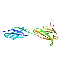
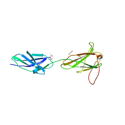
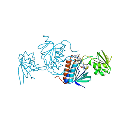
1Q7S
 | | Crystal structure of bit1 | | 分子名称: | bit1 | | 著者 | De Pereda, J.M, Waas, W.F, Jan, Y, Ruoslahti, E, Schimmel, P, Pascual, J. | | 登録日 | 2003-08-19 | | 公開日 | 2003-12-16 | | 最終更新日 | 2024-02-14 | | 実験手法 | X-RAY DIFFRACTION (2 Å) | | 主引用文献 | Crystal structure of a human peptidyl-tRNA hydrolase reveals a new fold and suggests basis for a bifunctional activity.
J.Biol.Chem., 279, 2004
|
|
3F7R
 | |
3F7P
 | |
3F7Q
 | |
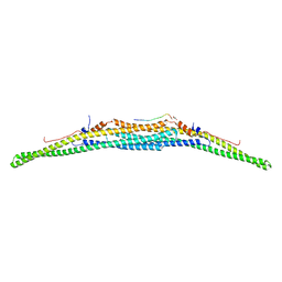
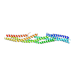
4GDO
 | | Structure of a fragment of the rod domain of plectin | | 分子名称: | Plectin | | 著者 | De Pereda, J.M, Buey, R.M, Uson, I, Sammito, M.D, De Marino, I. | | 登録日 | 2012-08-01 | | 公開日 | 2013-09-11 | | 最終更新日 | 2024-02-28 | | 実験手法 | X-RAY DIFFRACTION (1.7 Å) | | 主引用文献 | Exploiting tertiary structure through local folds for crystallographic phasing.
Nat.Methods, 10, 2013
|
|
2ODV
 | |
2ODU
 | |
1MB8
 | |
4WTX
 | | Crystal structure of the fourth FnIII domain of integrin beta4 | | 分子名称: | Integrin beta-4 | | 著者 | Alonso-Garcia, N, Urien, H, Buey, R.M, de Pereda, J.M. | | 登録日 | 2014-10-30 | | 公開日 | 2015-02-11 | | 最終更新日 | 2015-04-08 | | 実験手法 | X-RAY DIFFRACTION (1.5 Å) | | 主引用文献 | Combination of X-ray crystallography, SAXS and DEER to obtain the structure of the FnIII-3,4 domains of integrin alpha6beta4
Acta Crystallogr.,Sect.D, 71, 2015
|
|
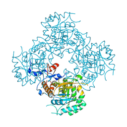
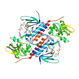
5TC3
 | | Structure of IMP dehydrogenase from Ashbya gossypii bound to ATP and GDP | | 分子名称: | ACETATE ION, ADENOSINE-5'-TRIPHOSPHATE, GUANOSINE-5'-DIPHOSPHATE, ... | | 著者 | Fernandez-Justel, D, de Pereda, J.M, Revuelta, J.L, Buey, R.M. | | 登録日 | 2016-09-14 | | 公開日 | 2017-06-14 | | 最終更新日 | 2024-01-17 | | 実験手法 | X-RAY DIFFRACTION (2.462 Å) | | 主引用文献 | A nucleotide-controlled conformational switch modulates the activity of eukaryotic IMP dehydrogenases.
Sci Rep, 7, 2017
|
|
7AAL
 | | Crystal structure of the F-BAR domain of PSTIPIP1, G258A mutant | | 分子名称: | Proline-serine-threonine phosphatase-interacting protein 1 | | 著者 | Manso, J.A, Alcon, P, Bayon, Y, Alonso, A, de Pereda, J.M. | | 登録日 | 2020-09-04 | | 公開日 | 2022-02-23 | | 最終更新日 | 2024-02-07 | | 実験手法 | X-RAY DIFFRACTION (1.97 Å) | | 主引用文献 | PSTPIP1-LYP phosphatase interaction: structural basis and implications for autoinflammatory disorders.
Cell.Mol.Life Sci., 79, 2022
|
|
7AAN
 | | Crystal structure of the F-BAR domain of PSTIPIP1 | | 分子名称: | Proline-serine-threonine phosphatase-interacting protein 1 | | 著者 | Manso, J.A, Alcon, P, Bayon, Y, Alonso, A, de Pereda, J.M. | | 登録日 | 2020-09-04 | | 公開日 | 2022-02-23 | | 最終更新日 | 2024-02-07 | | 実験手法 | X-RAY DIFFRACTION (2.14 Å) | | 主引用文献 | PSTPIP1-LYP phosphatase interaction: structural basis and implications for autoinflammatory disorders.
Cell.Mol.Life Sci., 79, 2022
|
|
7AAM
 | | Crystal structure of the F-BAR domain of PSTIPIP1 bound to the CTH domain of the phosphatase LYP | | 分子名称: | GLYCEROL, Proline-serine-threonine phosphatase-interacting protein 1, Tyrosine-protein phosphatase non-receptor type 22 | | 著者 | Manso, J.A, Alcon, P, Bayon, Y, Alonso, A, de Pereda, J.M. | | 登録日 | 2020-09-04 | | 公開日 | 2022-02-23 | | 最終更新日 | 2024-02-07 | | 実験手法 | X-RAY DIFFRACTION (2.15 Å) | | 主引用文献 | PSTPIP1-LYP phosphatase interaction: structural basis and implications for autoinflammatory disorders.
Cell.Mol.Life Sci., 79, 2022
|
|
5J1H
 | |
5J1F
 | |
5J1I
 | |
5J1G
 | |
5J60
 | | Structure of a thioredoxin reductase from Gloeobacter violaceus | | 分子名称: | CALCIUM ION, FLAVIN-ADENINE DINUCLEOTIDE, TETRAETHYLENE GLYCOL, ... | | 著者 | Buey, R.M, de Pereda, J.M, Balsera, M. | | 登録日 | 2016-04-04 | | 公開日 | 2016-07-27 | | 最終更新日 | 2024-01-10 | | 実験手法 | X-RAY DIFFRACTION (1.9 Å) | | 主引用文献 | A New Member of the Thioredoxin Reductase Family from Early Oxygenic Photosynthetic Organisms.
Mol Plant, 10, 2017
|
|
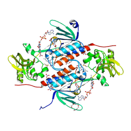
4XTD
 | | Structure of the covalent intermediate E-XMP* of the IMP dehydrogenase of Ashbya gossypii | | 分子名称: | INOSINIC ACID, Inosine-5'-monophosphate dehydrogenase,Inosine-5'-monophosphate dehydrogenase | | 著者 | Buey, R.M, Ledesma-Amaro, R, Balsera, M, de Pereda, J.M, Revuelta, J.L. | | 登録日 | 2015-01-23 | | 公開日 | 2015-07-22 | | 最終更新日 | 2024-01-10 | | 実験手法 | X-RAY DIFFRACTION (2.05 Å) | | 主引用文献 | Increased riboflavin production by manipulation of inosine 5'-monophosphate dehydrogenase in Ashbya gossypii.
Appl.Microbiol.Biotechnol., 99, 2015
|
|
4XTI
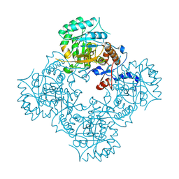 | | Structure of IMP dehydrogenase of Ashbya gossypii with IMP bound to the active site | | 分子名称: | INOSINIC ACID, Inosine-5'-monophosphate dehydrogenase,Inosine-5'-monophosphate dehydrogenase, POTASSIUM ION | | 著者 | Buey, R.M, Ledesma-Amaro, R, Balsera, M, de Pereda, J.M, Revuelta, J.L. | | 登録日 | 2015-01-23 | | 公開日 | 2015-07-22 | | 最終更新日 | 2024-01-10 | | 実験手法 | X-RAY DIFFRACTION (1.5 Å) | | 主引用文献 | Increased riboflavin production by manipulation of inosine 5'-monophosphate dehydrogenase in Ashbya gossypii.
Appl.Microbiol.Biotechnol., 99, 2015
|
|
4XWU
 | | Structure of the IMP dehydrogenase from Ashbya gossypii | | 分子名称: | Inosine-5'-monophosphate dehydrogenase,Inosine-5'-monophosphate dehydrogenase | | 著者 | Buey, R.M, Ledesma-Amaro, R, Balsera, M, de Pereda, J.M, Revuelta, J.L. | | 登録日 | 2015-01-29 | | 公開日 | 2015-07-22 | | 最終更新日 | 2024-01-10 | | 実験手法 | X-RAY DIFFRACTION (1.75 Å) | | 主引用文献 | Increased riboflavin production by manipulation of inosine 5'-monophosphate dehydrogenase in Ashbya gossypii.
Appl.Microbiol.Biotechnol., 99, 2015
|
|
5JRI
 | | Structure of an oxidoreductase SeMet-labelled from Synechocystis sp. PCC6803 | | 分子名称: | 1,2-ETHANEDIOL, FLAVIN-ADENINE DINUCLEOTIDE, Pyridine nucleotide-disulfide oxidoreductase, ... | | 著者 | Buey, R.M, de Pereda, J.M, Balsera, M. | | 登録日 | 2016-05-06 | | 公開日 | 2017-11-15 | | 最終更新日 | 2017-12-13 | | 実験手法 | X-RAY DIFFRACTION (1.952 Å) | | 主引用文献 | Unprecedented pathway of reducing equivalents in a diflavin-linked disulfide oxidoreductase.
Proc. Natl. Acad. Sci. U.S.A., 114, 2017
|
|
5K0A
 | | Structure of an oxidoreductase from Synechocystis sp. PCC6803 | | 分子名称: | FLAVIN-ADENINE DINUCLEOTIDE, NITRATE ION, PENTAETHYLENE GLYCOL, ... | | 著者 | Buey, R.M, de Pereda, J.M, Balsera, M. | | 登録日 | 2016-05-17 | | 公開日 | 2017-11-15 | | 最終更新日 | 2024-01-10 | | 実験手法 | X-RAY DIFFRACTION (1.706 Å) | | 主引用文献 | Unprecedented pathway of reducing equivalents in a diflavin-linked disulfide oxidoreductase.
Proc. Natl. Acad. Sci. U.S.A., 114, 2017
|
|


