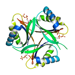
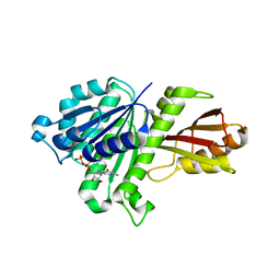
3ATM
 
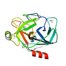 | | Crystal structure of trypsin complexed with 2-(1H-indol-3-yl)ethanamine | | 分子名称: | 2-(1H-INDOL-3-YL)ETHANAMINE, CALCIUM ION, Cationic trypsin, ... | | 著者 | Yamane, J, Yao, M, Tanaka, I. | | 登録日 | 2011-01-05 | | 公開日 | 2011-08-24 | | 最終更新日 | 2023-11-01 | | 実験手法 | X-RAY DIFFRACTION (1.72 Å) | | 主引用文献 | In-crystal affinity ranking of fragment hit compounds reveals a relationship with their inhibitory activities
J.Appl.Crystallogr., 44, 2011
|
|
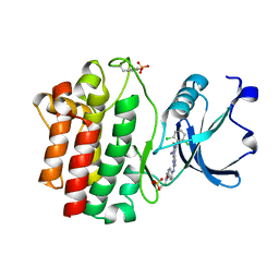
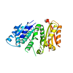
3ATL
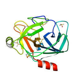 | | Crystal structure of trypsin complexed with benzamidine | | 分子名称: | BENZAMIDINE, CALCIUM ION, Cationic trypsin, ... | | 著者 | Yamane, J, Yao, M, Tanaka, I. | | 登録日 | 2011-01-05 | | 公開日 | 2011-08-24 | | 最終更新日 | 2023-11-01 | | 実験手法 | X-RAY DIFFRACTION (1.74 Å) | | 主引用文献 | In-crystal affinity ranking of fragment hit compounds reveals a relationship with their inhibitory activities
J.Appl.Crystallogr., 44, 2011
|
|
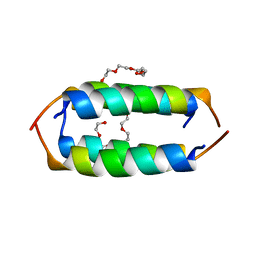
2GS0
 | | NMR structure of the complex between the PH domain of the Tfb1 subunit from TFIIH and the activation domain of p53 | | 分子名称: | Cellular tumor antigen p53, RNA polymerase II transcription factor B subunit 1 | | 著者 | Di Lello, P, Jones, T.N, Nguyen, B.D, Legault, P, Omichinski, J.G. | | 登録日 | 2006-04-25 | | 公開日 | 2006-10-31 | | 最終更新日 | 2021-10-20 | | 実験手法 | SOLUTION NMR | | 主引用文献 | Structure of the Tfb1/p53 complex: Insights into the interaction between the p62/Tfb1 subunit of TFIIH and the activation domain of p53.
Mol.Cell, 22, 2006
|
|
1UFL
 | | Crystal Structure of TT1020 from Thermus thermophilus HB8 | | 分子名称: | Nitrogen regulatory protein P-II | | 著者 | Wang, H, Sakai, H, Hori-Takemoto, C, Kaminishi, T, Terada, T, Kuramitsu, S, Shirouzu, M, Yokoyama, S, RIKEN Structural Genomics/Proteomics Initiative (RSGI) | | 登録日 | 2003-05-31 | | 公開日 | 2003-11-30 | | 最終更新日 | 2023-12-27 | | 実験手法 | X-RAY DIFFRACTION (2.7 Å) | | 主引用文献 | Crystal structures of the signal transducing protein GlnK from Thermus thermophilus HB8.
J.Struct.Biol., 149, 2005
|
|
1V3S
 | | Crystal structure of TT1020 from Thermus thermophilus HB8 | | 分子名称: | ADENOSINE-5'-TRIPHOSPHATE, Nitrogen regulatory protein P-II | | 著者 | Wang, H, Sakai, H, Takemoto-Hori, C, Kaminishi, T, Terada, T, Kuramitsu, S, Shirouzu, M, Yokoyama, S, RIKEN Structural Genomics/Proteomics Initiative (RSGI) | | 登録日 | 2003-11-05 | | 公開日 | 2004-11-23 | | 最終更新日 | 2023-12-27 | | 実験手法 | X-RAY DIFFRACTION (1.85 Å) | | 主引用文献 | Crystal structures of the signal transducing protein GlnK from Thermus thermophilus HB8.
J.Struct.Biol., 149, 2005
|
|
1V9O
 | | Crystal structure of TT1020 from Thermus thermophilus HB8 | | 分子名称: | ADENOSINE-5'-DIPHOSPHATE, NITROGEN REGULATORY PROTEIN PII | | 著者 | Wang, H, Sakai, H, Takemoto-Hori, C, Kaminishi, T, Terada, T, Kuramitsu, S, Shirouzu, M, Yokoyama, S, RIKEN Structural Genomics/Proteomics Initiative (RSGI) | | 登録日 | 2004-01-27 | | 公開日 | 2005-01-11 | | 最終更新日 | 2023-12-27 | | 実験手法 | X-RAY DIFFRACTION (2 Å) | | 主引用文献 | Crystal structures of the signal transducing protein GlnK from Thermus thermophilus HB8.
J.Struct.Biol., 149, 2005
|
|
1VFJ
 | | Crystal structure of TT1020 from Thermus thermophilus HB8 | | 分子名称: | nitrogen regulatory protein p-II | | 著者 | Wang, H, Sakai, H, Takemoto-Hori, C, Kaminishi, T, Terada, T, Kuramitsu, S, Shirouzu, M, Yokoyama, S, RIKEN Structural Genomics/Proteomics Initiative (RSGI) | | 登録日 | 2004-04-15 | | 公開日 | 2005-01-11 | | 最終更新日 | 2023-12-27 | | 実験手法 | X-RAY DIFFRACTION (1.7 Å) | | 主引用文献 | Crystal structures of the signal transducing protein GlnK from Thermus thermophilus HB8
J.STRUCT.BIOL., 149, 2005
|
|
3KMM
 | | Structure of human LCK kinase with a small molecule inhibitor | | 分子名称: | 3-(2,6-dichlorophenyl)-7-({4-[2-(diethylamino)ethoxy]phenyl}amino)-1-methyl-3,4-dihydropyrimido[4,5-d]pyrimidin-2(1H)-one, Proto-oncogene tyrosine-protein kinase LCK, SULFATE ION | | 著者 | Graves, B.J, Surgenor, A, Harris, W, Smith, I, Orchard, S, Flotow, H, Murray, E. | | 登録日 | 2009-11-10 | | 公開日 | 2010-12-08 | | 最終更新日 | 2023-11-22 | | 実験手法 | X-RAY DIFFRACTION (2.8 Å) | | 主引用文献 | Structure of human LCK kinase with a small molecule inhibitor
To be Published
|
|
1IUH
 | | Crystal structure of TT0787 of thermus thermophilus HB8 | | 分子名称: | 2'-5' RNA Ligase | | 著者 | Kato, M, Sakai, H, Shirouzu, M, Kuramitsu, S, Yokoyama, S, RIKEN Structural Genomics/Proteomics Initiative (RSGI) | | 登録日 | 2002-03-05 | | 公開日 | 2003-06-17 | | 最終更新日 | 2023-12-27 | | 実験手法 | X-RAY DIFFRACTION (2.5 Å) | | 主引用文献 | Crystal Structure of the 2'-5' RNA Ligase from Thermus thermophilus HB8
J.MOL.BIOL., 329, 2003
|
|
7VPC
 | | Neryl diphosphate synthase from Solanum lycopersicum | | 分子名称: | 1,2-ETHANEDIOL, D-MALATE, Neryl-diphosphate synthase 1 | | 著者 | Imaizumi, R, Misawa, S, Takeshita, K, Sakai, N, Yamamoto, M, Kataoka, K, Nakayama, T, Takahashi, S, Yamashita, S. | | 登録日 | 2021-10-15 | | 公開日 | 2022-05-18 | | 最終更新日 | 2023-11-29 | | 実験手法 | X-RAY DIFFRACTION (1.94 Å) | | 主引用文献 | Structure-based engineering of a short-chain cis-prenyltransferase to biosynthesize nonnatural all-cis-polyisoprenoids: molecular mechanisms for primer substrate recognition and ultimate product chain-length determination.
Febs J., 289, 2022
|
|
2E0M
 | | Mutant Human Ribonuclease 1 (T24L, Q28L, R31L, R32L) | | 分子名称: | CADMIUM ION, CHLORIDE ION, Ribonuclease | | 著者 | Yamada, H, Tamada, T, Kosaka, M, Kuroki, R. | | 登録日 | 2006-10-10 | | 公開日 | 2007-08-28 | | 最終更新日 | 2023-10-25 | | 実験手法 | X-RAY DIFFRACTION (1.7 Å) | | 主引用文献 | 'Crystal lattice engineering,' an approach to engineer protein crystal contacts by creating intermolecular symmetry: crystallization and structure determination of a mutant human RNase 1 with a hydrophobic interface of leucines
Protein Sci., 16, 2007
|
|
5ZA1
 | | Ligand complex of RORgt LBD | | 分子名称: | 1,2-ETHANEDIOL, 2-[4-({[4-(ethylsulfonyl)phenyl]acetyl}amino)phenyl]-2-methyl-N-phenylpropanamide, DIMETHYLFORMAMIDE, ... | | 著者 | Yamamoto, S, Yamaguchi, H. | | 登録日 | 2018-02-06 | | 公開日 | 2018-10-31 | | 最終更新日 | 2023-11-22 | | 実験手法 | X-RAY DIFFRACTION (2.52 Å) | | 主引用文献 | Discovery of a potent orally bioavailable retinoic acid receptor-related orphan receptor-gamma-t (ROR gamma t) inhibitor, S18-000003.
Bioorg. Med. Chem. Lett., 28, 2018
|
|
3VPA
 | | Staphylococcus aureus FtsZ apo-form | | 分子名称: | Cell division protein FtsZ | | 著者 | Matsui, T, Yamane, J, Mogi, N, Yao, M, Tanaka, I. | | 登録日 | 2012-02-28 | | 公開日 | 2012-08-29 | | 最終更新日 | 2023-11-08 | | 実験手法 | X-RAY DIFFRACTION (2.487 Å) | | 主引用文献 | Structural reorganization of the bacterial cell-division protein FtsZ from Staphylococcus aureus
Acta Crystallogr.,Sect.D, 68, 2012
|
|
3VO8
 | | Staphylococcus aureus FtsZ GDP-form | | 分子名称: | CALCIUM ION, Cell division protein FtsZ, GUANOSINE-5'-DIPHOSPHATE | | 著者 | Matsui, T, Mogi, N, Yao, M, Tanaka, I. | | 登録日 | 2012-01-20 | | 公開日 | 2012-08-29 | | 最終更新日 | 2023-11-08 | | 実験手法 | X-RAY DIFFRACTION (2.255 Å) | | 主引用文献 | Structural reorganization of the bacterial cell-division protein FtsZ from Staphylococcus aureus
Acta Crystallogr.,Sect.D, 68, 2012
|
|
3VO9
 | | Staphylococcus aureus FtsZ apo-form (SeMet) | | 分子名称: | Cell division protein FtsZ | | 著者 | Matsui, T, Yamane, J, Mogi, N, Yao, M, Tanaka, I. | | 登録日 | 2012-01-20 | | 公開日 | 2012-08-29 | | 最終更新日 | 2013-08-14 | | 実験手法 | X-RAY DIFFRACTION (2.706 Å) | | 主引用文献 | Structural reorganization of the bacterial cell-division protein FtsZ from Staphylococcus aureus
Acta Crystallogr.,Sect.D, 68, 2012
|
|
3VOA
 | | Staphylococcus aureus FtsZ 12-316 GDP-form | | 分子名称: | CALCIUM ION, Cell division protein FtsZ, GUANOSINE-5'-DIPHOSPHATE | | 著者 | Yamane, J, Matsui, T, Mogi, N, Yao, M, Tanaka, I. | | 登録日 | 2012-01-20 | | 公開日 | 2012-08-29 | | 最終更新日 | 2023-11-08 | | 実験手法 | X-RAY DIFFRACTION (1.73 Å) | | 主引用文献 | Structural reorganization of the bacterial cell-division protein FtsZ from Staphylococcus aureus
Acta Crystallogr.,Sect.D, 68, 2012
|
|
3P0B
 | |
3WGE
 | | Crystal structure of ERp46 Trx2 | | 分子名称: | GLYCEROL, Thioredoxin domain-containing protein 5 | | 著者 | Inaba, K, Suzuki, M, Kojima, R. | | 登録日 | 2013-08-04 | | 公開日 | 2014-06-25 | | 実験手法 | X-RAY DIFFRACTION (0.95 Å) | | 主引用文献 | Radically different thioredoxin domain arrangement of ERp46, an efficient disulfide bond introducer of the mammalian PDI family
Structure, 22, 2014
|
|
3WGD
 | | Crystal structure of ERp46 Trx1 | | 分子名称: | GLYCEROL, PHOSPHATE ION, POTASSIUM ION, ... | | 著者 | Inaba, K, Suzuki, M, Kojima, R. | | 登録日 | 2013-08-04 | | 公開日 | 2014-06-25 | | 実験手法 | X-RAY DIFFRACTION (2.5 Å) | | 主引用文献 | Radically different thioredoxin domain arrangement of ERp46, an efficient disulfide bond introducer of the mammalian PDI family
Structure, 22, 2014
|
|
3WGX
 | | Crystal structure of ERp46 Trx2 in a complex with Prx4 C-term | | 分子名称: | GLYCEROL, Peroxiredoxin-4, Thioredoxin domain-containing protein 5 | | 著者 | Inaba, K, Suzuki, M, Kojima, R. | | 登録日 | 2013-08-13 | | 公開日 | 2014-06-25 | | 実験手法 | X-RAY DIFFRACTION (0.92 Å) | | 主引用文献 | Radically different thioredoxin domain arrangement of ERp46, an efficient disulfide bond introducer of the mammalian PDI family
Structure, 22, 2014
|
|
8GOH
 | |
8GOI
 | |
3AGI
 | | High resolution X-ray analysis of Arg-lysozyme complex in the presence of 500 mM Arg | | 分子名称: | ACETATE ION, ARGININE, CHLORIDE ION, ... | | 著者 | Ito, L, Shiraki, K, Hasegawa, K, Baba, S, Kumasaka, T. | | 登録日 | 2010-03-31 | | 公開日 | 2011-03-23 | | 最終更新日 | 2023-11-01 | | 実験手法 | X-RAY DIFFRACTION (1.2 Å) | | 主引用文献 | High-resolution X-ray analysis reveals binding of arginine to aromatic residues of lysozyme surface: implication of suppression of protein aggregation by arginine
Protein Eng.Des.Sel., 24, 2011
|
|
3AGG
 | | X-ray analysis of lysozyme in the absence of Arg | | 分子名称: | ACETATE ION, CHLORIDE ION, Lysozyme C, ... | | 著者 | Ito, L, Shiraki, K, Hasegawa, K, Baba, S, Kumasaka, T. | | 登録日 | 2010-03-31 | | 公開日 | 2011-03-23 | | 最終更新日 | 2023-11-01 | | 実験手法 | X-RAY DIFFRACTION (1.6 Å) | | 主引用文献 | High-resolution X-ray analysis reveals binding of arginine to aromatic residues of lysozyme surface: implication of suppression of protein aggregation by arginine
Protein Eng.Des.Sel., 24, 2011
|
|
3AGH
 | | X-ray analysis of lysozyme in the presence of 200 mM Arg | | 分子名称: | ACETATE ION, CHLORIDE ION, Lysozyme C, ... | | 著者 | Ito, L, Shiraki, K, Hasegawa, K, Baba, S, Kumasaka, T. | | 登録日 | 2010-03-31 | | 公開日 | 2011-03-23 | | 最終更新日 | 2023-11-01 | | 実験手法 | X-RAY DIFFRACTION (1.49 Å) | | 主引用文献 | High-resolution X-ray analysis reveals binding of arginine to aromatic residues of lysozyme surface: implication of suppression of protein aggregation by arginine
Protein Eng.Des.Sel., 24, 2011
|
|


