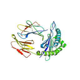
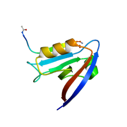
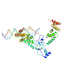
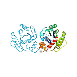
1H65
 
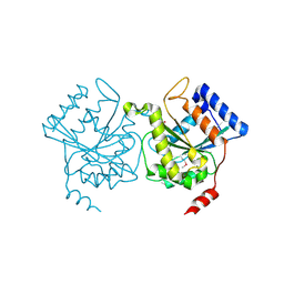 | | Crystal structure of pea Toc34 - a novel GTPase of the chloroplast protein translocon | | 分子名称: | CHLOROPLAST OUTER ENVELOPE PROTEIN OEP34, GUANOSINE-5'-DIPHOSPHATE, MAGNESIUM ION | | 著者 | Sun, Y.J, Forouhar, F, Li, H.M, Tu, S.L, Kao, S, Shr, H.L, Chou, C.C, Hsiao, C.D. | | 登録日 | 2001-06-06 | | 公開日 | 2002-01-29 | | 最終更新日 | 2019-06-12 | | 実験手法 | X-RAY DIFFRACTION (2 Å) | | 主引用文献 | Crystal Structure of Pea Toc34 - a Novel Gtpase of the Chloroplast Protein Translocon
Nat.Struct.Biol., 9, 2002
|
|
6NID
 | |
6NEK
 | |
6NH9
 | |
7CJQ
 | | Structure of DLA-88*001:04 | | 分子名称: | ARG-THR-ILE-SER-TYR-THR-TYR-PRO-PHE, Beta-2-microglobulin, MHC class I DLA-88 | | 著者 | Sun, Y.J, Ma, L.Z, Li, S. | | 登録日 | 2020-07-12 | | 公開日 | 2020-08-26 | | 最終更新日 | 2023-11-29 | | 実験手法 | X-RAY DIFFRACTION (2.7 Å) | | 主引用文献 | Structure of canine MHC class I at 2.7 Angstroms resolution
To Be Published
|
|
1E9L
 | |
5EZ1
 | |
6MYF
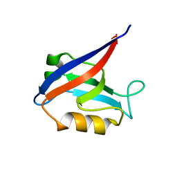 | |
6MYE
 | | Crystal structure of human Scribble PDZ1 domain in complex with internal PDZ binding motif of Src homology 3 domain-containing guanine nucleotide exchange factor (SGEF) | | 分子名称: | FORMIC ACID, Protein scribble homolog, Rho guanine nucleotide exchange factor 26, ... | | 著者 | Sun, Y.J, Hou, T, Gakhar, L, Fuentes, E.J. | | 登録日 | 2018-11-01 | | 公開日 | 2019-04-10 | | 最終更新日 | 2023-10-11 | | 実験手法 | X-RAY DIFFRACTION (1.1 Å) | | 主引用文献 | SGEF forms a complex with Scribble and Dlg1 and regulates epithelial junctions and contractility.
J.Cell Biol., 218, 2019
|
|
2VYF
 | | Crystal Structure of the DnaC | | 分子名称: | GOLD ION, REPLICATIVE DNA HELICASE | | 著者 | Lo, Y.H, Tsai, K.L, Sun, Y.J, Hsiao, C.D. | | 登録日 | 2008-07-23 | | 公開日 | 2008-12-30 | | 最終更新日 | 2011-07-13 | | 実験手法 | X-RAY DIFFRACTION (3.6 Å) | | 主引用文献 | The Crystal Structure of a Replicative Hexameric Helicase Dnac and its Complex with Single-Stranded DNA.
Nucleic Acids Res., 37, 2009
|
|
2W58
 | | Crystal Structure of the DnaI | | 分子名称: | ADENOSINE-5'-DIPHOSPHATE, MAGNESIUM ION, PHOSPHATE ION, ... | | 著者 | Tsai, K.L, Lo, Y.H, Sun, Y.J, Hsiao, C.D. | | 登録日 | 2008-12-08 | | 公開日 | 2009-12-22 | | 最終更新日 | 2019-10-16 | | 実験手法 | X-RAY DIFFRACTION (2.5 Å) | | 主引用文献 | Molecular Interplay between the Replicative Helicase Dnac and its Loader Protein Dnai from Geobacillus Kaustophilus.
J.Mol.Biol., 393, 2009
|
|
2VYE
 | | Crystal Structure of the DnaC-ssDNA complex | | 分子名称: | 5'-D(*TP*TP*TP*TP*TP*TP*TP*TP*TP)-3', REPLICATIVE DNA HELICASE | | 著者 | Lo, Y.H, Tsai, K.L, Sun, Y.J, Hsiao, C.D. | | 登録日 | 2008-07-23 | | 公開日 | 2008-12-30 | | 最終更新日 | 2023-12-13 | | 実験手法 | X-RAY DIFFRACTION (4.1 Å) | | 主引用文献 | The Crystal Structure of a Replicative Hexameric Helicase Dnac and its Complex with Single-Stranded DNA.
Nucleic Acids Res., 37, 2009
|
|
4UMK
 | | The complex of Spo0J and parS DNA in chromosomal partition system | | 分子名称: | DNA, PROBABLE CHROMOSOME-PARTITIONING PROTEIN PARB, SULFATE ION | | 著者 | Chen, B.W, Chu, C.H, Tung, J.Y, Hsu, C.E, Hsiao, C.D, Sun, Y.J. | | 登録日 | 2014-05-19 | | 公開日 | 2015-05-13 | | 最終更新日 | 2018-08-29 | | 実験手法 | X-RAY DIFFRACTION (3.096 Å) | | 主引用文献 | Insights into ParB spreading from the complex structure of Spo0J and parS.
Proc. Natl. Acad. Sci. U.S.A., 112, 2015
|
|
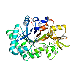
4A7W
 | | Crystal structure of uridylate kinase from Helicobacter pylori | | 分子名称: | GLYCEROL, GUANOSINE-5'-TRIPHOSPHATE, URIDYLATE KINASE | | 著者 | Chu, C.H, Chen, P.C, Liu, M.H, Sun, Y.J. | | 登録日 | 2011-11-15 | | 公開日 | 2012-06-27 | | 最終更新日 | 2023-12-20 | | 実験手法 | X-RAY DIFFRACTION (1.8 Å) | | 主引用文献 | Structures of Helicobacter Pylori Uridylate Kinase: Insight Into Release of the Product Udp
Acta Crystallogr.,Sect.D, 68, 2012
|
|
4A7X
 | | Crystal structure of uridylate kinase from Helicobacter pylori | | 分子名称: | URIDINE-5'-DIPHOSPHATE, URIDYLATE KINASE | | 著者 | Chu, C.H, Liu, M.H, Chen, P.C, Sun, Y.J. | | 登録日 | 2011-11-15 | | 公開日 | 2012-06-27 | | 最終更新日 | 2023-12-20 | | 実験手法 | X-RAY DIFFRACTION (2.49 Å) | | 主引用文献 | Structures of Helicobacter Pylori Uridylate Kinase: Insight Into Release of the Product Udp
Acta Crystallogr.,Sect.D, 68, 2012
|
|
6P3Q
 | | Calpain-5 (CAPN5) Protease Core (PC) | | 分子名称: | Calpain-5 | | 著者 | Velez, G, Sun, Y.J, Khan, S, Yang, J, Koster, H.J, Lokesh, G, Mahajan, V. | | 登録日 | 2019-05-24 | | 公開日 | 2020-02-05 | | 最終更新日 | 2024-04-03 | | 実験手法 | X-RAY DIFFRACTION (2.8 Å) | | 主引用文献 | Structural Insights into the Unique Activation Mechanisms of a Non-classical Calpain and Its Disease-Causing Variants.
Cell Rep, 30, 2020
|
|
5GPJ
 | |
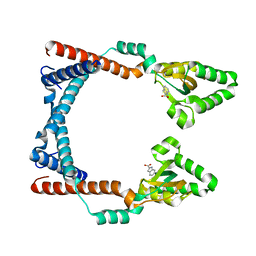
4GKG
 | | Crystal structure of the S-Helix Linker | | 分子名称: | C4-dicarboxylate transport sensor protein dctB, PHOSPHATE ION | | 著者 | Liu, J.W, Lu, D, Sun, Y.J, Wen, J, Yang, Y, Yang, J.G, Wei, X.L, Zhang, X.D, Wang, Y.P. | | 登録日 | 2012-08-11 | | 公開日 | 2013-08-28 | | 最終更新日 | 2024-04-03 | | 実験手法 | X-RAY DIFFRACTION (1.695 Å) | | 主引用文献 | Crystal structure of the S-Helix Linker
To be Published
|
|
6AEZ
 | | Crystal structure of human CCL5 trimer | | 分子名称: | C-C motif chemokine 5, SULFATE ION | | 著者 | Chen, Y.C, Li, K.M, Chen, P.J, Zarivach, R, Sun, Y.J, Sue, S.C. | | 登録日 | 2018-08-07 | | 公開日 | 2019-08-07 | | 最終更新日 | 2023-11-22 | | 実験手法 | X-RAY DIFFRACTION (1.63 Å) | | 主引用文献 | Integrative Model to Coordinate the Oligomerization and Aggregation Mechanisms of CCL5.
J.Mol.Biol., 432, 2020
|
|
7JL6
 | |
7DUV
 | | Structure of Sulfolobus solfataricus SegB protein | | 分子名称: | SULFATE ION, SegB | | 著者 | Yen, C.Y, Lin, M.G, Sun, Y.J, Hsiao, C.D. | | 登録日 | 2021-01-11 | | 公開日 | 2021-12-22 | | 最終更新日 | 2022-02-16 | | 実験手法 | X-RAY DIFFRACTION (3.2 Å) | | 主引用文献 | Chromosome segregation in Archaea: SegA- and SegB-DNA complex structures provide insights into segrosome assembly.
Nucleic Acids Res., 49, 2021
|
|
7DUT
 | | Structure of Sulfolobus solfataricus SegA protein | | 分子名称: | ADENOSINE-5'-DIPHOSPHATE, MAGNESIUM ION, SOJ protein (Soj) | | 著者 | Yen, C.Y, Lin, M.G, Hsiao, C.D, Sun, Y.J. | | 登録日 | 2021-01-11 | | 公開日 | 2021-12-22 | | 最終更新日 | 2023-11-29 | | 実験手法 | X-RAY DIFFRACTION (2.1 Å) | | 主引用文献 | Chromosome segregation in Archaea: SegA- and SegB-DNA complex structures provide insights into segrosome assembly.
Nucleic Acids Res., 49, 2021
|
|
7DV2
 | | Structure of Sulfolobus solfataricus SegB-DNA complex | | 分子名称: | DNA (5'-D(P*AP*CP*GP*TP*AP*GP*AP*AP*GP*AP*GP*TP*CP*TP*AP*GP*AP*CP*TP*G)-3'), DNA (5'-D(P*CP*AP*GP*TP*CP*TP*AP*GP*AP*CP*TP*CP*TP*TP*CP*TP*AP*CP*GP*TP*A)-3'), SegB | | 著者 | Yen, C.Y, Lin, M.G, Sun, Y.J, Hsiao, C.D. | | 登録日 | 2021-01-12 | | 公開日 | 2021-12-22 | | 最終更新日 | 2023-11-29 | | 実験手法 | X-RAY DIFFRACTION (3.1 Å) | | 主引用文献 | Chromosome segregation in Archaea: SegA- and SegB-DNA complex structures provide insights into segrosome assembly.
Nucleic Acids Res., 49, 2021
|
|
7DV3
 | | Structure of Sulfolobus solfataricus SegA-AMPPNP protein | | 分子名称: | MAGNESIUM ION, PHOSPHOAMINOPHOSPHONIC ACID-ADENYLATE ESTER, SOJ protein (Soj) | | 著者 | Yen, C.Y, Lin, M.G, Wu, C.T, Hsiao, C.D, Sun, Y.J. | | 登録日 | 2021-01-12 | | 公開日 | 2021-12-22 | | 最終更新日 | 2023-11-29 | | 実験手法 | X-RAY DIFFRACTION (2.6 Å) | | 主引用文献 | Chromosome segregation in Archaea: SegA- and SegB-DNA complex structures provide insights into segrosome assembly.
Nucleic Acids Res., 49, 2021
|
|
7DWR
 | | Structure of Sulfolobus solfataricus SegA-ADP complex bound to DNA | | 分子名称: | ADENOSINE-5'-DIPHOSPHATE, DNA (5'-D(P*AP*GP*GP*GP*TP*GP*TP*TP*CP*CP*AP*CP*GP*TP*GP*AP*AP*AP*CP*AP*GP*GP*GP*A)-3'), DNA (5'-D(P*TP*CP*CP*CP*TP*GP*TP*TP*TP*CP*AP*CP*GP*TP*GP*GP*AP*AP*CP*AP*CP*CP*CP*T)-3'), ... | | 著者 | Yen, C.Y, Lin, M.G, Hsiao, C.D, Sun, Y.J. | | 登録日 | 2021-01-17 | | 公開日 | 2021-12-22 | | 最終更新日 | 2023-11-29 | | 実験手法 | X-RAY DIFFRACTION (2.8 Å) | | 主引用文献 | Chromosome segregation in Archaea: SegA- and SegB-DNA complex structures provide insights into segrosome assembly.
Nucleic Acids Res., 49, 2021
|
|


