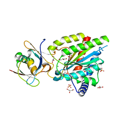
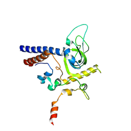
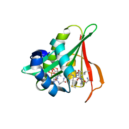
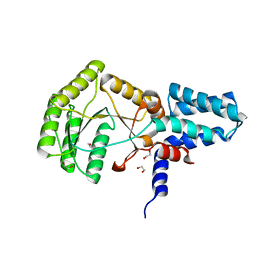
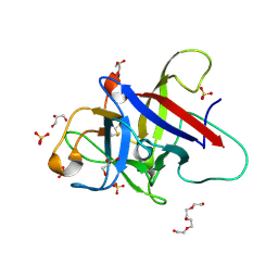
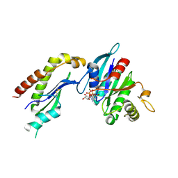
3ZRJ
 
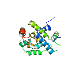 | | Complex of ClpV N-domain with VipB peptide | | 分子名称: | 1,2-ETHANEDIOL, CLPB PROTEIN, VIPB | | 著者 | Lenherr, E.D, Kopp, J, Sinning, I. | | 登録日 | 2011-06-16 | | 公開日 | 2011-07-06 | | 最終更新日 | 2019-05-15 | | 実験手法 | X-RAY DIFFRACTION (1.94 Å) | | 主引用文献 | Molecular Basis for the Unique Role of the Aaa+ Chaperone Clpv in Type Vi Protein Secretion.
J.Biol.Chem., 286, 2011
|
|
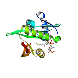
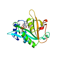
4B9Q
 | |
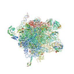
6EMF
 | |
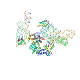
6EMG
 | |
6QTA
 | | Crystal structure of Rea1-MIDAS/Rsa4-UBL complex from Chaetomium thermophilum | | 分子名称: | GLYCEROL, MAGNESIUM ION, Midasin,Midasin, ... | | 著者 | Ahmed, Y.L, Thoms, M, Hurt, E, Sinning, I. | | 登録日 | 2019-02-22 | | 公開日 | 2019-08-07 | | 最終更新日 | 2024-01-24 | | 実験手法 | X-RAY DIFFRACTION (1.89 Å) | | 主引用文献 | Crystal structures of Rea1-MIDAS bound to its ribosome assembly factor ligands resembling integrin-ligand-type complexes.
Nat Commun, 10, 2019
|
|
6QTB
 | | Crystal structure of Rea1-MIDAS/Ytm1-UBL complex from Chaetomium thermophilum | | 分子名称: | GLYCEROL, MAGNESIUM ION, Midasin,Midasin, ... | | 著者 | Ahmed, Y.L, Thoms, M, Hurt, E, Sinning, I. | | 登録日 | 2019-02-22 | | 公開日 | 2019-08-07 | | 最終更新日 | 2024-01-24 | | 実験手法 | X-RAY DIFFRACTION (1.89 Å) | | 主引用文献 | Crystal structures of Rea1-MIDAS bound to its ribosome assembly factor ligands resembling integrin-ligand-type complexes.
Nat Commun, 10, 2019
|
|
6QT8
 | | Crystal structure of Rea1-MIDAS domain from Chaetomium thermophilum | | 分子名称: | GLYCEROL, IODIDE ION, Midasin, ... | | 著者 | Ahmed, Y.L, Thoms, M, Hurt, E, Sinning, I. | | 登録日 | 2019-02-22 | | 公開日 | 2019-08-07 | | 最終更新日 | 2024-05-15 | | 実験手法 | X-RAY DIFFRACTION (2.33 Å) | | 主引用文献 | Crystal structures of Rea1-MIDAS bound to its ribosome assembly factor ligands resembling integrin-ligand-type complexes.
Nat Commun, 10, 2019
|
|
6YGU
 | | Crystal structure of the minimal Mtr4-Red1 complex (single chain) from Chaetomium thermophilum | | 分子名称: | 1,2-ETHANEDIOL, ACETATE ION, ATP dependent RNA helicase (Dob1)-like protein, ... | | 著者 | Dobrev, N, Ahmed, Y.L, Sinning, I. | | 登録日 | 2020-03-27 | | 公開日 | 2021-05-05 | | 最終更新日 | 2024-05-15 | | 実験手法 | X-RAY DIFFRACTION (1.99 Å) | | 主引用文献 | The zinc-finger protein Red1 orchestrates MTREC submodules and binds the Mtl1 helicase arch domain.
Nat Commun, 12, 2021
|
|
6YFV
 | | Crystal structure of Mtr4-Red1 minimal complex from Chaetomium thermophilum | | 分子名称: | 4-(2-HYDROXYETHYL)-1-PIPERAZINE ETHANESULFONIC ACID, ATP dependent RNA helicase (Dob1)-like protein, Red1, ... | | 著者 | Dobrev, N, Ahmed, Y.L, Sinning, I. | | 登録日 | 2020-03-26 | | 公開日 | 2021-05-05 | | 最終更新日 | 2021-06-23 | | 実験手法 | X-RAY DIFFRACTION (2.75 Å) | | 主引用文献 | The zinc-finger protein Red1 orchestrates MTREC submodules and binds the Mtl1 helicase arch domain.
Nat Commun, 12, 2021
|
|
6RYI
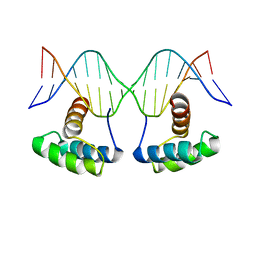 | | WUS-HD bound to G-Box DNA | | 分子名称: | DNA (5'-D(P*CP*CP*CP*AP*TP*CP*AP*CP*GP*TP*GP*AP*CP*GP*AP*C)-3'), DNA (5'-D(P*GP*TP*CP*GP*TP*CP*AP*CP*GP*TP*GP*AP*TP*GP*GP*G)-3'), Protein WUSCHEL | | 著者 | Sloan, J.J, Wild, K, Sinning, I. | | 登録日 | 2019-06-10 | | 公開日 | 2020-04-29 | | 最終更新日 | 2024-01-24 | | 実験手法 | X-RAY DIFFRACTION (2.691 Å) | | 主引用文献 | Structural basis for the complex DNA binding behavior of the plant stem cell regulator WUSCHEL.
Nat Commun, 11, 2020
|
|
6RYD
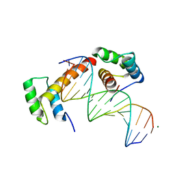 | | WUS-HD bound to TGAA DNA | | 分子名称: | DNA (5'-D(*AP*GP*TP*GP*TP*AP*TP*GP*AP*AP*TP*GP*AP*AP*CP*G)-3'), DNA (5'-D(*CP*GP*TP*TP*CP*AP*TP*TP*CP*AP*TP*AP*CP*AP*CP*T)-3'), MAGNESIUM ION, ... | | 著者 | Sloan, J.J, Wild, K, Sinning, I. | | 登録日 | 2019-06-10 | | 公開日 | 2020-04-29 | | 最終更新日 | 2024-01-24 | | 実験手法 | X-RAY DIFFRACTION (1.575 Å) | | 主引用文献 | Structural basis for the complex DNA binding behavior of the plant stem cell regulator WUSCHEL.
Nat Commun, 11, 2020
|
|
6RYL
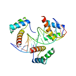 | | WUS-HD bound to TAAT DNA | | 分子名称: | DNA (5'-D(P*CP*AP*CP*AP*AP*CP*CP*CP*AP*TP*TP*AP*AP*CP*AP*C)-3'), DNA (5'-D(P*GP*TP*GP*TP*TP*AP*AP*TP*GP*GP*GP*TP*TP*GP*TP*G)-3'), Protein WUSCHEL | | 著者 | Sloan, J.J, Wild, K, Sinning, I. | | 登録日 | 2019-06-10 | | 公開日 | 2020-04-29 | | 最終更新日 | 2024-01-24 | | 実験手法 | X-RAY DIFFRACTION (2.63 Å) | | 主引用文献 | Structural basis for the complex DNA binding behavior of the plant stem cell regulator WUSCHEL.
Nat Commun, 11, 2020
|
|
6Z00
 | | Arabidopsis thaliana Naa50 in complex with bisubstrate analogue CoA-Ac-MVNAL | | 分子名称: | Acyl-CoA N-acyltransferases (NAT) superfamily protein, CARBOXYMETHYL COENZYME *A, MET-VAL-ASN-ALA-LEU | | 著者 | Weidenhausen, J, Kopp, J, Lapouge, K, Sinning, I. | | 登録日 | 2020-05-07 | | 公開日 | 2020-12-30 | | 最終更新日 | 2024-05-01 | | 実験手法 | X-RAY DIFFRACTION (1.42 Å) | | 主引用文献 | Structural and functional characterization of the N-terminal acetyltransferase Naa50.
Structure, 29, 2021
|
|
6YZZ
 | | Arabidopsis thaliana Naa50 in complex with AcCoA | | 分子名称: | ACETYL COENZYME *A, N-alpha-acetyltransferase 50 | | 著者 | Weidenhausen, J, Kopp, J, Lapouge, K, Sinning, I. | | 登録日 | 2020-05-07 | | 公開日 | 2020-12-30 | | 最終更新日 | 2024-01-24 | | 実験手法 | X-RAY DIFFRACTION (1.79 Å) | | 主引用文献 | Structural and functional characterization of the N-terminal acetyltransferase Naa50.
Structure, 29, 2021
|
|
6RY3
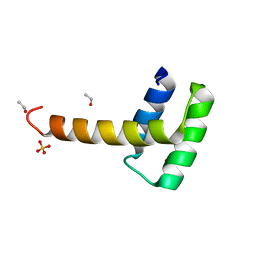 | | Structure of the WUS homeodomain | | 分子名称: | ACETYL GROUP, Protein WUSCHEL, SULFATE ION | | 著者 | Sloan, J.J, Wild, K, Sinning, I. | | 登録日 | 2019-06-10 | | 公開日 | 2020-04-29 | | 最終更新日 | 2024-01-24 | | 実験手法 | X-RAY DIFFRACTION (1.374 Å) | | 主引用文献 | Structural basis for the complex DNA binding behavior of the plant stem cell regulator WUSCHEL.
Nat Commun, 11, 2020
|
|
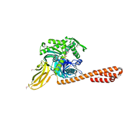
6SO5
 | | Homo sapiens WRB/CAML heterotetramer in complex with a TRC40 dimer | | 分子名称: | ATPase ASNA1, Calcium signal-modulating cyclophilin ligand, Tail-anchored protein insertion receptor WRB, ... | | 著者 | McDowell, M.A, Heimes, M, Wild, K, Flemming, D, Sinning, I. | | 登録日 | 2019-08-29 | | 公開日 | 2020-09-09 | | 最終更新日 | 2021-02-17 | | 実験手法 | ELECTRON MICROSCOPY (4.2 Å) | | 主引用文献 | Structural Basis of Tail-Anchored Membrane Protein Biogenesis by the GET Insertase Complex.
Mol.Cell, 80, 2020
|
|
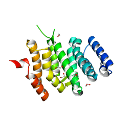
2YHS
 | | Structure of the E. coli SRP receptor FtsY | | 分子名称: | 1,2-ETHANEDIOL, CELL DIVISION PROTEIN FTSY | | 著者 | Stjepanovic, G, Bange, G, Wild, K, Sinning, I. | | 登録日 | 2011-05-05 | | 公開日 | 2011-05-18 | | 最終更新日 | 2023-12-20 | | 実験手法 | X-RAY DIFFRACTION (1.6 Å) | | 主引用文献 | Lipids Trigger a Conformational Switch that Regulates Signal Recognition Particle (Srp)-Mediated Protein Targeting.
J.Biol.Chem., 286, 2011
|
|
4IPA
 | | Structure of a thermophilic Arx1 | | 分子名称: | Putative curved DNA-binding protein, SULFATE ION | | 著者 | Bange, G, Sinning, I. | | 登録日 | 2013-01-09 | | 公開日 | 2013-01-30 | | 最終更新日 | 2023-09-20 | | 実験手法 | X-RAY DIFFRACTION (2.3 Å) | | 主引用文献 | Consistent mutational paths predict eukaryotic thermostability.
BMC Evol Biol, 13, 2013
|
|
3BS6
 | |
6ZQQ
 | | Structure of the Pmt3-MIR domain with bound ligands | | 分子名称: | GLYCEROL, PMT3 isoform 1 | | 著者 | Wild, K, Chiapparino, A, Hackmann, Y, Mortensen, S, Sinning, I. | | 登録日 | 2020-07-10 | | 公開日 | 2020-12-23 | | 最終更新日 | 2024-01-31 | | 実験手法 | X-RAY DIFFRACTION (1.9 Å) | | 主引用文献 | Functional implications of MIR domains in protein O -mannosylation.
Elife, 9, 2020
|
|
6ZQP
 | | Structure of the Pmt2-MIR domain with bound ligands | | 分子名称: | GLYCEROL, PMT2 isoform 1, SULFATE ION, ... | | 著者 | Wild, K, Chiapparino, A, Hackmann, Y, Mortensen, S, Sinning, I. | | 登録日 | 2020-07-10 | | 公開日 | 2020-12-23 | | 最終更新日 | 2024-01-31 | | 実験手法 | X-RAY DIFFRACTION (1.6 Å) | | 主引用文献 | Functional implications of MIR domains in protein O -mannosylation.
Elife, 9, 2020
|
|
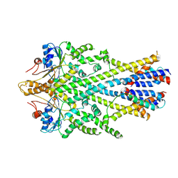
6ZMP
 | |
2J28
 | | MODEL OF E. COLI SRP BOUND TO 70S RNCS | | 分子名称: | 23S RIBOSOMAL RNA, 4.5S SIGNAL RECOGNITION PARTICLE RNA, 50S ribosomal protein L11, ... | | 著者 | Halic, M, Blau, M, Becker, T, Mielke, T, Pool, M.R, Wild, K, Sinning, I, Beckmann, R. | | 登録日 | 2006-08-16 | | 公開日 | 2006-11-08 | | 最終更新日 | 2024-05-08 | | 実験手法 | ELECTRON MICROSCOPY (9.5 Å) | | 主引用文献 | Following the Signal Sequence from Ribosomal Tunnel Exit to Signal Recognition Particle
Nature, 444, 2006
|
|
2J37
 | | MODEL OF MAMMALIAN SRP BOUND TO 80S RNCS | | 分子名称: | 60S RIBOSOMAL PROTEIN L23, RIBOSOMAL PROTEIN L31, RIBOSOMAL PROTEIN L35, ... | | 著者 | Halic, M, Blau, M, Becker, T, Mielke, T, Pool, M.R, Wild, K, Sinning, I, Beckmann, R. | | 登録日 | 2006-08-18 | | 公開日 | 2006-11-08 | | 最終更新日 | 2024-05-08 | | 実験手法 | ELECTRON MICROSCOPY (8.7 Å) | | 主引用文献 | Following the signal sequence from ribosomal tunnel exit to signal recognition particle.
Nature, 444, 2006
|
|
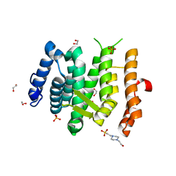
2FH5
 | | The Structure of the Mammalian SRP Receptor | | 分子名称: | GUANOSINE-5'-TRIPHOSPHATE, MAGNESIUM ION, Signal recognition particle receptor alpha subunit, ... | | 著者 | Schlenker, O, Wild, K, Sinning, I. | | 登録日 | 2005-12-23 | | 公開日 | 2006-01-31 | | 最終更新日 | 2011-07-13 | | 実験手法 | X-RAY DIFFRACTION (2.45 Å) | | 主引用文献 | The structure of the mammalian signal recognition particle (SRP) receptor as prototype for the interaction of small GTPases with Longin domains.
J.Biol.Chem., 281, 2006
|
|


