7VGW
 
 | | Yeast gid10 with Pro-peptide | | 分子名称: | BJ4_G0041530.mRNA.1.CDS.1 | | 著者 | Shin, J.S, Park, S.H, Kim, L, Heo, J, Song, H.K. | | 登録日 | 2021-09-19 | | 公開日 | 2022-07-27 | | 最終更新日 | 2023-11-29 | | 実験手法 | X-RAY DIFFRACTION (2.8 Å) | | 主引用文献 | Crystal structure of yeast Gid10 in complex with Pro/N-degron.
Biochem.Biophys.Res.Commun., 582, 2021
|
|
2MPS
 | |
2H8G
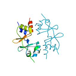
 | | 5'-Methylthioadenosine Nucleosidase from Arabidopsis thaliana | | 分子名称: | 5'-Methylthioadenosine Nucleosidase, ADENINE | | 著者 | Park, E.Y, Oh, S.I, Nam, M.J, Shin, J.S, Kim, K.N, Song, H.K. | | 登録日 | 2006-06-07 | | 公開日 | 2006-10-10 | | 最終更新日 | 2024-03-13 | | 実験手法 | X-RAY DIFFRACTION (1.5 Å) | | 主引用文献 | Crystal structure of 5'-methylthioadenosine nucleosidase from Arabidopsis thaliana at 1.5-A resolution
Proteins, 65, 2006
|
|
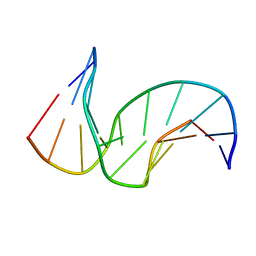
1PIB
 | | Solution structure of DNA containing CPD opposited by GA | | 分子名称: | 5'-D(*CP*GP*CP*AP*TP*TP*AP*CP*GP*C)-3', 5'-D(*GP*CP*GP*TP*GP*AP*TP*GP*CP*G)-3' | | 著者 | Lee, J.H, Park, C.J, Shin, J.S, Choi, B.S. | | 登録日 | 2003-05-30 | | 公開日 | 2004-05-18 | | 最終更新日 | 2022-02-23 | | 実験手法 | SOLUTION NMR | | 主引用文献 | NMR structure of the DNA decamer duplex containing double T*G mismatches of cis-syn cyclobutane pyrimidine dimer: implications for DNA damage recognition by the XPC-hHR23B complex.
Nucleic Acids Res., 32, 2004
|
|
1SNH
 | | Solution structure of the DNA Decamer Duplex Containing Double TG Mismatches of Cis-syn Cyclobutane Pyrimidine Dimer | | 分子名称: | 5'-D(*CP*GP*CP*AP*TP*TP*AP*CP*GP*C)-3', 5'-D(*GP*CP*GP*TP*GP*GP*TP*GP*CP*G)-3' | | 著者 | Lee, J.H, Park, C.J, Shin, J.S, Ikegami, T, Akutsu, H, Choi, B.S. | | 登録日 | 2004-03-11 | | 公開日 | 2004-05-18 | | 最終更新日 | 2022-03-02 | | 実験手法 | SOLUTION NMR | | 主引用文献 | NMR structure of the DNA decamer duplex containing double T*G mismatches of cis-syn cyclobutane pyrimidine dimer: implications for DNA damage recognition by the XPC-hHR23B complex.
Nucleic Acids Res., 32, 2004
|
|
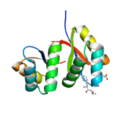
4GQV
 | | Crystal structure of CBS-pair protein, CBSX1 from Arabidopsis thaliana | | 分子名称: | CBS domain-containing protein CBSX1, chloroplastic | | 著者 | Jeong, B.-C, Park, S.H, Yoo, K.S, Shin, J.S, Song, H.K. | | 登録日 | 2012-08-24 | | 公開日 | 2013-01-16 | | 最終更新日 | 2024-03-20 | | 実験手法 | X-RAY DIFFRACTION (2.392 Å) | | 主引用文献 | Crystal structure of the single cystathionine beta-synthase domain-containing protein CBSX1 from Arabidopsis thaliana
Biochem.Biophys.Res.Commun., 430, 2013
|
|
7D34
 | | AtClpS1-peptide complex | | 分子名称: | ACETIC ACID, ALANINE, ATP-dependent Clp protease adapter protein CLPS1, ... | | 著者 | Heo, J, Kim, L, Kwon, D.H, Song, H.K. | | 登録日 | 2020-09-18 | | 公開日 | 2021-04-28 | | 最終更新日 | 2023-11-29 | | 実験手法 | X-RAY DIFFRACTION (2.007 Å) | | 主引用文献 | Structural basis for the N-degron specificity of ClpS1 from Arabidopsis thaliana.
Protein Sci., 30, 2021
|
|
3SL7
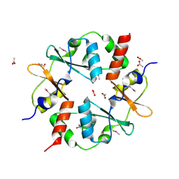 | | Crystal structure of CBS-pair protein, CBSX2 from Arabidopsis thaliana | | 分子名称: | ACETATE ION, CBS domain-containing protein CBSX2, GLYCEROL | | 著者 | Jeong, B.-C, Lee, M.-R, Song, H.K. | | 登録日 | 2011-06-24 | | 公開日 | 2011-11-09 | | 最終更新日 | 2013-10-09 | | 実験手法 | X-RAY DIFFRACTION (1.905 Å) | | 主引用文献 | Single cystathionine beta-synthase domain-containing proteins modulate development by regulating the thioredoxin system in Arabidopsis
Plant Cell, 23, 2011
|
|
5H60
 | | Structure of Transferase mutant-C23S,C199S | | 分子名称: | MANGANESE (II) ION, Transferase, URIDINE-5'-DIPHOSPHATE | | 著者 | Park, J.B, Yoo, Y, Kim, J. | | 登録日 | 2016-11-10 | | 公開日 | 2017-12-20 | | 最終更新日 | 2018-10-31 | | 実験手法 | X-RAY DIFFRACTION (3.64 Å) | | 主引用文献 | Structural basis for arginine glycosylation of host substrates by bacterial effector proteins.
Nat Commun, 9, 2018
|
|
5H61
 | |
5H5Y
 | |
5H63
 | | Structure of Transferase mutant-C23S,C199S | | 分子名称: | MANGANESE (II) ION, Transferase, URIDINE-DIPHOSPHATE-N-ACETYLGLUCOSAMINE | | 著者 | Park, J.B, Yoo, Y, Kim, J. | | 登録日 | 2016-11-10 | | 公開日 | 2017-12-20 | | 最終更新日 | 2024-03-20 | | 実験手法 | X-RAY DIFFRACTION (1.92 Å) | | 主引用文献 | Structural basis for arginine glycosylation of host substrates by bacterial effector proteins.
Nat Commun, 9, 2018
|
|
5H62
 | | Structure of Transferase mutant-C23S,C199S | | 分子名称: | 1,2-ETHANEDIOL, MANGANESE (II) ION, Transferase, ... | | 著者 | Park, J.B, Yoo, Y, Kim, J. | | 登録日 | 2016-11-10 | | 公開日 | 2017-12-27 | | 最終更新日 | 2024-03-20 | | 実験手法 | X-RAY DIFFRACTION (1.66 Å) | | 主引用文献 | Structural basis for arginine glycosylation of host substrates by bacterial effector proteins.
Nat Commun, 9, 2018
|
|
3KYG
 | | Crystal structure of VCA0042 (L135R) complexed with c-di-GMP | | 分子名称: | GUANOSINE-5'-MONOPHOSPHATE, Putative uncharacterized protein VCA0042 | | 著者 | Ryu, K.S, Ko, J, Kim, H, Choi, B.S. | | 登録日 | 2009-12-06 | | 公開日 | 2010-04-14 | | 最終更新日 | 2021-11-10 | | 実験手法 | X-RAY DIFFRACTION (2.1 Å) | | 主引用文献 | Structure of PP4397 Reveals the Molecular Basis for Different c-di-GMP Binding Modes by Pilz Domain Proteins.
J.Mol.Biol., 398, 2010
|
|
3KYF
 | | Crystal structure of P4397 complexed with c-di-GMP | | 分子名称: | GUANOSINE-5'-MONOPHOSPHATE, Putative uncharacterized protein | | 著者 | Ryu, K.S, Ko, J, Kim, H, Choi, B.S. | | 登録日 | 2009-12-06 | | 公開日 | 2010-04-14 | | 最終更新日 | 2024-03-20 | | 実験手法 | X-RAY DIFFRACTION (2.1 Å) | | 主引用文献 | Structure of PP4397 Reveals the Molecular Basis for Different c-di-GMP Binding Modes by Pilz Domain Proteins.
J.Mol.Biol., 398, 2010
|
|
6K3L
 | | Crystal structure of CX-4945 bound Cka1 from C. neoformans | | 分子名称: | 5-[(3-chlorophenyl)amino]benzo[c][2,6]naphthyridine-8-carboxylic acid, CMGC/CK2 protein kinase, SULFATE ION | | 著者 | Cho, H.S, Yoo, Y. | | 登録日 | 2019-05-20 | | 公開日 | 2019-11-06 | | 最終更新日 | 2023-11-22 | | 実験手法 | X-RAY DIFFRACTION (2.09 Å) | | 主引用文献 | Structural analysis of fungal pathogenicity-related casein kinase alpha subunit, Cka1, in the human fungal pathogen Cryptococcus neoformans.
Sci Rep, 9, 2019
|
|
6KO6
 | | Crystal structure of AMPPNP bound Cka1 from C. neoformans | | 分子名称: | CMGC/CK2 protein kinase, MAGNESIUM ION, PHOSPHOAMINOPHOSPHONIC ACID-ADENYLATE ESTER, ... | | 著者 | Cho, H.S, Yoo, Y. | | 登録日 | 2019-08-08 | | 公開日 | 2019-11-06 | | 最終更新日 | 2023-11-22 | | 実験手法 | X-RAY DIFFRACTION (2.4 Å) | | 主引用文献 | Structural analysis of fungal pathogenicity-related casein kinase alpha subunit, Cka1, in the human fungal pathogen Cryptococcus neoformans.
Sci Rep, 9, 2019
|
|
4GQW
 | |
4GQY
 | | Crystal structure of CBSX2 in complex with AMP | | 分子名称: | ADENOSINE MONOPHOSPHATE, CBS domain-containing protein CBSX2, chloroplastic | | 著者 | Jeong, B.C, Song, H.K. | | 登録日 | 2012-08-24 | | 公開日 | 2013-07-24 | | 最終更新日 | 2024-03-20 | | 実験手法 | X-RAY DIFFRACTION (2.193 Å) | | 主引用文献 | Change in single cystathionine beta-synthase domain-containing protein from a bent to flat conformation upon adenosine monophosphate binding
J.Struct.Biol., 183, 2013
|
|


