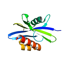
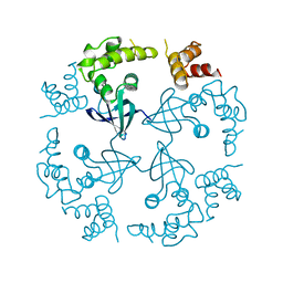
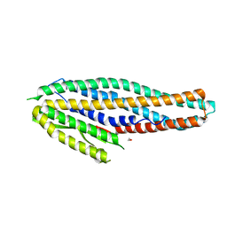
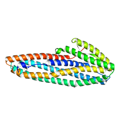
1MJU
 
 | | 1.22 ANGSTROM RESOLUTION CRYSTAL STRUCTURE OF THE FAB FRAGMENT OF ESTEROLYTIC ANTIBODY MS6-12 | | 分子名称: | GLYCEROL, IMMUNOGLOBULIN MS6-12 | | 著者 | Ruzheinikov, S.N, Muranova, T.A, Sedelnikova, S.E, Partridge, L.J, Blackburn, G.M, Murray, I.A, Kakinuma, H, Takashi, N, Shimazaki, K, Sun, J, Nishi, Y, Rice, D.W. | | 登録日 | 2002-08-28 | | 公開日 | 2003-09-23 | | 最終更新日 | 2019-12-25 | | 実験手法 | X-RAY DIFFRACTION (1.22 Å) | | 主引用文献 | High-resolution crystal structure of the Fab-fragments of a family of mouse catalytic antibodies with esterase activity
J.Mol.Biol., 332, 2003
|
|
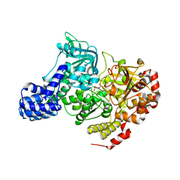
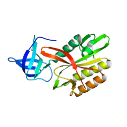
1MJ7
 | | Crystal Structure Of The Complex Of The Fab fragment of Esterolytic Antibody MS5-393 and A Transition-State Analog | | 分子名称: | IMMUNOGLOBULIN MS5-393, N-{[2-({[1-(4-CARBOXYBUTANOYL)AMINO]-2-PHENYLETHYL}-HYDROXYPHOSPHINYL)OXY]ACETYL}-2-PHENYLETHYLAMINE | | 著者 | Ruzheinikov, S.N, Muranova, T.A, Sedelnikova, S.E, Partridge, L.J, Blackburn, G.M, Murray, I.A, Kakinuma, H, Takashi, N, Shimazaki, K, Sun, J, Nishi, Y, Rice, D.W. | | 登録日 | 2002-08-27 | | 公開日 | 2003-09-23 | | 最終更新日 | 2011-11-16 | | 実験手法 | X-RAY DIFFRACTION (2.25 Å) | | 主引用文献 | High-resolution crystal structure of the Fab-fragments of a family of mouse catalytic antibodies with esterase activity
J.Mol.Biol., 332, 2003
|
|
1KT9
 | | Crystal Structure of C. elegans Ap4A Hydrolase | | 分子名称: | Diadenosine Tetraphosphate Hydrolase | | 著者 | Bailey, S, Sedelnikova, S.E, Blackburn, G.M, Abdelghany, H.M, Baker, P.J, McLennan, A.G, Rafferty, J.B. | | 登録日 | 2002-01-15 | | 公開日 | 2002-05-08 | | 最終更新日 | 2024-02-14 | | 実験手法 | X-RAY DIFFRACTION (1.98 Å) | | 主引用文献 | The crystal structure of diadenosine tetraphosphate hydrolase from Caenorhabditis elegans in free and binary complex forms
Structure, 10, 2002
|
|
1L5J
 | | CRYSTAL STRUCTURE OF E. COLI ACONITASE B. | | 分子名称: | ACONITATE ION, Aconitate hydratase 2, FE3-S4 CLUSTER | | 著者 | Williams, C.H, Stillman, T.J, Barynin, V.V, Sedelnikova, S.E, Tang, Y, Green, J, Guest, J.R, Artymiuk, P.J. | | 登録日 | 2002-03-07 | | 公開日 | 2002-06-12 | | 最終更新日 | 2024-02-14 | | 実験手法 | X-RAY DIFFRACTION (2.4 Å) | | 主引用文献 | E. coli aconitase B structure reveals a HEAT-like domain with implications for protein-protein recognition.
Nat.Struct.Biol., 9, 2002
|
|
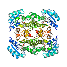
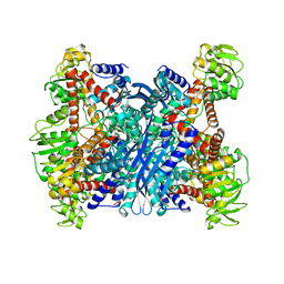
1LEH
 | | LEUCINE DEHYDROGENASE FROM BACILLUS SPHAERICUS | | 分子名称: | LEUCINE DEHYDROGENASE | | 著者 | Baker, P.J, Turnbull, A.P, Sedelnikova, S.E, Stillman, T.J, Rice, D.W. | | 登録日 | 1995-06-09 | | 公開日 | 1996-12-23 | | 最終更新日 | 2024-02-14 | | 実験手法 | X-RAY DIFFRACTION (2.2 Å) | | 主引用文献 | A role for quaternary structure in the substrate specificity of leucine dehydrogenase.
Structure, 3, 1995
|
|
1NO1
 | | Structure of truncated variant of B.subtilis SPP1 phage G39P helicase loader/inhibitor protein | | 分子名称: | replisome organizer | | 著者 | Bailey, S, Sedelnikova, S.E, Mesa, P, Ayora, S, Waltho, J.P, Ashcroft, A.E, Baron, A.J, Alonso, J.C, Rafferty, J.B. | | 登録日 | 2003-01-15 | | 公開日 | 2003-05-06 | | 最終更新日 | 2011-07-13 | | 実験手法 | X-RAY DIFFRACTION (2.4 Å) | | 主引用文献 | Structural analysis of Bacillus subtilis SPP1 phage helicase loader protein G39P
J.Biol.Chem., 278, 2003
|
|
1DFI
 | |
1DFG
 | | X-RAY STRUCTURE OF ESCHERICHIA COLI ENOYL REDUCTASE WITH BOUND NAD AND BENZO-DIAZABORINE | | 分子名称: | 2-(TOLUENE-4-SULFONYL)-2H-BENZO[D][1,2,3]DIAZABORININ-1-OL, ENOYL ACYL CARRIER PROTEIN REDUCTASE, NICOTINAMIDE-ADENINE-DINUCLEOTIDE | | 著者 | Baldock, C, Rafferty, J.B, Rice, D.W. | | 登録日 | 1997-01-16 | | 公開日 | 1998-01-28 | | 最終更新日 | 2024-04-03 | | 実験手法 | X-RAY DIFFRACTION (2.5 Å) | | 主引用文献 | A mechanism of drug action revealed by structural studies of enoyl reductase.
Science, 274, 1996
|
|
1DFH
 | | X-RAY STRUCTURE OF ESCHERICHIA COLI ENOYL REDUCTASE WITH BOUND NAD AND THIENO-DIAZABORINE | | 分子名称: | 6-METHYL-2(PROPANE-1-SULFONYL)-2H-THIENO[3,2-D][1,2,3]DIAZABORININ-1-OL, ENOYL ACYL CARRIER PROTEIN REDUCTASE, NICOTINAMIDE-ADENINE-DINUCLEOTIDE | | 著者 | Baldock, C, Rafferty, J.B, Rice, D.W. | | 登録日 | 1997-01-16 | | 公開日 | 1998-01-28 | | 最終更新日 | 2024-04-03 | | 実験手法 | X-RAY DIFFRACTION (2.2 Å) | | 主引用文献 | A mechanism of drug action revealed by structural studies of enoyl reductase.
Science, 274, 1996
|
|
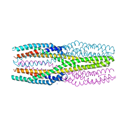
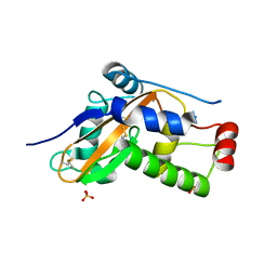
5LBM
 | | The asymmetric tetrameric structure of the formaldehyde sensing transcriptional repressor FrmR from Escherichia coli | | 分子名称: | FORMYL GROUP, Transcriptional repressor FrmR | | 著者 | Bisson, C, Baker, P.J, Green, J, Chivers, P.T. | | 登録日 | 2016-06-16 | | 公開日 | 2016-12-21 | | 最終更新日 | 2017-08-30 | | 実験手法 | X-RAY DIFFRACTION (2.7 Å) | | 主引用文献 | The mechanism of a formaldehyde-sensing transcriptional regulator.
Sci Rep, 6, 2016
|
|
1CUK
 | |
6R1J
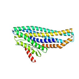
 | | Structure of the soluble AhlC triple head mutant of the tripartite alpha-pore forming toxin, AHL, from Aeromonas hydrophila. | | 分子名称: | SODIUM ION, Uncharacterized protein | | 著者 | Churchill-Angus, A.M, Wilson, J.S, Baker, P.J. | | 登録日 | 2019-03-14 | | 公開日 | 2019-07-10 | | 最終更新日 | 2024-01-24 | | 実験手法 | X-RAY DIFFRACTION (1.92 Å) | | 主引用文献 | Identification and structural analysis of the tripartite alpha-pore forming toxin of Aeromonas hydrophila.
Nat Commun, 10, 2019
|
|
6EZM
 | | Imidazoleglycerol-phosphate dehydratase from Saccharomyces cerevisiae | | 分子名称: | Imidazoleglycerol-phosphate dehydratase, MANGANESE (II) ION, [(2R)-2-hydroxy-3-(1H-1,2,4-triazol-1-yl)propyl]phosphonic acid | | 著者 | Rawson, S, Bisson, C, Hurdiss, D.L, Muench, S.P. | | 登録日 | 2017-11-15 | | 公開日 | 2018-02-07 | | 最終更新日 | 2024-05-15 | | 実験手法 | ELECTRON MICROSCOPY (3.2 Å) | | 主引用文献 | Elucidating the structural basis for differing enzyme inhibitor potency by cryo-EM.
Proc. Natl. Acad. Sci. U.S.A., 115, 2018
|
|
6EZJ
 | | Imidazoleglycerol-phosphate dehydratase | | 分子名称: | Imidazoleglycerol-phosphate dehydratase 2, chloroplastic, MANGANESE (II) ION, ... | | 著者 | Rawson, S, Bisson, C, Hurdiss, D.L, Muench, S.P. | | 登録日 | 2017-11-15 | | 公開日 | 2018-02-07 | | 最終更新日 | 2024-05-15 | | 実験手法 | ELECTRON MICROSCOPY (3.1 Å) | | 主引用文献 | Elucidating the structural basis for differing enzyme inhibitor potency by cryo-EM.
Proc. Natl. Acad. Sci. U.S.A., 115, 2018
|
|
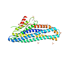
4K1P
 | | Structure of the NheA component of the Nhe toxin from Bacillus cereus | | 分子名称: | 1,2-ETHANEDIOL, NheA, SULFATE ION | | 著者 | Ganash, M, Phung, D, Artymiuk, P.J. | | 登録日 | 2013-04-05 | | 公開日 | 2013-09-18 | | 最終更新日 | 2024-02-28 | | 実験手法 | X-RAY DIFFRACTION (2.05 Å) | | 主引用文献 | Structure of the NheA Component of the Nhe Toxin from Bacillus cereus: Implications for Function.
Plos One, 8, 2013
|
|
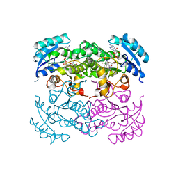
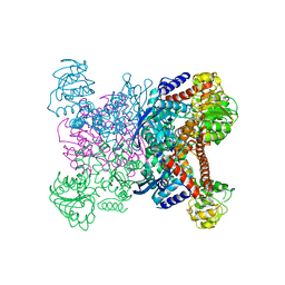
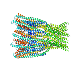
1BVU
 | | GLUTAMATE DEHYDROGENASE FROM THERMOCOCCUS LITORALIS | | 分子名称: | PROTEIN (GLUTAMATE DEHYDROGENASE) | | 著者 | Baker, P.J, Britton, K.L, Yip, K.S, Stillman, T.J, Rice, D.W. | | 登録日 | 1999-07-20 | | 公開日 | 1999-09-18 | | 最終更新日 | 2023-12-27 | | 実験手法 | X-RAY DIFFRACTION (2.5 Å) | | 主引用文献 | Structure determination of the glutamate dehydrogenase from the hyperthermophile Thermococcus litoralis and its comparison with that from Pyrococcus furiosus
J.Mol.Biol., 293, 1999
|
|
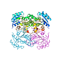
1GTM
 | | STRUCTURE OF GLUTAMATE DEHYDROGENASE | | 分子名称: | GLUTAMATE DEHYDROGENASE, SULFATE ION | | 著者 | Yip, K.S.P, Stillman, T.J, Britton, K.L, Pasquo, A, Rice, D.W. | | 登録日 | 1996-08-22 | | 公開日 | 1997-01-11 | | 最終更新日 | 2024-02-07 | | 実験手法 | X-RAY DIFFRACTION (2.2 Å) | | 主引用文献 | The structure of Pyrococcus furiosus glutamate dehydrogenase reveals a key role for ion-pair networks in maintaining enzyme stability at extreme temperatures.
Structure, 3, 1995
|
|
6ZZB
 | |
6ZZH
 | |
7A27
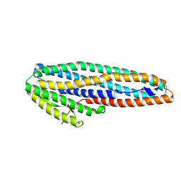 | |
6ZZ5
 | | Structure of soluble SmhB of the tripartite alpha-pore forming toxin, Smh, from Serratia marcescens. | | 分子名称: | 1,2-ETHANEDIOL, SULFATE ION, SmhB | | 著者 | Churchill-Angus, A.M, Baker, P.J. | | 登録日 | 2020-08-03 | | 公開日 | 2021-03-31 | | 最終更新日 | 2024-01-31 | | 実験手法 | X-RAY DIFFRACTION (1.84 Å) | | 主引用文献 | Characterisation of a tripartite alpha-pore forming toxin from Serratia marcescens
Sci Rep, 11, 2021
|
|
7A0G
 | |
7A26
 | |
4DAP
 | | The structure of Escherichia coli SfsA | | 分子名称: | SODIUM ION, Sugar fermentation stimulation protein A | | 著者 | Baker, P.J, Allen, F.L. | | 登録日 | 2012-01-13 | | 公開日 | 2013-08-14 | | 最終更新日 | 2024-02-28 | | 実験手法 | X-RAY DIFFRACTION (2.2 Å) | | 主引用文献 | The structure of SfsA and its DNA complex; A DNA/RNA nuclease with a novel domain combination
To be Published
|
|
4DAV
 | |


