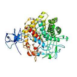
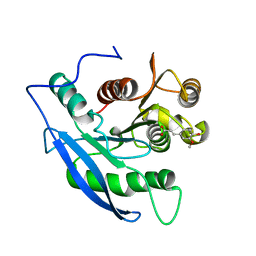
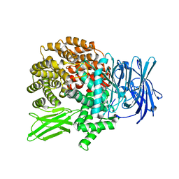
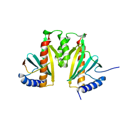
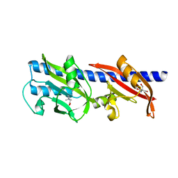
1Z21
 
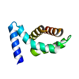 | | Crystal structure of the core domain of Yersinia pestis virulence factor YopR | | 分子名称: | Yop proteins translocation protein H | | 著者 | Schubot, F.D, Cherry, S, Tropea, J.E, Austin, B.P, Waugh, D.S. | | 登録日 | 2005-03-07 | | 公開日 | 2005-06-07 | | 最終更新日 | 2024-02-14 | | 実験手法 | X-RAY DIFFRACTION (1.499 Å) | | 主引用文献 | Crystal structure of the protease-resistant core domain of Yersinia pestis virulence factor YopR.
Protein Sci., 14, 2005
|
|
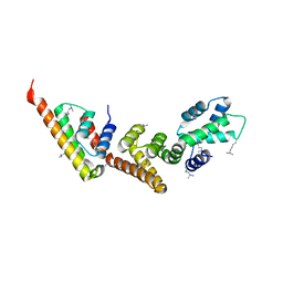
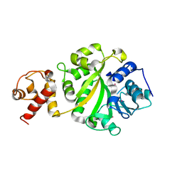
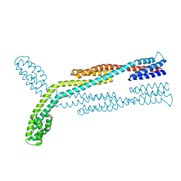
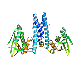
1XKP
 | | Crystal structure of the virulence factor YopN in complex with its heterodimeric chaperone SycN-YscB | | 分子名称: | Chaperone protein sycN, Chaperone protein yscB, putative membrane-bound Yop targeting protein YopN | | 著者 | Schubot, F.D, Jackson, M.W, Penrose, K.J, Cherry, S, Tropea, J.E, Plano, G.V, Waugh, D.S. | | 登録日 | 2004-09-29 | | 公開日 | 2005-03-22 | | 最終更新日 | 2021-10-20 | | 実験手法 | X-RAY DIFFRACTION (1.7 Å) | | 主引用文献 | Three-dimensional structure of a macromolecular assembly that regulates type III secretion in Yersinia pestis.
J.Mol.Biol., 346, 2005
|
|
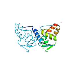
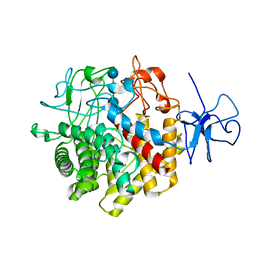
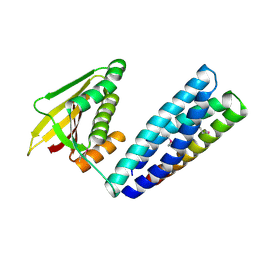
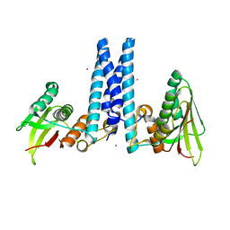
1XL3
 | | Complex structure of Y.pestis virulence Factors YopN and TyeA | | 分子名称: | Secretion control protein, protein type A | | 著者 | Schubot, F.D, Jackson, M.W, Penrose, K.J, Cherry, S, Tropea, J.E, Plano, G.V, Waugh, D.S. | | 登録日 | 2004-09-30 | | 公開日 | 2005-03-22 | | 最終更新日 | 2023-08-23 | | 実験手法 | X-RAY DIFFRACTION (2.2 Å) | | 主引用文献 | Three-dimensional structure of a macromolecular assembly that regulates type III secretion in Yersinia pestis.
J.Mol.Biol., 346, 2005
|
|
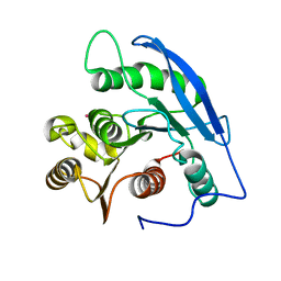
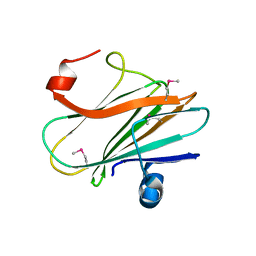
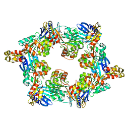
2IF5
 | | Structure of the POZ domain of human LRF, a master regulator of oncogenesis | | 分子名称: | PRASEODYMIUM ION, Zinc finger and BTB domain-containing protein 7A | | 著者 | Schubot, F.D, Waugh, D.S, Tropea, J. | | 登録日 | 2006-09-20 | | 公開日 | 2006-11-21 | | 最終更新日 | 2023-08-30 | | 実験手法 | X-RAY DIFFRACTION (2 Å) | | 主引用文献 | Structure of the POZ domain of human LRF, a master regulator of oncogenesis.
Biochem.Biophys.Res.Commun., 351, 2006
|
|
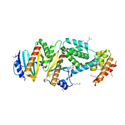
1UT9
 | | Structural Basis for the Exocellulase Activity of the Cellobiohydrolase CbhA from C. thermocellum | | 分子名称: | CELLULOSE 1,4-BETA-CELLOBIOSIDASE | | 著者 | Schubot, F.D, Kataeva, I.A, Chang, J, Shah, A.K, Ljungdahl, L.G, Rose, J.P, Wang, B.C. | | 登録日 | 2003-12-04 | | 公開日 | 2004-02-12 | | 最終更新日 | 2018-06-13 | | 実験手法 | X-RAY DIFFRACTION (2.1 Å) | | 主引用文献 | Structural basis for the exocellulase activity of the cellobiohydrolase CbhA from Clostridium thermocellum.
Biochemistry, 43, 2004
|
|
1I4W
 | | THE CRYSTAL STRUCTURE OF THE TRANSCRIPTION FACTOR SC-MTTFB OFFERS INTRIGUING INSIGHTS INTO MITOCHONDRIAL TRANSCRIPTION | | 分子名称: | MITOCHONDRIAL REPLICATION PROTEIN MTF1, XENON | | 著者 | Schubot, F.D, Chen, C.-J, Rose, J.P, Dailey, T.A, Dailey, H.A, Wang, B.-C. | | 登録日 | 2001-02-23 | | 公開日 | 2001-10-03 | | 最終更新日 | 2024-02-07 | | 実験手法 | X-RAY DIFFRACTION (2.6 Å) | | 主引用文献 | Crystal structure of the transcription factor sc-mtTFB offers insights into mitochondrial transcription.
Protein Sci., 10, 2001
|
|
1RQ5
 | | Structural Basis for the Exocellulase Activity of the Cellobiohydrolase CbhA from C. thermocellum | | 分子名称: | CALCIUM ION, Cellobiohydrolase, beta-D-glucopyranose-(1-4)-beta-D-glucopyranose-(1-4)-beta-D-glucopyranose-(1-4)-beta-D-glucopyranose | | 著者 | Schubot, F.D, Kataeva, I.A, Chang, J, Shah, A.K, Ljungdahl, L.G, Rose, J.P, Wang, B.C. | | 登録日 | 2003-12-04 | | 公開日 | 2004-03-30 | | 最終更新日 | 2024-04-03 | | 実験手法 | X-RAY DIFFRACTION (2.4 Å) | | 主引用文献 | Structural basis for the exocellulase activity of the cellobiohydrolase CbhA from Clostridium thermocellum
Biochemistry, 43, 2004
|
|
1JJF
 | | STRUCTURAL BASIS FOR THE SUBSTRATE SPECIFICITY OF THE FERULOYL ESTERASE DOMAIN OF THE CELLULOSOMAL XYLANASE Z OF CLOSTRIDIUM THERMOCELLUM | | 分子名称: | ENDO-1,4-BETA-XYLANASE Z, PLATINUM (II) ION | | 著者 | Schubot, F.D, Kataeva, I.A, Blum, D.L, Shah, A.K, Ljungdahl, L.G, Rose, J.P, Wang, B.C, Southeast Collaboratory for Structural Genomics (SECSG) | | 登録日 | 2001-07-05 | | 公開日 | 2001-10-31 | | 最終更新日 | 2024-02-07 | | 実験手法 | X-RAY DIFFRACTION (1.75 Å) | | 主引用文献 | Structural basis for the substrate specificity of the feruloyl esterase domain of the cellulosomal xylanase Z from Clostridium thermocellum.
Biochemistry, 40, 2001
|
|
1JT2
 | | STRUCTURAL BASIS FOR THE SUBSTRATE SPECIFICITY OF THE FERUL DOMAIN OF THE CELLULOSOMAL XYLANASE Z FROM C. THERMOCELLUM | | 分子名称: | 3-(4-HYDROXY-3-METHOXYPHENYL)-2-PROPENOIC ACID, PROTEIN (ENDO-1,4-BETA-XYLANASE Z) | | 著者 | Schubot, F.D, Kataeva, I.A, Blum, D.L, Shah, A.K, Ljungdahl, L.G, Rose, J.P, Wang, B.-C. | | 登録日 | 2001-08-20 | | 公開日 | 2002-03-27 | | 最終更新日 | 2023-08-16 | | 実験手法 | X-RAY DIFFRACTION (1.8 Å) | | 主引用文献 | Structural basis for the substrate specificity of the feruloyl esterase domain of the cellulosomal xylanase Z from Clostridium thermocellum.
Biochemistry, 40, 2001
|
|
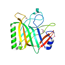
3FD9
 | |
4ZUA
 | | Crystal structure of the ExsA regulatory domain | | 分子名称: | Exoenzyme S synthesis regulatory protein ExsA | | 著者 | Schubot, F.D. | | 登録日 | 2015-05-15 | | 公開日 | 2016-02-03 | | 最終更新日 | 2024-03-06 | | 実験手法 | X-RAY DIFFRACTION (2.5 Å) | | 主引用文献 | Structural Analysis of the Regulatory Domain of ExsA, a Key Transcriptional Regulator of the Type Three Secretion System in Pseudomonas aeruginosa.
Plos One, 10, 2015
|
|
4FET
 | | Catalytic domain of germination-specific lytic tansglycosylase SleB from Bacillus anthracis | | 分子名称: | SODIUM ION, Spore cortex-lytic enzyme prepeptide | | 著者 | Jing, X, Heffron, J, Popham, D.L, Schubot, F.D. | | 登録日 | 2012-05-30 | | 公開日 | 2012-07-25 | | 最終更新日 | 2017-11-15 | | 実験手法 | X-RAY DIFFRACTION (1.909 Å) | | 主引用文献 | The catalytic domain of the germination-specific lytic transglycosylase SleB from Bacillus anthracis displays a unique active site topology.
Proteins, 80, 2012
|
|
4J3B
 | | A naturally variable residue in the S1 subsite of M1-family aminopeptidases modulates catalytic properties and promotes functional specialization | | 分子名称: | ARGININE, M1 family aminopeptidase, MAGNESIUM ION, ... | | 著者 | Dalal, S, Ragheb, D.R.T, Schubot, F.D, Klemba, M. | | 登録日 | 2013-02-05 | | 公開日 | 2013-08-07 | | 最終更新日 | 2023-09-20 | | 実験手法 | X-RAY DIFFRACTION (2.2 Å) | | 主引用文献 | A naturally variable residue in the s1 subsite of m1 family aminopeptidases modulates catalytic properties and promotes functional specialization.
J.Biol.Chem., 288, 2013
|
|
8F7N
 | | Crystal structure of chemoreceptor McpZ ligand sensing domain | | 分子名称: | Methyl-accepting chemotaxis protein, YTTERBIUM (III) ION | | 著者 | Salar, S, Schubot, F.D. | | 登録日 | 2022-11-19 | | 公開日 | 2023-07-05 | | 最終更新日 | 2023-10-04 | | 実験手法 | X-RAY DIFFRACTION (2.7 Å) | | 主引用文献 | The structural analysis of the periplasmic domain of Sinorhizobium meliloti chemoreceptor McpZ reveals a novel fold and suggests a complex mechanism of transmembrane signaling.
Proteins, 91, 2023
|
|
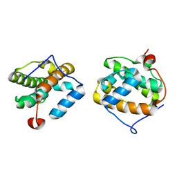
7N0E
 | |
3JYB
 | |
3KXY
 | | Crystal Structure of the ExsC-ExsE Complex | | 分子名称: | Exoenzyme S synthesis protein C, ExsE | | 著者 | Vogelaar, N.J, Robinson, H.H, Schubot, F.D. | | 登録日 | 2009-12-04 | | 公開日 | 2010-06-30 | | 最終更新日 | 2024-02-21 | | 実験手法 | X-RAY DIFFRACTION (2.804 Å) | | 主引用文献 | Analysis of the Crystal Structure of the ExsC.ExsE Complex Reveals Distinctive Binding Interactions of the Pseudomonas aeruginosa Type III Secretion Chaperone ExsC with ExsE and ExsD.
Biochemistry, 49, 2010
|
|
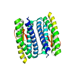
2QWU
 | |
1VKA
 | | Southeast Collaboratory for Structural Genomics: Hypothetical Human Protein Q15691 N-Terminal Fragment | | 分子名称: | Microtubule-associated protein RP/EB family member 1 | | 著者 | Liu, Z.-J, Tempel, W, Schubot, F.D, Shah, A, Dailey, T.A, Mayer, M.R, Rose, J.P, Dailey, H.A, Wang, B.-C, Southeast Collaboratory for Structural Genomics (SECSG) | | 登録日 | 2004-05-10 | | 公開日 | 2004-07-13 | | 最終更新日 | 2023-12-27 | | 実験手法 | X-RAY DIFFRACTION (1.6 Å) | | 主引用文献 | Southeast Collaboratory for Structural Genomics: Hypothetical Human Protein Q15691 N-Terminal Fragment
TO BE PUBLISHED
|
|
1NNQ
 | | rubrerythrin from Pyrococcus furiosus Pfu-1210814 | | 分子名称: | Rubrerythrin, ZINC ION | | 著者 | Liu, Z.-J, Tempel, W, Schubot, F.D, Shah, A, Arendall III, W.B, Rose, J.P, Richardson, D.C, Richardson, J.S, Wang, B.-C, Southeast Collaboratory for Structural Genomics (SECSG) | | 登録日 | 2003-01-14 | | 公開日 | 2004-03-02 | | 最終更新日 | 2024-02-14 | | 実験手法 | X-RAY DIFFRACTION (2.35 Å) | | 主引用文献 | Structural genomics of Pyrococcus furiosus: X-ray crystallography reveals 3D domain swapping in rubrerythrin
Proteins, 57, 2004
|
|
6D8V
 | | Methyl-accepting Chemotaxis protein X | | 分子名称: | 1,1-DIMETHYL-PROLINIUM, 2-AMINO-2-HYDROXYMETHYL-PROPANE-1,3-DIOL, Probable chemoreceptor (Methyl-accepting chemotaxis) transmembrane protein | | 著者 | Shrestha, M, Schubot, F.D. | | 登録日 | 2018-04-27 | | 公開日 | 2019-04-17 | | 最終更新日 | 2024-03-13 | | 実験手法 | X-RAY DIFFRACTION (2.8 Å) | | 主引用文献 | Structure of the sensory domain of McpX fromSinorhizobium meliloti, the first known bacterial chemotactic sensor for quaternary ammonium compounds.
Biochem. J., 475, 2018
|
|
6DK8
 | | RetS kinase region without cobalt | | 分子名称: | NICKEL (II) ION, RetS (Regulator of Exopolysaccharide and Type III Secretion) | | 著者 | Mancl, J.M, Schubot, F.D. | | 登録日 | 2018-05-29 | | 公開日 | 2019-03-20 | | 最終更新日 | 2023-10-11 | | 実験手法 | X-RAY DIFFRACTION (3.8 Å) | | 主引用文献 | Helix Cracking Regulates the Critical Interaction between RetS and GacS in Pseudomonas aeruginosa.
Structure, 27, 2019
|
|
6DK7
 | | RetS histidine kinase region with cobalt | | 分子名称: | COBALT (II) ION, RetS (Regulator of Exopolysaccharide and Type III Secretion) | | 著者 | Mancl, J.M, Schubot, F.D. | | 登録日 | 2018-05-29 | | 公開日 | 2019-03-20 | | 最終更新日 | 2024-03-13 | | 実験手法 | X-RAY DIFFRACTION (2.6 Å) | | 主引用文献 | Helix Cracking Regulates the Critical Interaction between RetS and GacS in Pseudomonas aeruginosa.
Structure, 27, 2019
|
|
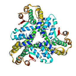
5IT5
 | | Thermus thermophilus PilB core ATPase region | | 分子名称: | ADENOSINE-5'-TRIPHOSPHATE, ATP binding motif-containing protein PilF, MAGNESIUM ION, ... | | 著者 | Mancl, J, Robinson, H, Black, W, Yang, Z, Schubot, F. | | 登録日 | 2016-03-16 | | 公開日 | 2016-10-19 | | 最終更新日 | 2023-09-27 | | 実験手法 | X-RAY DIFFRACTION (2.648 Å) | | 主引用文献 | Crystal Structure of a Type IV Pilus Assembly ATPase: Insights into the Molecular Mechanism of PilB from Thermus thermophilus.
Structure, 24, 2016
|
|
1NNH
 | | Hypothetical protein from Pyrococcus furiosus Pfu-1801964 | | 分子名称: | SODIUM ION, asparaginyl-tRNA synthetase-related peptide | | 著者 | Tempel, W, Liu, Z.-J, Schubot, F.D, Shah, A, Arendall III, W.B, Rose, J.P, Richardson, D.C, Richardson, J.S, Wang, B.-C, Southeast Collaboratory for Structural Genomics (SECSG) | | 登録日 | 2003-01-13 | | 公開日 | 2004-02-03 | | 最終更新日 | 2024-02-14 | | 実験手法 | X-RAY DIFFRACTION (1.65 Å) | | 主引用文献 | Hypothetical protein from Pyrococcus furiosus Pfu-1801964
To be published
|
|


