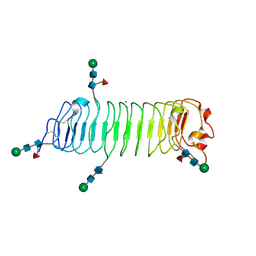
3M9G
 
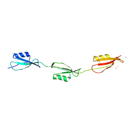 | | Crystal structure of the three-PASTA-domain of a Ser/Thr kinase from Staphylococcus aureus | | 分子名称: | Protein kinase, ZINC ION | | 著者 | Paracuellos, P, Ballandras, A, Robert, X, Creze, C, Cozzone, A.J, Duclos, B, Gouet, P. | | 登録日 | 2010-03-22 | | 公開日 | 2010-11-03 | | 最終更新日 | 2024-03-20 | | 実験手法 | X-RAY DIFFRACTION (2.9 Å) | | 主引用文献 | The Extended Conformation of the 2.9-A Crystal Structure of the Three-PASTA Domain of a Ser/Thr Kinase from the Human Pathogen Staphylococcus aureus
J.Mol.Biol., 404, 2010
|
|
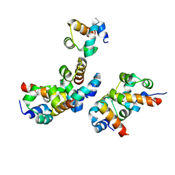
5AO5
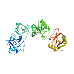 | | Endo180 D1-4, monoclinic form | | 分子名称: | C-TYPE MANNOSE RECEPTOR 2, SODIUM ION, SULFATE ION | | 著者 | Paracuellos, P, Briggs, D.C, Carafoli, F, Loncar, T, Hohenester, E. | | 登録日 | 2015-09-09 | | 公開日 | 2015-10-28 | | 最終更新日 | 2024-01-10 | | 実験手法 | X-RAY DIFFRACTION (2.48 Å) | | 主引用文献 | Insights Into Collagen Uptake by C-Type Mannose Receptors from the Crystal Structure of Endo180 Domains 1-4.
Structure, 23, 2015
|
|
5AO6
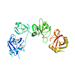 | | Endo180 D1-4, trigonal form | | 分子名称: | C-TYPE MANNOSE RECEPTOR 2 | | 著者 | Paracuellos, P, Briggs, D.C, Carafoli, F, Loncar, T, Hohenester, E. | | 登録日 | 2015-09-09 | | 公開日 | 2015-10-28 | | 最終更新日 | 2024-01-10 | | 実験手法 | X-RAY DIFFRACTION (3.36 Å) | | 主引用文献 | Insights Into Collagen Uptake by C-Type Mannose Receptors from the Crystal Structure of Endo180 Domains 1-4.
Structure, 23, 2015
|
|
5MX1
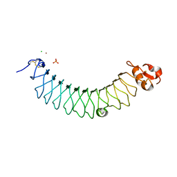 | |
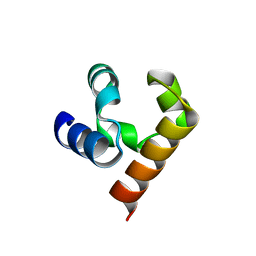
5MX0
 | | Crystal structure of human fibromodulin | | 分子名称: | CHLORIDE ION, Fibromodulin, NICKEL (II) ION, ... | | 著者 | Paracuellos, P, Hohenester, E. | | 登録日 | 2017-01-20 | | 公開日 | 2017-03-01 | | 最終更新日 | 2024-01-17 | | 実験手法 | X-RAY DIFFRACTION (2.21 Å) | | 主引用文献 | Structural and functional analysis of two small leucine-rich repeat proteoglycans, fibromodulin and chondroadherin.
Matrix Biol., 63, 2017
|
|
5JU5
 | |
5JTI
 | |
5JRT
 | |


