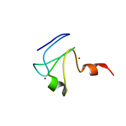
4HCZ
 
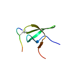 | | PHF1 Tudor in complex with H3K36me3 | | 分子名称: | H3L-like histone, PHD finger protein 1 | | 著者 | Musselman, C.A, Roy, S, Nunez, J, Kutateladze, T.G. | | 登録日 | 2012-10-01 | | 公開日 | 2012-11-14 | | 最終更新日 | 2023-09-20 | | 実験手法 | X-RAY DIFFRACTION (1.85 Å) | | 主引用文献 | Molecular basis for H3K36me3 recognition by the Tudor domain of PHF1.
Nat.Struct.Mol.Biol., 19, 2012
|
|
3LH0
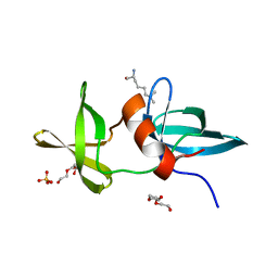 | |
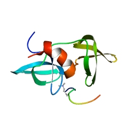
3MDF
 | | Crystal structure of the RRM domain of Cyclophilin 33 | | 分子名称: | Peptidyl-prolyl cis-trans isomerase E | | 著者 | Hom, R.A, Chang, P.Y, Roy, S, Mussleman, C.A, Glass, K.C, Seleznevia, A.I, Gozani, O, Ismagilov, R.F, Cleary, M.L, Kutateladze, T.G. | | 登録日 | 2010-03-30 | | 公開日 | 2010-05-12 | | 最終更新日 | 2023-09-06 | | 実験手法 | X-RAY DIFFRACTION (1.85 Å) | | 主引用文献 | Molecular mechanism of MLL PHD3 and RNA recognition by the Cyp33 RRM domain.
J.Mol.Biol., 400, 2010
|
|
2L75
 | |
2L5U
 | |
2MWO
 | |
2MWP
 | |
3LGF
 | |
3LGL
 | |


