7NZZ
 
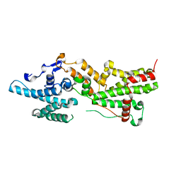 | |
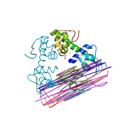
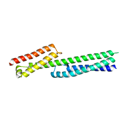
7AAL
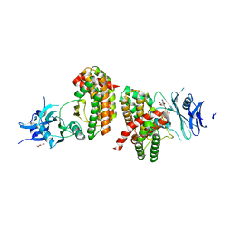
 | | Crystal structure of the F-BAR domain of PSTIPIP1, G258A mutant | | 分子名称: | Proline-serine-threonine phosphatase-interacting protein 1 | | 著者 | Manso, J.A, Alcon, P, Bayon, Y, Alonso, A, de Pereda, J.M. | | 登録日 | 2020-09-04 | | 公開日 | 2022-02-23 | | 最終更新日 | 2024-02-07 | | 実験手法 | X-RAY DIFFRACTION (1.97 Å) | | 主引用文献 | PSTPIP1-LYP phosphatase interaction: structural basis and implications for autoinflammatory disorders.
Cell.Mol.Life Sci., 79, 2022
|
|
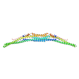
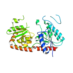
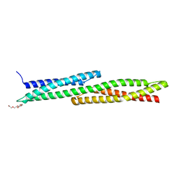
7AAN
 | | Crystal structure of the F-BAR domain of PSTIPIP1 | | 分子名称: | Proline-serine-threonine phosphatase-interacting protein 1 | | 著者 | Manso, J.A, Alcon, P, Bayon, Y, Alonso, A, de Pereda, J.M. | | 登録日 | 2020-09-04 | | 公開日 | 2022-02-23 | | 最終更新日 | 2024-02-07 | | 実験手法 | X-RAY DIFFRACTION (2.14 Å) | | 主引用文献 | PSTPIP1-LYP phosphatase interaction: structural basis and implications for autoinflammatory disorders.
Cell.Mol.Life Sci., 79, 2022
|
|
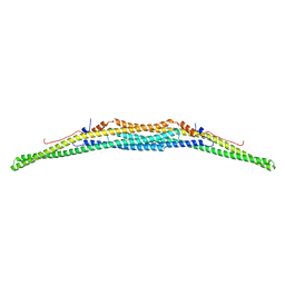
7AAM
 | | Crystal structure of the F-BAR domain of PSTIPIP1 bound to the CTH domain of the phosphatase LYP | | 分子名称: | GLYCEROL, Proline-serine-threonine phosphatase-interacting protein 1, Tyrosine-protein phosphatase non-receptor type 22 | | 著者 | Manso, J.A, Alcon, P, Bayon, Y, Alonso, A, de Pereda, J.M. | | 登録日 | 2020-09-04 | | 公開日 | 2022-02-23 | | 最終更新日 | 2024-02-07 | | 実験手法 | X-RAY DIFFRACTION (2.15 Å) | | 主引用文献 | PSTPIP1-LYP phosphatase interaction: structural basis and implications for autoinflammatory disorders.
Cell.Mol.Life Sci., 79, 2022
|
|
6GVL
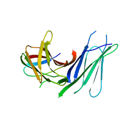 | | Second pair of Fibronectin type III domains of integrin beta4 bound to the bullous pemphigoid antigen BP230 (BPAG1e) | | 分子名称: | Dystonin, Integrin beta-4 | | 著者 | Manso, J.A, Gomez-Hernandez, M, Alonso-Garcia, N, de Pereda, J.M. | | 登録日 | 2018-06-21 | | 公開日 | 2019-03-20 | | 最終更新日 | 2024-05-01 | | 実験手法 | X-RAY DIFFRACTION (2.05 Å) | | 主引用文献 | Integrin alpha 6 beta 4 Recognition of a Linear Motif of Bullous Pemphigoid Antigen BP230 Controls Its Recruitment to Hemidesmosomes.
Structure, 27, 2019
|
|
6GVK
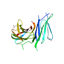 | | Second pair of Fibronectin type III domains of integrin beta4 (T1663R mutant) bound to the bullous pemphigoid antigen BP230 (BPAG1e) | | 分子名称: | Dystonin, GLYCEROL, Integrin beta-4 | | 著者 | Manso, J.A, Gomez-Hernandez, M, Alonso-Garcia, N, de Pereda, J.M. | | 登録日 | 2018-06-21 | | 公開日 | 2019-03-20 | | 最終更新日 | 2024-01-17 | | 実験手法 | X-RAY DIFFRACTION (1.55 Å) | | 主引用文献 | Integrin alpha 6 beta 4 Recognition of a Linear Motif of Bullous Pemphigoid Antigen BP230 Controls Its Recruitment to Hemidesmosomes.
Structure, 27, 2019
|
|
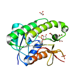
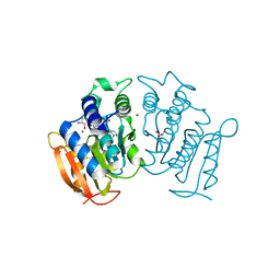
6HWK
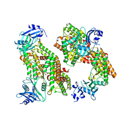 | | Glucosamine kinase (crystal form B) | | 分子名称: | CHLORIDE ION, Glucosamine kinase, MAGNESIUM ION | | 著者 | Manso, J.A, Pereira, P.J.B. | | 登録日 | 2018-10-12 | | 公開日 | 2019-05-01 | | 最終更新日 | 2024-05-01 | | 実験手法 | X-RAY DIFFRACTION (2.688 Å) | | 主引用文献 | Molecular Fingerprints for a Novel Enzyme Family in Actinobacteria with Glucosamine Kinase Activity.
Mbio, 10, 2019
|
|
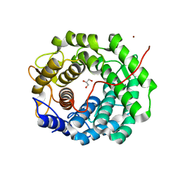
6HWJ
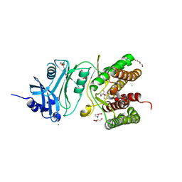 | | Glucosamine kinase (crystal form A) | | 分子名称: | 2-[BIS-(2-HYDROXY-ETHYL)-AMINO]-2-HYDROXYMETHYL-PROPANE-1,3-DIOL, CHLORIDE ION, DI(HYDROXYETHYL)ETHER, ... | | 著者 | Manso, J.A, Pereira, P.J.B. | | 登録日 | 2018-10-12 | | 公開日 | 2019-05-01 | | 最終更新日 | 2024-01-24 | | 実験手法 | X-RAY DIFFRACTION (1.979 Å) | | 主引用文献 | Molecular Fingerprints for a Novel Enzyme Family in Actinobacteria with Glucosamine Kinase Activity.
Mbio, 10, 2019
|
|
6HWL
 | | Glucosamine kinase in complex with glucosamine, ADP and inorganic phosphate | | 分子名称: | 2-[BIS-(2-HYDROXY-ETHYL)-AMINO]-2-HYDROXYMETHYL-PROPANE-1,3-DIOL, 2-amino-2-deoxy-beta-D-glucopyranose, ADENOSINE-5'-DIPHOSPHATE, ... | | 著者 | Manso, J.A, Pereira, P.J.B. | | 登録日 | 2018-10-12 | | 公開日 | 2019-05-01 | | 最終更新日 | 2024-05-01 | | 実験手法 | X-RAY DIFFRACTION (2.148 Å) | | 主引用文献 | Molecular Fingerprints for a Novel Enzyme Family in Actinobacteria with Glucosamine Kinase Activity.
Mbio, 10, 2019
|
|
7QSG
 | |
5OOG
 | |
5OOH
 | |
7QSJ
 | | Methylmannose polysaccharide hydrolase MmpH from M. hassiacum | | 分子名称: | GLYCEROL, MAGNESIUM ION, Methylmannose polysaccharide hydrolase (MmpH), ... | | 著者 | Ripoll-Rozada, J, Manso, J.A, Pereira, P.J.B. | | 登録日 | 2022-01-13 | | 公開日 | 2023-01-25 | | 最終更新日 | 2023-02-08 | | 実験手法 | X-RAY DIFFRACTION (1.35 Å) | | 主引用文献 | Self-recycling and partially conservative replication of mycobacterial methylmannose polysaccharides.
Commun Biol, 6, 2023
|
|
5NFG
 | | Structure of recombinant cardosin B from Cynara cardunculus | | 分子名称: | Procardosin-B,Procardosin-B, alpha-D-mannopyranose-(1-2)-[alpha-D-mannopyranose-(1-6)]alpha-D-mannopyranose-(1-3)-beta-D-mannopyranose-(1-4)-2-acetamido-2-deoxy-beta-D-glucopyranose-(1-4)-2-acetamido-2-deoxy-beta-D-glucopyranose, alpha-D-mannopyranose-(1-6)-beta-D-mannopyranose-(1-4)-2-acetamido-2-deoxy-beta-D-glucopyranose-(1-4)-2-acetamido-2-deoxy-beta-D-glucopyranose | | 著者 | Pereira, P.J.B, Figueiredo, A.C, Manso, J.A, Almeida, C.M, Simoes, I. | | 登録日 | 2017-03-14 | | 公開日 | 2017-10-11 | | 最終更新日 | 2024-01-17 | | 実験手法 | X-RAY DIFFRACTION (2.375 Å) | | 主引用文献 | Functional and structural characterization of synthetic cardosin B-derived rennet.
Appl. Microbiol. Biotechnol., 101, 2017
|
|
6Q5T
 | |
8PVZ
 | |
8PVT
 | |
8PW0
 | |
4WTX
 | | Crystal structure of the fourth FnIII domain of integrin beta4 | | 分子名称: | Integrin beta-4 | | 著者 | Alonso-Garcia, N, Urien, H, Buey, R.M, de Pereda, J.M. | | 登録日 | 2014-10-30 | | 公開日 | 2015-02-11 | | 最終更新日 | 2024-05-08 | | 実験手法 | X-RAY DIFFRACTION (1.5 Å) | | 主引用文献 | Combination of X-ray crystallography, SAXS and DEER to obtain the structure of the FnIII-3,4 domains of integrin alpha6beta4
Acta Crystallogr.,Sect.D, 71, 2015
|
|
4WTW
 | | Crystal structure of the third FnIII domain of integrin beta4 | | 分子名称: | 1,2-ETHANEDIOL, Integrin beta-4, PENTAETHYLENE GLYCOL, ... | | 著者 | Alonso-Garcia, N, Urien, H, de Pereda, J.M. | | 登録日 | 2014-10-30 | | 公開日 | 2015-02-11 | | 最終更新日 | 2024-01-10 | | 実験手法 | X-RAY DIFFRACTION (1.606 Å) | | 主引用文献 | Combination of X-ray crystallography, SAXS and DEER to obtain the structure of the FnIII-3,4 domains of integrin alpha6beta4
Acta Crystallogr.,Sect.D, 71, 2015
|
|
5J1F
 | |
5J1I
 | |
5J1H
 | |
5J1G
 | |
6G7D
 | |


