2YNB
 
 | |
2YNA
 | |
2CGH
 | |
2CHC
 | |
2CGQ
 | |
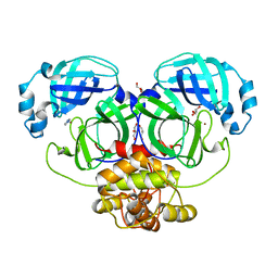
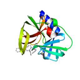
1OVN
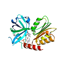
 | | Crystal Structure and Functional Analysis of Drosophila Wind-- a PDI-Related Protein | | 分子名称: | CESIUM ION, Windbeutel | | 著者 | Ma, Q, Guo, C, Barnewitz, K, Sheldrick, G.M, Soling, H.D, Uson, I, Ferrari, D.M. | | 登録日 | 2003-03-27 | | 公開日 | 2004-02-24 | | 最終更新日 | 2017-10-11 | | 実験手法 | X-RAY DIFFRACTION (1.9 Å) | | 主引用文献 | Crystal structure and functional analysis of Drosophila Wind, a protein-disulfide isomerase-related protein.
J.Biol.Chem., 278, 2003
|
|
1W66
 | |
1RQW
 | |
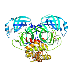
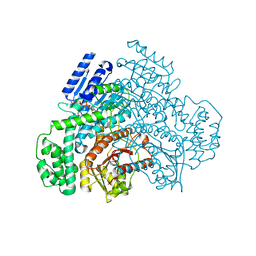
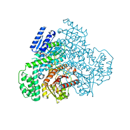
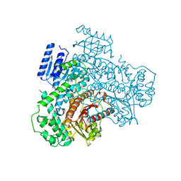
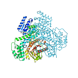
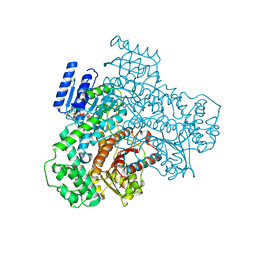
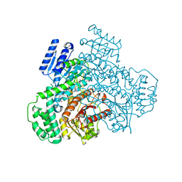
4OP0
 | | Crystal structure of biotin protein ligase (RV3279C) of Mycobacterium tuberculosis, complexed with biotinyl-5'-AMP | | 分子名称: | BIOTINYL-5-AMP, BirA bifunctional protein, SULFATE ION | | 著者 | Ma, Q, Wilmanns, M, Akhter, Y. | | 登録日 | 2014-02-04 | | 公開日 | 2014-04-30 | | 最終更新日 | 2023-09-20 | | 実験手法 | X-RAY DIFFRACTION (1.7 Å) | | 主引用文献 | Active site conformational changes upon reaction intermediate biotinyl-5'-AMP binding in biotin protein ligase from Mycobacterium tuberculosis.
Protein Sci., 23, 2014
|
|
8J2B
 | |
8J2E
 | |
8J29
 | |
8J2D
 | |
8J2A
 | |
8J2C
 | |
7XJN
 | | Structure of VcPotD1 in complex with norspermidine | | 分子名称: | HEXAETHYLENE GLYCOL, N-(3-aminopropyl)propane-1,3-diamine, Putrescine-binding periplasmic protein, ... | | 著者 | Ma, Q, Liu, C. | | 登録日 | 2022-04-18 | | 公開日 | 2023-04-26 | | 最終更新日 | 2023-11-29 | | 実験手法 | X-RAY DIFFRACTION (1.79 Å) | | 主引用文献 | Structure of VcPotD1 in complex with norspermidine
To Be Published
|
|
7XJM
 | |
7XLZ
 | |
7XYK
 | |
7XYJ
 | |
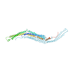
2X3V
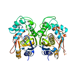
 | | Structure of The F-BAR Domain of Mouse Syndapin I | | 分子名称: | PROTEIN KINASE C AND CASEIN KINASE SUBSTRATE IN NEURONS PROTEIN 1 | | 著者 | Ma, Q, Rao, Y, Vahedi-Faridi, A, Saenger, W, Haucke, V. | | 登録日 | 2010-01-27 | | 公開日 | 2010-04-07 | | 最終更新日 | 2011-07-13 | | 実験手法 | X-RAY DIFFRACTION (2.45 Å) | | 主引用文献 | Molecular Basis for SH3 Domain Regulation of F-Bar-Mediated Membrane Deformation.
Proc.Natl.Acad.Sci.USA, 107, 2010
|
|
2X3W
 | |
2X3X
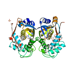
 | | structure of mouse syndapin I (crystal form 1) | | 分子名称: | PROTEIN KINASE C AND CASEIN KINASE SUBSTRATE IN NEURONS PROTEIN 1 | | 著者 | Ma, Q, Rao, Y, Vahedi-Faridi, A, Saenger, W, Haucke, V. | | 登録日 | 2010-01-28 | | 公開日 | 2010-04-07 | | 最終更新日 | 2011-07-13 | | 実験手法 | X-RAY DIFFRACTION (3.35 Å) | | 主引用文献 | Molecular Basis for SH3 Domain Regulation of F-Bar-Mediated Membrane Deformation.
Proc.Natl.Acad.Sci.USA, 107, 2010
|
|
5NFS
 | |
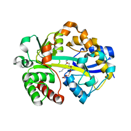
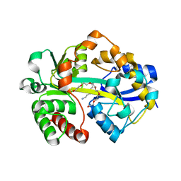
2FGH
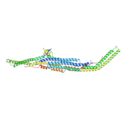
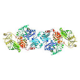 | | ATP bound gelsolin | | 分子名称: | ADENOSINE-5'-TRIPHOSPHATE, gelsolin | | 著者 | Ma, Q, Robinson, R.C, Burtnick, L.D, Urosev, D. | | 登録日 | 2005-12-22 | | 公開日 | 2006-04-18 | | 最終更新日 | 2017-12-20 | | 実験手法 | X-RAY DIFFRACTION (2.8 Å) | | 主引用文献 | The structure of gelsolin bound to ATP
J.Mol.Biol., 357, 2006
|
|


