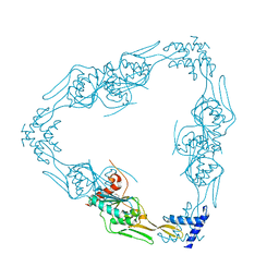
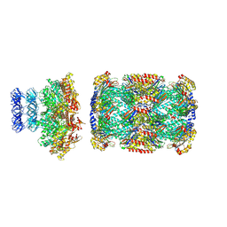
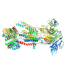
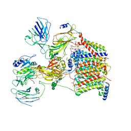
3CWD
 
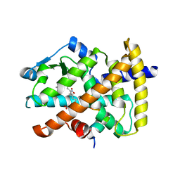 | | Molecular recognition of nitro-fatty acids by PPAR gamma | | 分子名称: | (9E,12Z)-10-nitrooctadeca-9,12-dienoic acid, (9Z,12E)-12-nitrooctadeca-9,12-dienoic acid, Peroxisome proliferator-activated receptor gamma, ... | | 著者 | Martynowski, D, Li, Y. | | 登録日 | 2008-04-21 | | 公開日 | 2008-07-08 | | 最終更新日 | 2024-02-21 | | 実験手法 | X-RAY DIFFRACTION (2.4 Å) | | 主引用文献 | Molecular recognition of nitrated fatty acids by PPAR gamma.
Nat.Struct.Mol.Biol., 15, 2008
|
|
6DJV
 | | Mtb ClpB in complex with ATPgammaS and casein, Conformer 2 | | 分子名称: | ADENOSINE-5'-DIPHOSPHATE, Chaperone protein ClpB, PHOSPHOTHIOPHOSPHORIC ACID-ADENYLATE ESTER, ... | | 著者 | Yu, H.J, Li, H.L. | | 登録日 | 2018-05-26 | | 公開日 | 2018-09-26 | | 最終更新日 | 2024-03-13 | | 実験手法 | ELECTRON MICROSCOPY (3.9 Å) | | 主引用文献 | ATP hydrolysis-coupled peptide translocation mechanism ofMycobacterium tuberculosisClpB.
Proc. Natl. Acad. Sci. U.S.A., 115, 2018
|
|
6DJU
 | | Mtb ClpB in complex with ATPgammaS and casein, Conformer 1 | | 分子名称: | ADENOSINE-5'-DIPHOSPHATE, Chaperone protein ClpB, PHOSPHOTHIOPHOSPHORIC ACID-ADENYLATE ESTER, ... | | 著者 | Yu, H.J, Li, H.L. | | 登録日 | 2018-05-26 | | 公開日 | 2018-09-26 | | 最終更新日 | 2024-03-13 | | 実験手法 | ELECTRON MICROSCOPY (3.8 Å) | | 主引用文献 | ATP hydrolysis-coupled peptide translocation mechanism ofMycobacterium tuberculosisClpB.
Proc. Natl. Acad. Sci. U.S.A., 115, 2018
|
|
7RQH
 | |
7RQF
 | |
7RPQ
 | |
6ED3
 | | Mtb ClpB in complex with AMPPNP | | 分子名称: | Chaperone protein ClpB | | 著者 | Yu, H.J, Li, H.L. | | 登録日 | 2018-08-08 | | 公開日 | 2018-09-26 | | 最終更新日 | 2024-03-13 | | 実験手法 | ELECTRON MICROSCOPY (6.3 Å) | | 主引用文献 | ATP hydrolysis-coupled peptide translocation mechanism ofMycobacterium tuberculosisClpB.
Proc. Natl. Acad. Sci. U.S.A., 115, 2018
|
|
8D6Y
 | |
8D6W
 | |
8D6X
 | |
8D6V
 | |
6PSY
 | | Cryo-EM structure of S. cerevisiae Drs2p-Cdc50p in the autoinhibited apo form | | 分子名称: | 2-acetamido-2-deoxy-beta-D-glucopyranose, 2-acetamido-2-deoxy-beta-D-glucopyranose-(1-4)-2-acetamido-2-deoxy-beta-D-glucopyranose, Cell division control protein 50, ... | | 著者 | Bai, L, Li, H. | | 登録日 | 2019-07-14 | | 公開日 | 2019-09-25 | | 最終更新日 | 2020-07-29 | | 実験手法 | ELECTRON MICROSCOPY (2.8 Å) | | 主引用文献 | Autoinhibition and activation mechanisms of the eukaryotic lipid flippase Drs2p-Cdc50p.
Nat Commun, 10, 2019
|
|
6PSX
 | | Cryo-EM structure of S. cerevisiae Drs2p-Cdc50p in the PI4P-activated form | | 分子名称: | 2-acetamido-2-deoxy-beta-D-glucopyranose, 2-acetamido-2-deoxy-beta-D-glucopyranose-(1-4)-2-acetamido-2-deoxy-beta-D-glucopyranose, Cell division control protein 50, ... | | 著者 | Bai, L, Li, H. | | 登録日 | 2019-07-14 | | 公開日 | 2019-09-25 | | 最終更新日 | 2020-07-29 | | 実験手法 | ELECTRON MICROSCOPY (3.3 Å) | | 主引用文献 | Autoinhibition and activation mechanisms of the eukaryotic lipid flippase Drs2p-Cdc50p.
Nat Commun, 10, 2019
|
|
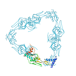
6WB9
 | | Structure of the S. cerevisiae ER membrane complex | | 分子名称: | (2S)-3-(hexadecanoyloxy)-2-[(9Z)-octadec-9-enoyloxy]propyl 2-(trimethylammonio)ethyl phosphate, 2-acetamido-2-deoxy-beta-D-glucopyranose, ER membrane protein complex subunit 1, ... | | 著者 | Bai, L, Li, H. | | 登録日 | 2020-03-26 | | 公開日 | 2020-06-03 | | 最終更新日 | 2020-09-02 | | 実験手法 | ELECTRON MICROSCOPY (3 Å) | | 主引用文献 | Structure of the ER membrane complex, a transmembrane-domain insertase.
Nature, 584, 2020
|
|
7L6N
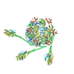 | | The Mycobacterium tuberculosis ClpB disaggregase hexamer structure with three locally refined ClpB middle domains and three DnaK nucleotide binding domains | | 分子名称: | ADENOSINE-5'-DIPHOSPHATE, Chaperone protein ClpB, Chaperone protein DnaK, ... | | 著者 | Yin, Y.Y, Feng, X, Li, H. | | 登録日 | 2020-12-23 | | 公開日 | 2021-05-26 | | 最終更新日 | 2021-12-08 | | 実験手法 | ELECTRON MICROSCOPY (7 Å) | | 主引用文献 | Structural basis for aggregate dissolution and refolding by the Mycobacterium tuberculosis ClpB-DnaK bi-chaperone system.
Cell Rep, 35, 2021
|
|
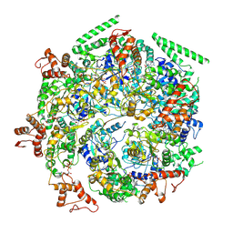
6BGL
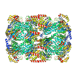 | |
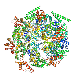
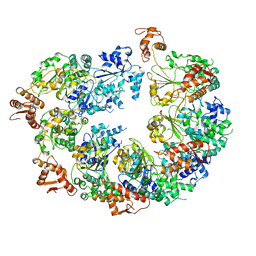
6BGO
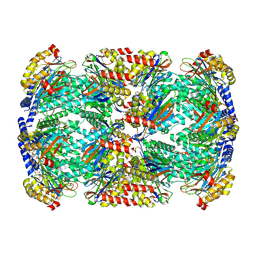 | | Singly PafE-capped 20S CP in Mycobacterium tuberculosis | | 分子名称: | Bacterial proteasome activator, Proteasome subunit alpha, Proteasome subunit beta | | 著者 | Li, H, Hu, K. | | 登録日 | 2017-10-29 | | 公開日 | 2018-02-14 | | 最終更新日 | 2024-03-13 | | 実験手法 | ELECTRON MICROSCOPY (4.2 Å) | | 主引用文献 | Proteasome substrate capture and gate opening by the accessory factor PafE fromMycobacterium tuberculosis.
J. Biol. Chem., 293, 2018
|
|
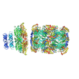
6C26
 | | The Cryo-EM structure of a eukaryotic oligosaccharyl transferase complex | | 分子名称: | (4R,7R)-4-hydroxy-N,N,N-trimethyl-4,9-dioxo-7-[(undecanoyloxy)methyl]-3,5,8-trioxa-4lambda~5~-phosphadocosan-1-aminium, 2-acetamido-2-deoxy-beta-D-glucopyranose, Dolichyl-diphosphooligosaccharide--protein glycosyltransferase subunit 1, ... | | 著者 | Bai, L, Li, H. | | 登録日 | 2018-01-06 | | 公開日 | 2018-01-31 | | 最終更新日 | 2020-07-29 | | 実験手法 | ELECTRON MICROSCOPY (3.5 Å) | | 主引用文献 | The atomic structure of a eukaryotic oligosaccharyltransferase complex.
Nature, 555, 2018
|
|


