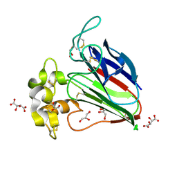
5YYP
 
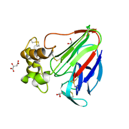 | | Structure K137A thaumatin | | 分子名称: | GLYCEROL, L(+)-TARTARIC ACID, Preprothaumatin I | | 著者 | Masuda, T, Kigo, S, Mitsumoto, M, Ohta, K, Suzuki, M, Mikami, B, Kitabatake, N, Tani, F. | | 登録日 | 2017-12-10 | | 公開日 | 2018-03-21 | | 最終更新日 | 2023-11-22 | | 実験手法 | X-RAY DIFFRACTION (1.01 Å) | | 主引用文献 | Positive Charges on the Surface of Thaumatin Are Crucial for the Multi-Point Interaction with the Sweet Receptor.
Front Mol Biosci, 5, 2018
|
|
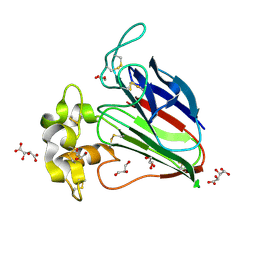
5YYQ
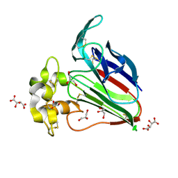 | | Structure K78A thaumatin | | 分子名称: | GLYCEROL, L(+)-TARTARIC ACID, Preprothaumatin I | | 著者 | Masuda, T, Kigo, S, Mitsumoto, M, Ohta, K, Suzuki, M, Mikami, B, Kitabatake, N, Tani, F. | | 登録日 | 2017-12-10 | | 公開日 | 2018-03-21 | | 最終更新日 | 2023-11-22 | | 実験手法 | X-RAY DIFFRACTION (1.07 Å) | | 主引用文献 | Positive Charges on the Surface of Thaumatin Are Crucial for the Multi-Point Interaction with the Sweet Receptor.
Front Mol Biosci, 5, 2018
|
|
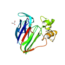
5YYR
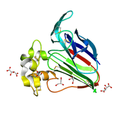 | | Structure K106A thaumatin | | 分子名称: | GLYCEROL, L(+)-TARTARIC ACID, Preprothaumatin I | | 著者 | Masuda, T, Kigo, S, Ohta, K, Mitsumoto, M, Mikami, B, Suzuki, M, Kitabatake, N, Tani, F. | | 登録日 | 2017-12-10 | | 公開日 | 2018-03-21 | | 最終更新日 | 2023-11-22 | | 実験手法 | X-RAY DIFFRACTION (1.07 Å) | | 主引用文献 | Positive Charges on the Surface of Thaumatin Are Crucial for the Multi-Point Interaction with the Sweet Receptor.
Front Mol Biosci, 5, 2018
|
|
3AOK
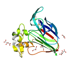 | |
3ALD
 | | Crystal structure of sweet-tasting protein Thaumatin I at 1.10 A | | 分子名称: | GLYCEROL, L(+)-TARTARIC ACID, Thaumatin I | | 著者 | Masuda, T, Mikami, B, Kitabatake, N. | | 登録日 | 2010-07-29 | | 公開日 | 2011-06-08 | | 最終更新日 | 2011-11-02 | | 実験手法 | X-RAY DIFFRACTION (1.1 Å) | | 主引用文献 | High-resolution structure of the recombinant sweet-tasting protein thaumatin I
Acta Crystallogr.,Sect.F, 67, 2011
|
|
3AL7
 | | Recombinant thaumatin I at 1.1 A | | 分子名称: | GLYCEROL, L(+)-TARTARIC ACID, Thaumatin I | | 著者 | Masuda, T, Mikami, B, Kitabatake, N. | | 登録日 | 2010-07-27 | | 公開日 | 2011-06-08 | | 最終更新日 | 2023-11-01 | | 実験手法 | X-RAY DIFFRACTION (1.1 Å) | | 主引用文献 | High-resolution structure of the recombinant sweet-tasting protein thaumatin I
Acta Crystallogr.,Sect.F, 67, 2011
|
|
3VHG
 | |
3VHF
 | | plant thaumatin I at pH 8.0 | | 分子名称: | GLYCEROL, Thaumatin I | | 著者 | Masuda, T, Mikami, B, Kitabatake, N, Tani, F. | | 登録日 | 2011-08-24 | | 公開日 | 2012-05-16 | | 最終更新日 | 2023-11-08 | | 実験手法 | X-RAY DIFFRACTION (1.39 Å) | | 主引用文献 | Atomic structure of the sweet-tasting protein thaumatin I at pH 8.0 reveals the large disulfide-rich region in domain II to be sensitive to a pH change
Biochem.Biophys.Res.Commun., 419, 2012
|
|
3VJQ
 | |


