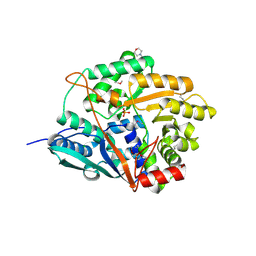
Loading
PDBj
MenuPDBj@FacebookPDBj@TwitterPDBj@YouTubewwPDB FoundationwwPDB
RCSB PDBPDBeBMRBAdv. SearchSearch help
Search by PDB author
3OE9
DownloadVisualize
BU of 3oe9 by Molmil
Crystal structure of the chemokine CXCR4 receptor in complex with a small molecule antagonist IT1t in P1 spacegroup
Descriptor: (6,6-dimethyl-5,6-dihydroimidazo[2,1-b][1,3]thiazol-3-yl)methyl N,N'-dicyclohexylimidothiocarbamate, C-X-C chemokine receptor type 4, Lysozyme Chimera
Authors:Wu, B, Mol, C.D, Han, G.W, Katritch, V, Chien, E.Y.T, Liu, W, Cherezov, V, Stevens, R.C, Accelerated Technologies Center for Gene to 3D Structure (ATCG3D), GPCR Network (GPCR)
Deposit date:2010-08-12
Release date:2010-10-27
Last modified:2021-10-06
Method:X-RAY DIFFRACTION (3.1 Å)
Cite:Structures of the CXCR4 chemokine GPCR with small-molecule and cyclic peptide antagonists.
Science, 330, 2010
1VPZ
DownloadVisualize
BU of 1vpz by Molmil
Crystal structure of a putative carbon storage regulator protein (csra, pa0905) from pseudomonas aeruginosa at 2.05 A resolution
Descriptor: Carbon storage regulator homolog
Authors:Joint Center for Structural Genomics (JCSG)
Deposit date:2004-11-24
Release date:2004-12-14
Last modified:2023-01-25
Method:X-RAY DIFFRACTION (2.05 Å)
Cite:Crystal structure of the global regulatory protein CsrA from Pseudomonas putida at 2.05 A resolution reveals a new fold.
Proteins, 61, 2005
1VQR
DownloadVisualize
BU of 1vqr by Molmil
Crystal structure of a virulence factor (cj0248) from campylobacter jejuni subsp. jejuni at 2.25 A resolution
Descriptor: hypothetical protein Cj0248
Authors:Joint Center for Structural Genomics (JCSG)
Deposit date:2004-12-17
Release date:2004-12-28
Last modified:2023-01-25
Method:X-RAY DIFFRACTION (2.25 Å)
Cite:Crystal structure of virulence factor CJ0248 from Campylobacter jejuni at 2.25 A resolution reveals a new fold.
Proteins, 62, 2006
1VR8
DownloadVisualize
BU of 1vr8 by Molmil
Crystal structure of GTP binding regulator (TM1622) from Thermotoga Maritima at 1.75 A resolution
Descriptor: AZIDE ION, GLYCEROL, GTP binding regulator
Authors:Joint Center for Structural Genomics (JCSG)
Deposit date:2005-02-16
Release date:2005-03-15
Last modified:2023-01-25
Method:X-RAY DIFFRACTION (1.75 Å)
Cite:Crystal structure of an ORFan protein (TM1622) from Thermotoga maritima at 1.75 A resolution reveals a fold similar to the Ran-binding protein Mog1p.
Proteins, 65, 2006
1VK3
DownloadVisualize
BU of 1vk3 by Molmil
Crystal structure of Phosphoribosylformylglycinamidine synthase II (TM1246) from Thermotoga maritima at 2.15 A resolution
Descriptor: CHLORIDE ION, Phosphoribosylformylglycinamidine synthase II
Authors:Joint Center for Structural Genomics (JCSG)
Deposit date:2004-04-23
Release date:2004-05-11
Last modified:2023-01-25
Method:X-RAY DIFFRACTION (2.15 Å)
Cite:Crystal structure of phosphoribosylformylglycinamidine synthase II (smPurL) from Thermotoga maritima at 2.15 A resolution.
Proteins, 63, 2006
1VL4
DownloadVisualize
BU of 1vl4 by Molmil
CRYSTAL STRUCTURE OF A PUTATIVE MODULATOR OF A DNA GYRASE (TM0727) FROM THERMOTOGA MARITIMA MSB8 AT 1.95 A RESOLUTION
Descriptor: pmbA-related protein
Authors:Joint Center for Structural Genomics (JCSG)
Deposit date:2004-07-09
Release date:2004-09-21
Last modified:2024-05-22
Method:X-RAY DIFFRACTION (1.95 Å)
Cite:Crystal structure of a putative modulator of DNA gyrase (pmbA) from Thermotoga maritima at 1.95 A resolution reveals a new fold.
Proteins, 61, 2005
1VLP
DownloadVisualize
BU of 1vlp by Molmil
Crystal structure of a putative nicotinate phosphoribosyltransferase (yor209c, npt1) from saccharomyces cerevisiae at 1.75 A resolution
Descriptor: 1,2-ETHANEDIOL, 2-(N-MORPHOLINO)-ETHANESULFONIC ACID, CHLORIDE ION, ...
Authors:Joint Center for Structural Genomics (JCSG)
Deposit date:2004-08-06
Release date:2004-08-24
Last modified:2023-01-25
Method:X-RAY DIFFRACTION (1.75 Å)
Cite:The structure of a eukaryotic nicotinic acid phosphoribosyltransferase reveals structural heterogeneity among type II PRTases.
Structure, 13, 2005
1VKH
DownloadVisualize
BU of 1vkh by Molmil
CRYSTAL STRUCTURE OF A PUTATIVE SERINE HYDROLASE (YDR428C) FROM SACCHAROMYCES CEREVISIAE AT 1.85 A RESOLUTION
Descriptor: CHLORIDE ION, GLYCEROL, putative serine hydrolase
Authors:Joint Center for Structural Genomics (JCSG)
Deposit date:2004-05-20
Release date:2004-06-08
Last modified:2023-01-25
Method:X-RAY DIFFRACTION (1.85 Å)
Cite:Crystal structure of an alpha/beta serine hydrolase (YDR428C) from Saccharomyces cerevisiae at 1.85 A resolution
Proteins, 58, 2005
3ODU
DownloadVisualize
BU of 3odu by Molmil
The 2.5 A structure of the CXCR4 chemokine receptor in complex with small molecule antagonist IT1t
Descriptor: (2R)-2,3-dihydroxypropyl (9Z)-octadec-9-enoate, (6,6-dimethyl-5,6-dihydroimidazo[2,1-b][1,3]thiazol-3-yl)methyl N,N'-dicyclohexylimidothiocarbamate, C-X-C chemokine receptor type 4, ...
Authors:Wu, B, Mol, C.D, Han, G.W, Katritch, V, Chien, E.Y.T, Liu, W, Cherezov, V, Stevens, R.C, Accelerated Technologies Center for Gene to 3D Structure (ATCG3D), GPCR Network (GPCR)
Deposit date:2010-08-11
Release date:2010-10-27
Last modified:2021-10-06
Method:X-RAY DIFFRACTION (2.5 Å)
Cite:Structures of the CXCR4 chemokine GPCR with small-molecule and cyclic peptide antagonists.
Science, 330, 2010
3OE6
DownloadVisualize
BU of 3oe6 by Molmil
Crystal structure of the CXCR4 chemokine receptor in complex with a small molecule antagonist IT1t in I222 spacegroup
Descriptor: (2R)-2,3-dihydroxypropyl (9Z)-octadec-9-enoate, (6,6-dimethyl-5,6-dihydroimidazo[2,1-b][1,3]thiazol-3-yl)methyl N,N'-dicyclohexylimidothiocarbamate, C-X-C chemokine receptor type 4, ...
Authors:Wu, B, Mol, C.D, Han, G.W, Katritch, V, Chien, E.Y.T, Liu, W, Cherezov, V, Stevens, R.C, Accelerated Technologies Center for Gene to 3D Structure (ATCG3D), GPCR Network (GPCR)
Deposit date:2010-08-12
Release date:2010-10-27
Last modified:2021-10-06
Method:X-RAY DIFFRACTION (3.2 Å)
Cite:Structures of the CXCR4 chemokine GPCR with small-molecule and cyclic peptide antagonists.
Science, 330, 2010
3OE8
DownloadVisualize
BU of 3oe8 by Molmil
Crystal structure of the CXCR4 chemokine receptor in complex with a small molecule antagonist IT1t in P1 spacegroup
Descriptor: (6,6-dimethyl-5,6-dihydroimidazo[2,1-b][1,3]thiazol-3-yl)methyl N,N'-dicyclohexylimidothiocarbamate, C-X-C chemokine receptor type 4, Lysozyme Chimera
Authors:Wu, B, Mol, C.D, Han, G.W, Katritch, V, Chien, E.Y.T, Liu, W, Cherezov, V, Stevens, R.C, Accelerated Technologies Center for Gene to 3D Structure (ATCG3D), GPCR Network (GPCR)
Deposit date:2010-08-12
Release date:2010-10-27
Last modified:2021-10-06
Method:X-RAY DIFFRACTION (3.1 Å)
Cite:Structures of the CXCR4 chemokine GPCR with small-molecule and cyclic peptide antagonists.
Science, 330, 2010
1ZCZ
DownloadVisualize
BU of 1zcz by Molmil
Crystal structure of Phosphoribosylaminoimidazolecarboxamide formyltransferase / IMP cyclohydrolase (TM1249) from THERMOTOGA MARITIMA at 1.88 A resolution
Descriptor: Bifunctional purine biosynthesis protein purH, POTASSIUM ION, TETRAETHYLENE GLYCOL
Authors:Joint Center for Structural Genomics (JCSG)
Deposit date:2005-04-13
Release date:2005-04-26
Last modified:2023-09-20
Method:X-RAY DIFFRACTION (1.88 Å)
Cite:Crystal structure of AICAR transformylase IMP cyclohydrolase (TM1249) from Thermotoga maritima at 1.88 A resolution.
Proteins, 71, 2008
1ZKG
DownloadVisualize
BU of 1zkg by Molmil
Crystal structure of Transcriptional regulator, TETR family (tm1030) from Thermotoga maritima at 2.30 A resolution
Descriptor: UNKNOWN LIGAND, transcriptional regulator, TetR family
Authors:Joint Center for Structural Genomics (JCSG)
Deposit date:2005-05-02
Release date:2005-07-26
Last modified:2023-01-25
Method:X-RAY DIFFRACTION (2.3 Å)
Cite:Crystal structure of TM1030 from Thermotoga maritima at 2.3 A resolution reveals molecular details of its transcription repressor function.
Proteins, 68, 2007
1ZX8
DownloadVisualize
BU of 1zx8 by Molmil
CRYSTAL STRUCTURE OF an atypical cyclophilin (peptidylprolyl cis-trans isomerase) (TM1367) FROM THERMOTOGA MARITIMA AT 1.90 A RESOLUTION
Descriptor: NICKEL (II) ION, PENTAETHYLENE GLYCOL, hypothetical protein TM1367
Authors:Joint Center for Structural Genomics (JCSG)
Deposit date:2005-06-07
Release date:2005-07-26
Last modified:2023-01-25
Method:X-RAY DIFFRACTION (1.9 Å)
Cite:Crystal structure of TM1367 from Thermotoga maritima at 1.90 A resolution reveals an atypical member of the cyclophilin (peptidylprolyl isomerase) fold.
Proteins, 63, 2006
3OE0
DownloadVisualize
BU of 3oe0 by Molmil
Crystal structure of the CXCR4 chemokine receptor in complex with a cyclic peptide antagonist CVX15
Descriptor: C-X-C chemokine receptor type 4, Lysozyme Chimera, Polyphemusin analog, ...
Authors:Wu, B, Mol, C.D, Han, G.W, Katritch, V, Chien, E.Y.T, Liu, W, Cherezov, V, Stevens, R.C, Accelerated Technologies Center for Gene to 3D Structure (ATCG3D), GPCR Network (GPCR)
Deposit date:2010-08-12
Release date:2010-10-27
Last modified:2023-11-15
Method:X-RAY DIFFRACTION (2.9 Å)
Cite:Structures of the CXCR4 chemokine GPCR with small-molecule and cyclic peptide antagonists.
Science, 330, 2010
3H41
DownloadVisualize
BU of 3h41 by Molmil
CRYSTAL STRUCTURE OF A NLPC/P60 FAMILY PROTEIN (BCE_2878) FROM BACILLUS CEREUS ATCC 10987 AT 1.79 A RESOLUTION
Descriptor: ALANINE, D-GLUTAMIC ACID, NLP/P60 family protein, ...
Authors:Joint Center for Structural Genomics (JCSG)
Deposit date:2009-04-17
Release date:2009-05-05
Last modified:2023-02-01
Method:X-RAY DIFFRACTION (1.79 Å)
Cite:Structure of the gamma-D-glutamyl-L-diamino acid endopeptidase YkfC from Bacillus cereus in complex with L-Ala-[gamma]-D-Glu: insights into substrate recognition by NlpC/P60 cysteine peptidases
Acta Crystallogr.,Sect.F, 66, 2010
3H50
DownloadVisualize
BU of 3h50 by Molmil
CRYSTAL STRUCTURE OF A TETRACENOMYCIN POLYKETIDE SYNTHESIS PROTEIN (TCMJ) FROM XANTHOMONAS CAMPESTRIS PV. CAMPESTRIS AT 1.60 A RESOLUTION
Descriptor: ACETATE ION, Tetracenomycin polyketide synthesis protein, ZINC ION
Authors:Joint Center for Structural Genomics (JCSG)
Deposit date:2009-04-21
Release date:2009-05-05
Last modified:2023-02-01
Method:X-RAY DIFFRACTION (1.6 Å)
Cite:Conformational changes associated with the binding of zinc acetate at the putative active site of XcTcmJ, a cupin from Xanthomonas campestris pv. campestris.
Acta Crystallogr.,Sect.F, 66, 2010
3HSA
DownloadVisualize
BU of 3hsa by Molmil
Crystal structure of pleckstrin homology domain (YP_926556.1) from SHEWANELLA AMAZONENSIS SB2B at 1.99 A resolution
Descriptor: DI(HYDROXYETHYL)ETHER, GLYCEROL, pleckstrin homology domain
Authors:Joint Center for Structural Genomics (JCSG)
Deposit date:2009-06-10
Release date:2009-06-23
Last modified:2023-02-01
Method:X-RAY DIFFRACTION (1.99 Å)
Cite:Bacterial pleckstrin homology domains: a prokaryotic origin for the PH domain.
J.Mol.Biol., 396, 2010
3IRB
DownloadVisualize
BU of 3irb by Molmil
Crystal structure of protein with unknown function from DUF35 family (13815350) from SULFOLOBUS SOLFATARICUS at 1.80 A resolution
Descriptor: ACETIC ACID, SULFATE ION, ZINC ION, ...
Authors:Joint Center for Structural Genomics (JCSG)
Deposit date:2009-08-21
Release date:2009-09-01
Last modified:2023-02-01
Method:X-RAY DIFFRACTION (1.8 Å)
Cite:The structure of SSO2064, the first representative of Pfam family PF01796, reveals a novel two-domain zinc-ribbon OB-fold architecture with a potential acyl-CoA-binding role.
Acta Crystallogr.,Sect.F, 66, 2010
3H0N
DownloadVisualize
BU of 3h0n by Molmil
Crystal structure of a duf1470 family protein (jann_2411) from jannaschia sp. ccs1 at 1.45 A resolution
Descriptor: ACETATE ION, GLYCEROL, NICKEL (II) ION, ...
Authors:Joint Center for Structural Genomics (JCSG)
Deposit date:2009-04-09
Release date:2009-04-21
Last modified:2023-02-01
Method:X-RAY DIFFRACTION (1.45 Å)
Cite:The structure of Jann_2411 (DUF1470) from Jannaschia sp. at 1.45  resolution reveals a new fold (the ABATE domain) and suggests its possible role as a transcription regulator.
Acta Crystallogr.,Sect.F, 66, 2010
3HBZ
DownloadVisualize
BU of 3hbz by Molmil
Crystal structure of a putative glycoside hydrolase (bt_2081) from bacteroides thetaiotaomicron vpi-5482 at 2.05 A resolution
Descriptor: ACETATE ION, CACODYLATE ION, CALCIUM ION, ...
Authors:Joint Center for Structural Genomics (JCSG)
Deposit date:2009-05-05
Release date:2009-05-26
Last modified:2023-02-01
Method:X-RAY DIFFRACTION (2.05 Å)
Cite:Structure of Bacteroides thetaiotaomicron BT2081 at 2.05 A resolution: the first structural representative of a new protein family that may play a role in carbohydrate metabolism.
Acta Crystallogr.,Sect.F, 66, 2010
3K5J
DownloadVisualize
BU of 3k5j by Molmil
Crystal structure of Putative SUFU (suppressor of fused protein) homolog (YP_208451.1) from Neisseria gonorrhoeae FA 1090 at 1.40 A resolution
Descriptor: GLYCEROL, SULFATE ION, Suppressor of fused family protein
Authors:Joint Center for Structural Genomics (JCSG)
Deposit date:2009-10-07
Release date:2010-01-26
Last modified:2019-07-17
Method:X-RAY DIFFRACTION (1.4 Å)
Cite:The crystal structure of a bacterial Sufu-like protein defines a novel group of bacterial proteins that are similar to the N-terminal domain of human Sufu.
Protein Sci., 19, 2010
3BOS
DownloadVisualize
BU of 3bos by Molmil
Crystal structure of a putative dna replication regulator HDA (SAMA_1916) from Shewanella amazonensis sb2b at 1.75 A resolution
Descriptor: 1,2-ETHANEDIOL, CHLORIDE ION, CYTIDINE-5'-DIPHOSPHATE, ...
Authors:Joint Center for Structural Genomics (JCSG)
Deposit date:2007-12-17
Release date:2008-01-15
Last modified:2023-01-25
Method:X-RAY DIFFRACTION (1.75 Å)
Cite:A structural basis for the regulatory inactivation of DnaA.
J.Mol.Biol., 385, 2009
3CM1
DownloadVisualize
BU of 3cm1 by Molmil
Crystal structure of SsgA-like sporulation-specific cell division protein (YP_290167.1) from Thermobifida fusca YX-ER1 at 2.60 A resolution
Descriptor: SsgA-like sporulation-specific cell division protein
Authors:Joint Center for Structural Genomics (JCSG), Chruszcz, M, Minor, W, Wang, S.
Deposit date:2008-03-20
Release date:2008-04-01
Last modified:2023-02-01
Method:X-RAY DIFFRACTION (2.6 Å)
Cite:Structural and functional characterizations of SsgB, a conserved activator of developmental cell division in morphologically complex actinomycetes.
J.Biol.Chem., 284, 2009
3DEE
DownloadVisualize
BU of 3dee by Molmil
CRYSTAL STRUCTURE OF A PUTATIVE REGULATORY PROTEIN INVOLVED IN TRANSCRIPTION (NGO1945) FROM NEISSERIA GONORRHOEAE FA 1090 AT 2.25 A RESOLUTION
Descriptor: CHLORIDE ION, IMIDAZOLE, Putative regulatory protein
Authors:Joint Center for Structural Genomics (JCSG)
Deposit date:2008-06-09
Release date:2008-08-05
Last modified:2023-02-01
Method:X-RAY DIFFRACTION (2.1 Å)
Cite:The structure of the first representative of Pfam family PF09836 reveals a two-domain organization and suggests involvement in transcriptional regulation.
Acta Crystallogr.,Sect.F, 66, 2010

225158

PDB entries from 2024-09-18

PDB statisticsPDBj update infoContact PDBjnumon