5PCY
 
 | |
4PCY
 | |
6PCY
 | |
1PLC
 | |
2CBP
 | |
1JUG
 | |
1N9E
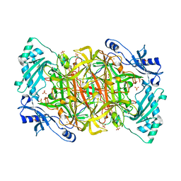 | | Crystal structure of Pichia pastoris Lysyl Oxidase PPLO | | 分子名称: | 2-acetamido-2-deoxy-beta-D-glucopyranose, 2-acetamido-2-deoxy-beta-D-glucopyranose-(1-4)-2-acetamido-2-deoxy-beta-D-glucopyranose, CALCIUM ION, ... | | 著者 | Guss, J.M, Duff, A.P. | | 登録日 | 2002-11-24 | | 公開日 | 2004-01-13 | | 最終更新日 | 2023-10-25 | | 実験手法 | X-RAY DIFFRACTION (1.65 Å) | | 主引用文献 | The Crystal Structure of Pichia pastoris Lysyl Oxidase
Biochemistry, 42, 2003
|
|
1RJO
 | | AGAO + Xe | | 分子名称: | COPPER (II) ION, GLYCEROL, Phenylethylamine oxidase, ... | | 著者 | Guss, J.M, Trambaiolo, D.M, Duff, A.P. | | 登録日 | 2003-11-19 | | 公開日 | 2004-12-07 | | 最終更新日 | 2024-04-03 | | 実験手法 | X-RAY DIFFRACTION (1.67 Å) | | 主引用文献 | Using Xenon as a Probe for Dioxygen-binding Sites in Copper Amine Oxidases
J.Mol.Biol., 344, 2004
|
|
1RKY
 | | PPLO + Xe | | 分子名称: | 2-acetamido-2-deoxy-beta-D-glucopyranose, CALCIUM ION, CHLORIDE ION, ... | | 著者 | Guss, J.M, Duff, A.P. | | 登録日 | 2003-11-24 | | 公開日 | 2004-12-07 | | 最終更新日 | 2023-10-25 | | 実験手法 | X-RAY DIFFRACTION (1.68 Å) | | 主引用文献 | Using Xenon as a Probe for Dioxygen-binding Sites in Copper Amine Oxidases
J.Mol.Biol., 344, 2004
|
|
1SII
 | | AGAO in covalent complex with the inhibitor NOBA ("4-(2-naphthyloxy)-2-butyn-1-amine") | | 分子名称: | COPPER (II) ION, GLYCEROL, Phenylethylamine oxidase, ... | | 著者 | Guss, J.M, Langley, D.B, Duff, A.P. | | 登録日 | 2004-02-29 | | 公開日 | 2004-09-07 | | 最終更新日 | 2017-10-11 | | 実験手法 | X-RAY DIFFRACTION (1.7 Å) | | 主引用文献 | Differential Inhibition of Six Copper Amine Oxidases by a Family of 4-(Aryloxy)-2-butynamines: Evidence for a New Mode of Inactivation.
Biochemistry, 43, 2004
|
|
1SIH
 | | AGAO in covalent complex with the inhibitor MOBA ("4-(4-methylphenoxy)-2-butyn-1-amine") | | 分子名称: | COPPER (II) ION, GLYCEROL, Phenylethylamine oxidase, ... | | 著者 | Guss, J.M, Langley, D.B, Duff, A.P. | | 登録日 | 2004-02-29 | | 公開日 | 2004-09-07 | | 最終更新日 | 2017-10-11 | | 実験手法 | X-RAY DIFFRACTION (1.73 Å) | | 主引用文献 | Differential Inhibition of Six Copper Amine Oxidases by a Family of 4-(Aryloxy)-2-butynamines: Evidence for a New Mode of Inactivation.
Biochemistry, 43, 2004
|
|
7PCY
 | | THE CRYSTAL STRUCTURE OF PLASTOCYANIN FROM A GREEN ALGA, ENTEROMORPHA PROLIFERA | | 分子名称: | COPPER (II) ION, PLASTOCYANIN | | 著者 | Collyer, C.A, Guss, J.M, Freeman, H.C. | | 登録日 | 1989-09-22 | | 公開日 | 1990-07-15 | | 最終更新日 | 2024-03-06 | | 実験手法 | X-RAY DIFFRACTION (1.8 Å) | | 主引用文献 | Crystal structure of plastocyanin from a green alga, Enteromorpha prolifera.
J.Mol.Biol., 211, 1990
|
|
1AQT
 | |
4XS0
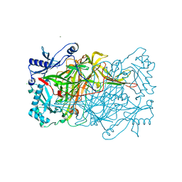
 | | Human methemoglobin in complex with the second and third NEAT domains of IsdH(F365Y/A369F/Y642A) from Staphylococcus aureus | | 分子名称: | CHLORIDE ION, Hemoglobin subunit alpha, Hemoglobin subunit beta, ... | | 著者 | Dickson, C.F, Jacques, D.A, Guss, J.M, Gell, D.A. | | 登録日 | 2015-01-21 | | 公開日 | 2015-06-03 | | 最終更新日 | 2024-01-10 | | 実験手法 | X-RAY DIFFRACTION (2.55 Å) | | 主引用文献 | The structure of haemoglobin bound to the haemoglobin receptor IsdH from Staphylococcus aureus shows disruption of the native alpha-globin haem pocket.
Acta Crystallogr.,Sect.D, 71, 2015
|
|
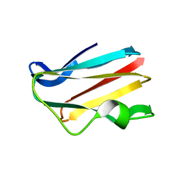
5K6P
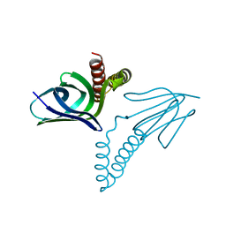
 | | The NMR structure of the m domain tri-helix bundle and C2 of human cardiac Myosin Binding Protein C | | 分子名称: | Myosin-binding protein C, cardiac-type | | 著者 | Michie, K.A, Kwan, A.H, Tung, C.S, Guss, J.M, Trewhella, J. | | 登録日 | 2016-05-25 | | 公開日 | 2016-11-09 | | 最終更新日 | 2023-06-14 | | 実験手法 | SOLUTION NMR | | 主引用文献 | A Highly Conserved Yet Flexible Linker Is Part of a Polymorphic Protein-Binding Domain in Myosin-Binding Protein C.
Structure, 24, 2016
|
|
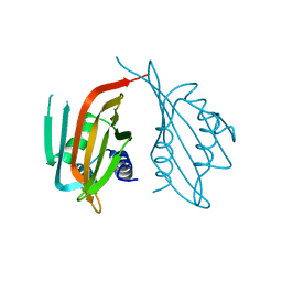
6CME
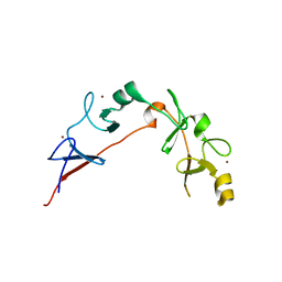 | | Structure of wild-type ISL2-LID in complex with LHX4-LIM1+2 | | 分子名称: | LIM/homeobox protein Lhx4,Insulin gene enhancer protein ISL-2, ZINC ION | | 著者 | Stokes, P.H, Silva, A, Guss, J.M, Matthews, J.M. | | 登録日 | 2018-03-04 | | 公開日 | 2019-04-10 | | 最終更新日 | 2023-10-04 | | 実験手法 | X-RAY DIFFRACTION (1.92 Å) | | 主引用文献 | Mutation in a flexible linker modulates binding affinity for modular complexes.
Proteins, 87, 2019
|
|
4MJ3
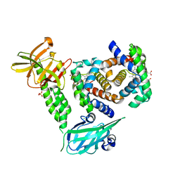
 | | Haloalkane dehalogenase DmrA from Mycobacterium rhodesiae JS60 | | 分子名称: | CHLORIDE ION, Haloalkane dehalogenase, POTASSIUM ION | | 著者 | Fung, H, Gadd, M.S, Guss, J.M, Matthews, J.M. | | 登録日 | 2013-09-03 | | 公開日 | 2015-02-25 | | 最終更新日 | 2015-08-19 | | 実験手法 | X-RAY DIFFRACTION (1.7 Å) | | 主引用文献 | Biochemical and biophysical characterisation of haloalkane dehalogenases DmrA and DmrB in Mycobacterium strain JS60 and their role in growth on haloalkanes.
Mol.Microbiol., 97, 2015
|
|
465D
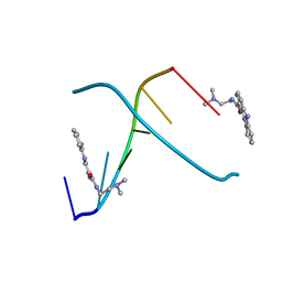 | | STRUCTURE OF THE TOPOISOMERASE II POISON BOUND TO DNA | | 分子名称: | 9-AMINO-(N-(2-DIMETHYLAMINO)ETHYL)ACRIDINE-4-CARBOXAMIDE, DNA (5'-D(*CP*GP*TP*AP*CP*G)-3') | | 著者 | Adams, A, Guss, J.M, Collyer, C.A, Denny, W.A, Wakelin, L.P. | | 登録日 | 1999-04-14 | | 公開日 | 1999-08-25 | | 最終更新日 | 2024-04-03 | | 実験手法 | X-RAY DIFFRACTION (1.6 Å) | | 主引用文献 | Crystal structure of the topoisomerase II poison 9-amino-[N-(2-dimethylamino)ethyl]acridine-4-carboxamide bound to the DNA hexanucleotide d(CGTACG)2.
Biochemistry, 38, 1999
|
|
1A16
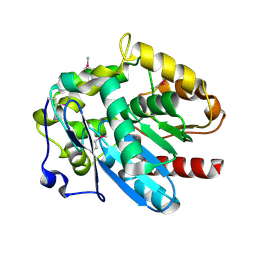
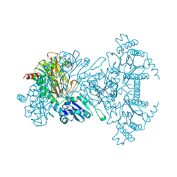 | | AMINOPEPTIDASE P FROM E. COLI WITH THE INHIBITOR PRO-LEU | | 分子名称: | AMINOPEPTIDASE P, LEUCINE, MANGANESE (II) ION, ... | | 著者 | Wilce, M.C, Bond, C.S, Lilley, P.E, Dixon, N.E, Freeman, H.C, Guss, J.M. | | 登録日 | 1997-12-22 | | 公開日 | 1999-04-06 | | 最終更新日 | 2024-02-07 | | 実験手法 | X-RAY DIFFRACTION (2.3 Å) | | 主引用文献 | Structure and mechanism of a proline-specific aminopeptidase from Escherichia coli.
Proc.Natl.Acad.Sci.USA, 95, 1998
|
|
1B13
 | | CLOSTRIDIUM PASTEURIANUM RUBREDOXIN G10A MUTANT | | 分子名称: | FE (III) ION, PROTEIN (RUBREDOXIN) | | 著者 | Maher, M.J, Guss, J.M, Wilce, M.C.J, Wedd, A.G. | | 登録日 | 1998-11-26 | | 公開日 | 1999-05-27 | | 最終更新日 | 2023-08-09 | | 実験手法 | X-RAY DIFFRACTION (1.5 Å) | | 主引用文献 | Rubredoxin from Clostridium pasteurianum. Structures of G10A, G43A and G10VG43A mutant proteins. Mutation of conserved glycine 10 to valine causes the 9-10 peptide link to invert.
Acta Crystallogr.,Sect.D, 55, 1999
|
|
1B2O
 | | CLOSTRIDIUM PASTEURIANUM RUBREDOXIN G10VG43A MUTANT | | 分子名称: | FE (III) ION, PROTEIN (RUBREDOXIN) | | 著者 | Maher, M.J, Guss, J.M, Wilce, M.C.J, Wedd, A.G. | | 登録日 | 1998-11-30 | | 公開日 | 1999-05-27 | | 最終更新日 | 2023-08-09 | | 実験手法 | X-RAY DIFFRACTION (1.9 Å) | | 主引用文献 | Rubredoxin from Clostridium pasteurianum. Structures of G10A, G43A and G10VG43A mutant proteins. Mutation of conserved glycine 10 to valine causes the 9-10 peptide link to invert.
Acta Crystallogr.,Sect.D, 55, 1999
|
|
1B2J
 | | CLOSTRIDIUM PASTEURIANUM RUBREDOXIN G43A MUTANT | | 分子名称: | FE (III) ION, PROTEIN (RUBREDOXIN) | | 著者 | Maher, M.J, Guss, J.M, Wilce, M.C.J, Wedd, A.G. | | 登録日 | 1998-11-27 | | 公開日 | 1999-05-27 | | 最終更新日 | 2023-08-09 | | 実験手法 | X-RAY DIFFRACTION (1.6 Å) | | 主引用文献 | Rubredoxin from Clostridium pasteurianum. Structures of G10A, G43A and G10VG43A mutant proteins. Mutation of conserved glycine 10 to valine causes the 9-10 peptide link to invert.
Acta Crystallogr.,Sect.D, 55, 1999
|
|
2PCY
 | |
3PCY
 | | THE CRYSTAL STRUCTURE OF MERCURY-SUBSTITUTED POPLAR PLASTOCYANIN AT 1.9-ANGSTROMS RESOLUTION | | 分子名称: | MERCURY (II) ION, PLASTOCYANIN | | 著者 | Church, W.B, Guss, J.M, Potter, J.J, Freeman, H.C. | | 登録日 | 1985-12-10 | | 公開日 | 1986-01-21 | | 最終更新日 | 2024-02-21 | | 実験手法 | X-RAY DIFFRACTION (1.9 Å) | | 主引用文献 | The crystal structure of mercury-substituted poplar plastocyanin at 1.9-A resolution.
J.Biol.Chem., 261, 1986
|
|
6PTL
 | |


