Loading
PDBj
MenuPDBj@FacebookPDBj@TwitterPDBj@YouTubewwPDB FoundationwwPDB
RCSB PDBPDBeBMRBAdv. SearchSearch help
Search by PDB author
6MQZ
DownloadVisualize
BU of 6mqz by Molmil
Crystal Structure of the 13-cis Product of the All-trans Retinal-Bound R111K:Y134F:T54V:R132Q:P39Y:R59Y:L121E Mutant of Human Cellular Retinoic Acid Binding Protein II Irradiated with 400 nm Laser (5 minutes) at 2.07 Angstrom
Descriptor: Cellular retinoic acid-binding protein 2, GLYCEROL, RETINAL
Authors:Ghanbarpour, A, Geiger, J.
Deposit date:2018-10-11
Release date:2019-01-09
Last modified:2023-10-11
Method:X-RAY DIFFRACTION (2.07 Å)
Cite:Mimicking Microbial Rhodopsin Isomerization in a Single Crystal.
J. Am. Chem. Soc., 141, 2019
6MOR
DownloadVisualize
BU of 6mor by Molmil
Crystal Structure of the All-Trans Retinal-Bound R111K:Y134F:T54V:R132Q:P39Y:R59Y:L121Y Mutant of Human Cellular Retinoic Acid Binding Protein II in the Dark at 1.79 Angstrom Resolution
Descriptor: Cellular retinoic acid-binding protein 2, RETINAL
Authors:Ghanbarpour, A, Geiger, J.
Deposit date:2018-10-04
Release date:2019-01-09
Last modified:2023-10-11
Method:X-RAY DIFFRACTION (1.79 Å)
Cite:Mimicking Microbial Rhodopsin Isomerization in a Single Crystal.
J. Am. Chem. Soc., 141, 2019
6VIT
DownloadVisualize
BU of 6vit by Molmil
The Crystal Structure of Apo Domain-Swapped dimer Q108K:T51D:I32C Variant of HCRBPII with an Engineered Disulfide Bond
Descriptor: Retinol-binding protein 2
Authors:Ghanbarpour, A, Geiger, J.
Deposit date:2020-01-13
Release date:2020-08-19
Last modified:2023-10-11
Method:X-RAY DIFFRACTION (3.2 Å)
Cite:Human Cellular Retinol Binding Protein II Forms a Domain-Swapped Trimer Representing a Novel Fold and a New Template for Protein Engineering.
Chembiochem, 21, 2020
6VIS
DownloadVisualize
BU of 6vis by Molmil
The Crystal Structure of Domain-Swapped Trimer Q108K:K40D:T53A:R58L:Q38F:Q4F:V62E Variant of HCRBPII
Descriptor: GLYCEROL, Retinol-binding protein 2
Authors:Ghanbarpour, A, Geiger, J.
Deposit date:2020-01-13
Release date:2020-09-09
Last modified:2023-10-11
Method:X-RAY DIFFRACTION (2.79 Å)
Cite:Human Cellular Retinol Binding Protein II Forms a Domain-Swapped Trimer Representing a Novel Fold and a New Template for Protein Engineering.
Chembiochem, 21, 2020
6WNF
DownloadVisualize
BU of 6wnf by Molmil
The Crystal Structure of Apo Domain-Swapped Dimer Q108K:K40D:T53A:R58L:Q38F:Q4F:F57H Variant of HCRBPII
Descriptor: ACETATE ION, GLYCEROL, Retinol-binding protein 2
Authors:Ghanbarpour, A, Geiger, J.
Deposit date:2020-04-22
Release date:2020-08-19
Last modified:2023-10-18
Method:X-RAY DIFFRACTION (1.67 Å)
Cite:Human Cellular Retinol Binding Protein II Forms a Domain-Swapped Trimer Representing a Novel Fold and a New Template for Protein Engineering.
Chembiochem, 21, 2020
6WNJ
DownloadVisualize
BU of 6wnj by Molmil
The Crystal Structure of Apo Domain-Swapped Trimer Q108K:T51D:A28C:I32C of HCRBPII
Descriptor: GLYCEROL, Retinol-binding protein 2, SULFATE ION, ...
Authors:Ghanbarpour, A, Geiger, J.
Deposit date:2020-04-22
Release date:2020-08-19
Last modified:2023-10-18
Method:X-RAY DIFFRACTION (2.1 Å)
Cite:Human Cellular Retinol Binding Protein II Forms a Domain-Swapped Trimer Representing a Novel Fold and a New Template for Protein Engineering.
Chembiochem, 21, 2020
6WP1
DownloadVisualize
BU of 6wp1 by Molmil
The Crystal Structure of Apo Domain-Swapped Trimer Q108K:K40L:T51K Variant of HCRBPII
Descriptor: ACETATE ION, GLYCEROL, Retinol-binding protein 2
Authors:Ghanbarpour, A, Geiger, J.
Deposit date:2020-04-26
Release date:2020-08-19
Last modified:2023-10-18
Method:X-RAY DIFFRACTION (2.99 Å)
Cite:Human Cellular Retinol Binding Protein II Forms a Domain-Swapped Trimer Representing a Novel Fold and a New Template for Protein Engineering.
Chembiochem, 21, 2020
6WP2
DownloadVisualize
BU of 6wp2 by Molmil
The Crystal Structure of Apo Zinc-Bound Domain Swapped-Trimer Q108K:K40D:T53A:R58L:Q38F:Q4F:F57H Variant of HCRBPII
Descriptor: ACETATE ION, GLYCEROL, Retinol-binding protein 2, ...
Authors:Ghanbarpour, A, Geiger, J.
Deposit date:2020-04-26
Release date:2020-08-19
Last modified:2023-10-18
Method:X-RAY DIFFRACTION (2.48 Å)
Cite:Human Cellular Retinol Binding Protein II Forms a Domain-Swapped Trimer Representing a Novel Fold and a New Template for Protein Engineering.
Chembiochem, 21, 2020
5DG4
DownloadVisualize
BU of 5dg4 by Molmil
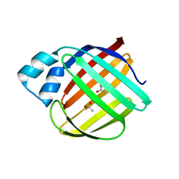
Crystal structure of monomer human cellular retinol binding protein II-Y60L
Descriptor: ACETATE ION, Retinol-binding protein 2
Authors:Assar, Z, Nossoni, Z, Wang, W, Gieger, J.H, Borhan, B.
Deposit date:2015-08-27
Release date:2016-09-21
Last modified:2024-03-06
Method:X-RAY DIFFRACTION (1.5 Å)
Cite:Domain-Swapped Dimers of Intracellular Lipid-Binding Proteins: Evidence for Ordered Folding Intermediates.
Structure, 24, 2016
4ZH6
DownloadVisualize
BU of 4zh6 by Molmil
Crystal Structure of the Domain-Swapped Dimer Y60L mutant of Human Cellular Retinol Binding Protein II
Descriptor: ACETATE ION, Retinol-binding protein 2
Authors:Assar, Z, Nossoni, Z, Geiger, J.H.
Deposit date:2015-04-24
Release date:2016-06-08
Last modified:2023-09-27
Method:X-RAY DIFFRACTION (1.5497 Å)
Cite:Domain-Swapped Dimers of Intracellular Lipid-Binding Proteins: Evidence for Ordered Folding Intermediates.
Structure, 24, 2016
4ZH9
DownloadVisualize
BU of 4zh9 by Molmil
Crystal Structure of the Domain-Swapped Dimer Wild-Type of Human Cellular Retinol Binding Protein II
Descriptor: Retinol-binding protein 2
Authors:Assar, Z, Nossoni, Z, Geiger, J.H.
Deposit date:2015-04-24
Release date:2016-06-08
Last modified:2023-09-27
Method:X-RAY DIFFRACTION (2.66 Å)
Cite:Domain-Swapped Dimers of Intracellular Lipid-Binding Proteins: Evidence for Ordered Folding Intermediates.
Structure, 24, 2016
4ZR2
DownloadVisualize
BU of 4zr2 by Molmil
Crystal Structure of the Domain-Swapped Dimer K40L:Q108K:Y60W mutant of Human Cellular Retinol Binding Protein II
Descriptor: ACETATE ION, RETINAL, Retinol-binding protein 2
Authors:Assar, Z, Nossoni, Z, Geiger, J.H.
Deposit date:2015-05-11
Release date:2016-06-08
Last modified:2023-09-27
Method:X-RAY DIFFRACTION (1.8024 Å)
Cite:Domain-Swapped Dimers of Intracellular Lipid-Binding Proteins: Evidence for Ordered Folding Intermediates.
Structure, 24, 2016
6E5E
DownloadVisualize
BU of 6e5e by Molmil
Crystal structure of the apo domain-swapped dimer Q108K:T51D mutant of human cellular retinol binding protein II
Descriptor: ACETATE ION, Retinol-binding protein 2
Authors:Ghanbarpour, A, Geiger, J.
Deposit date:2018-07-20
Release date:2019-10-16
Last modified:2023-10-11
Method:X-RAY DIFFRACTION (1.696 Å)
Cite:Engineering the hCRBPII Domain-Swapped Dimer into a New Class of Protein Switches.
J.Am.Chem.Soc., 141, 2019
6E5Q
DownloadVisualize
BU of 6e5q by Molmil
Crystal structure of the apo domain-swapped dimer Q108K:T51D:A28H mutant of human Cellular Retinol Binding Protein II
Descriptor: ACETATE ION, Retinol-binding protein 2
Authors:Ghanbarpour, A, Geiger, J.
Deposit date:2018-07-21
Release date:2019-10-16
Last modified:2023-10-11
Method:X-RAY DIFFRACTION (1.99 Å)
Cite:Engineering the hCRBPII Domain-Swapped Dimer into a New Class of Protein Switches.
J.Am.Chem.Soc., 141, 2019
6E51
DownloadVisualize
BU of 6e51 by Molmil
Crystal structure of the apo domain-swapped dimer Q108K:K40L:T51W mutant of human cellular retinol binding protein II
Descriptor: Retinol-binding protein 2
Authors:Ghanbarpour, A, Geiger, J.
Deposit date:2018-07-19
Release date:2019-10-16
Last modified:2023-10-11
Method:X-RAY DIFFRACTION (2.256 Å)
Cite:Engineering the hCRBPII Domain-Swapped Dimer into a New Class of Protein Switches.
J.Am.Chem.Soc., 141, 2019
6E50
DownloadVisualize
BU of 6e50 by Molmil
Crystal structure of the apo domain-swapped dimer Q108K:K40L:T51F mutant of human Cellular Retinol Binding Protein II
Descriptor: ACETATE ION, Retinol-binding protein 2
Authors:Ghanbarpour, A, Geiger, J.
Deposit date:2018-07-18
Release date:2019-10-16
Last modified:2023-10-11
Method:X-RAY DIFFRACTION (1.965 Å)
Cite:Engineering the hCRBPII Domain-Swapped Dimer into a New Class of Protein Switches.
J.Am.Chem.Soc., 141, 2019
6E5R
DownloadVisualize
BU of 6e5r by Molmil
Crystal structure of the apo domain-swapped dimer Q108K:T51D:A28C mutant of human Cellular Retinol Binding Protein II
Descriptor: ACETATE ION, GLYCEROL, Retinol-binding protein 2
Authors:Ghanbarpour, A, Geiger, J.
Deposit date:2018-07-22
Release date:2019-08-07
Last modified:2023-10-11
Method:X-RAY DIFFRACTION (2.592 Å)
Cite:Engineering the hCRBPII Domain-Swapped Dimer into a New Class of Protein Switches.
J.Am.Chem.Soc., 141, 2019
6E7M
DownloadVisualize
BU of 6e7m by Molmil
Crystal structure of the holo retinal-bound domain-swapped dimer Q108K:T51D:A28C mutant of human Cellular Retinol Binding Protein II
Descriptor: ACETATE ION, GLYCEROL, RETINAL, ...
Authors:Ghanbarpour, A, Geiger, J.
Deposit date:2018-07-26
Release date:2019-10-16
Last modified:2023-10-11
Method:X-RAY DIFFRACTION (2.7 Å)
Cite:Engineering the hCRBPII Domain-Swapped Dimer into a New Class of Protein Switches.
J.Am.Chem.Soc., 141, 2019
6E5S
DownloadVisualize
BU of 6e5s by Molmil
Crystal structure of holo retinal-bound domain-swapped dimer Q108K:T51D mutant of human Cellular Retinol Binding Protein II
Descriptor: RETINAL, Retinol-binding protein 2
Authors:Nosrati, M, Ghanbarpour, A, Geiger, J.
Deposit date:2018-07-22
Release date:2019-07-24
Last modified:2023-10-11
Method:X-RAY DIFFRACTION (2.061 Å)
Cite:Engineering the hCRBPII Domain-Swapped Dimer into a New Class of Protein Switches.
J.Am.Chem.Soc., 141, 2019
6E6L
DownloadVisualize
BU of 6e6l by Molmil
Crystal structure of the holo retinal-bound domain-swapped dimer Q108K:K40L:T51F:Y60A mutant of human cellular retinol binding protein II
Descriptor: ACETATE ION, RETINAL, Retinol-binding protein 2
Authors:Ghanbarpour, A, Geiger, J.
Deposit date:2018-07-25
Release date:2019-10-16
Last modified:2023-10-11
Method:X-RAY DIFFRACTION (2.08 Å)
Cite:Engineering the hCRBPII Domain-Swapped Dimer into a New Class of Protein Switches.
J.Am.Chem.Soc., 141, 2019
4YKO
DownloadVisualize
BU of 4yko by Molmil
Crystal structure of the R111K:Y134F:T54V:R132Q:P39Q:R59Y:A32Y:F3Q mutant of human Cellular Retinoic Acid Binding Protein II with Retinal at 1.58 angstrom resolution
Descriptor: Cellular retinoic acid-binding protein 2, RETINAL
Authors:Nosrati, M, Geiger, J.
Deposit date:2015-03-04
Release date:2016-03-09
Last modified:2023-09-27
Method:X-RAY DIFFRACTION (1.57 Å)
Cite:A Photoisomerizing Rhodopsin Mimic Observed at Atomic Resolution.
J.Am.Chem.Soc., 138, 2016
6MCU
DownloadVisualize
BU of 6mcu by Molmil
Crystal structure of the holo retinal-bound domain-swapped dimer Q108K:T51D:A28H mutant of human Cellular Retinol Binding Protein II
Descriptor: GLYCEROL, RETINAL, Retinol-binding protein 2
Authors:Ghanbarpour, A, Geiger, J.
Deposit date:2018-09-02
Release date:2019-10-16
Last modified:2023-10-11
Method:X-RAY DIFFRACTION (2.572 Å)
Cite:Engineering the hCRBPII Domain-Swapped Dimer into a New Class of Protein Switches.
J.Am.Chem.Soc., 141, 2019
6MCV
DownloadVisualize
BU of 6mcv by Molmil
Crystal Structure of Holo Retinal-Bound Domain-Swapped Dimer of Wild Type Human Cellular Retinol Binding Protein II
Descriptor: RETINAL, Retinol-binding protein 2
Authors:Ghanbarpour, A, Geiger, J.
Deposit date:2018-09-02
Release date:2019-10-16
Last modified:2023-10-11
Method:X-RAY DIFFRACTION (3.3 Å)
Cite:Engineering the hCRBPII Domain-Swapped Dimer into a New Class of Protein Switches.
J.Am.Chem.Soc., 141, 2019
6MLB
DownloadVisualize
BU of 6mlb by Molmil
Crystal structure of the holo retinal-bound domain-swapped dimer Q108K:K40L:T51F mutant of human cellular retinol binding protein II
Descriptor: ACETATE ION, GLYCEROL, RETINAL, ...
Authors:Ghanbarpour, A, Geiger, J.
Deposit date:2018-09-27
Release date:2019-10-16
Last modified:2023-10-11
Method:X-RAY DIFFRACTION (2.15 Å)
Cite:Engineering the hCRBPII Domain-Swapped Dimer into a New Class of Protein Switches.
J.Am.Chem.Soc., 141, 2019
6MKV
DownloadVisualize
BU of 6mkv by Molmil
Crystal structure of Retinal-bound holo Q108K:K40L:T51W domain-swapped dimer of human cellular retinol binding protein 2
Descriptor: ACETATE ION, GLYCEROL, RETINAL, ...
Authors:Ghanbarpour, A, Geiger, J.
Deposit date:2018-09-26
Release date:2019-10-16
Last modified:2023-10-11
Method:X-RAY DIFFRACTION (2.107 Å)
Cite:Engineering the hCRBPII Domain-Swapped Dimer into a New Class of Protein Switches.
J.Am.Chem.Soc., 141, 2019
<123


218853

PDB entries from 2024-04-24

PDB statisticsPDBj update infoContact PDBjnumon