Loading
PDBj
MenuPDBj@FacebookPDBj@TwitterPDBj@YouTubewwPDB FoundationwwPDB
RCSB PDBPDBeBMRBAdv. SearchSearch help
Search by PDB author
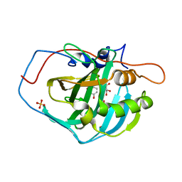
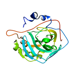
3MNK
DownloadVisualize
BU of 3mnk by Molmil
Human Carbonic Anhydrase II Mutant K170H
Descriptor: Carbonic anhydrase 2, SODIUM ION, ZINC ION
Authors:Domsic, J.F, McKenna, R.
Deposit date:2010-04-21
Release date:2010-07-14
Last modified:2023-09-06
Method:X-RAY DIFFRACTION (1.75 Å)
Cite:Structural and kinetic study of the extended active site for proton transfer in human carbonic anhydrase II.
Biochemistry, 49, 2010
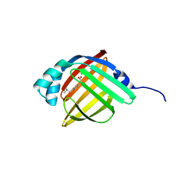
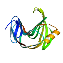
3P6G
DownloadVisualize
BU of 3p6g by Molmil
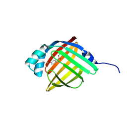
Human adipocyte lipid-binding protein FABP4 in complex with (R)-ibuprofen
Descriptor: (2R)-2-[4-(2-methylpropyl)phenyl]propanoic acid, Fatty acid-binding protein, adipocyte
Authors:Gonzalez, J.M, Pozharski, E.
Deposit date:2010-10-11
Release date:2011-04-13
Last modified:2024-02-21
Method:X-RAY DIFFRACTION (1.2 Å)
Cite:Structural analysis of ibuprofen binding to human adipocyte fatty-acid binding protein (FABP4).
Acta Crystallogr F Struct Biol Commun, 71, 2015
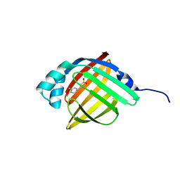
3P6D
DownloadVisualize
BU of 3p6d by Molmil
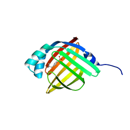
Human adipocyte lipid-binding protein FABP4 in complex with 3-(4-methoxy-3-methylphenyl) propionic acid
Descriptor: 3-(4-methoxy-3-methylphenyl)propanoic acid, Fatty acid-binding protein, adipocyte
Authors:Gonzalez, J.M, Pozharski, E.
Deposit date:2010-10-11
Release date:2011-04-13
Last modified:2024-02-21
Method:X-RAY DIFFRACTION (1.06 Å)
Cite:Structural analysis of ibuprofen binding to human adipocyte fatty-acid binding protein (FABP4).
Acta Crystallogr F Struct Biol Commun, 71, 2015
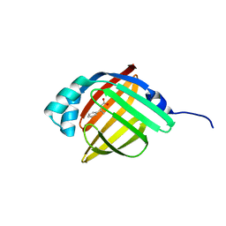
3P6F
DownloadVisualize
BU of 3p6f by Molmil
Human adipocyte lipid-binding protein FABP4 in complex with (S)-3-phenyl butyric acid
Descriptor: (3S)-3-phenylbutanoic acid, Fatty acid-binding protein, adipocyte
Authors:Gonzalez, J.M, Pozharski, E.
Deposit date:2010-10-11
Release date:2011-04-13
Last modified:2024-02-21
Method:X-RAY DIFFRACTION (1.2 Å)
Cite:Structural analysis of ibuprofen binding to human adipocyte fatty-acid binding protein (FABP4).
Acta Crystallogr F Struct Biol Commun, 71, 2015
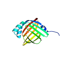
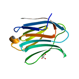
3P6C
DownloadVisualize
BU of 3p6c by Molmil
Human adipocyte lipid-binding protein FABP4 in complex with citric acid
Descriptor: CITRIC ACID, Fatty acid-binding protein, adipocyte
Authors:Gonzalez, J.M, Pozharski, E.
Deposit date:2010-10-11
Release date:2011-04-13
Last modified:2024-02-21
Method:X-RAY DIFFRACTION (1.25 Å)
Cite:Structural analysis of ibuprofen binding to human adipocyte fatty-acid binding protein (FABP4).
Acta Crystallogr F Struct Biol Commun, 71, 2015
3P6E
DownloadVisualize
BU of 3p6e by Molmil
Human adipocyte lipid-binding protein FABP4 in complex with 3-(4-methoxyphenyl) propionic acid
Descriptor: 3-(4-methoxyphenyl)propanoic acid, Fatty acid-binding protein, adipocyte
Authors:Gonzalez, J.M, Pozharski, E.
Deposit date:2010-10-11
Release date:2011-04-13
Last modified:2024-02-21
Method:X-RAY DIFFRACTION (1.08 Å)
Cite:Structural analysis of ibuprofen binding to human adipocyte fatty-acid binding protein (FABP4).
Acta Crystallogr F Struct Biol Commun, 71, 2015
3P6H
DownloadVisualize
BU of 3p6h by Molmil
Human adipocyte lipid-binding protein FABP4 in complex with (S)-ibuprofen
Descriptor: Fatty acid-binding protein, adipocyte, IBUPROFEN
Authors:Gonzalez, J.M, Pozharski, E.
Deposit date:2010-10-11
Release date:2011-04-13
Last modified:2024-02-21
Method:X-RAY DIFFRACTION (1.15 Å)
Cite:Structural analysis of ibuprofen binding to human adipocyte fatty-acid binding protein (FABP4).
Acta Crystallogr F Struct Biol Commun, 71, 2015
3QYS
DownloadVisualize
BU of 3qys by Molmil
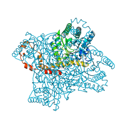
Room Temperature X-ray Structure of D-Xylose Isomerase in complex with 0.6Ni2+ cation bound in M2 metal binding site at pH=5.8
Descriptor: NICKEL (II) ION, Xylose isomerase
Authors:Kovalevsky, A.Y, Hanson, L, Langan, P.
Deposit date:2011-03-03
Release date:2011-08-17
Last modified:2024-02-21
Method:X-RAY DIFFRACTION (1.85 Å)
Cite:Identification of the Elusive Hydronium Ion Exchanging Roles with a Proton in an Enzyme at Lower pH Values.
Angew.Chem.Int.Ed.Engl., 50, 2011
3QZA
DownloadVisualize
BU of 3qza by Molmil
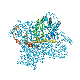
Joint neutron and X-ray structure of apo-D-Xylose Isomerase at pH=5.9
Descriptor: Xylose isomerase, deuterium(1+)
Authors:Kovalevsky, A.Y, Hanson, L, Langan, P.
Deposit date:2011-03-04
Release date:2011-08-17
Last modified:2023-09-13
Method:NEUTRON DIFFRACTION (2 Å), X-RAY DIFFRACTION
Cite:Identification of the Elusive Hydronium Ion Exchanging Roles with a Proton in an Enzyme at Lower pH Values.
Angew.Chem.Int.Ed.Engl., 50, 2011
4FIK
DownloadVisualize
BU of 4fik by Molmil
Human carbonic anhydrase II H64A complexed with thioxolone hydrolysis products
Descriptor: 2,4-dihydroxybenzenesulfenic acid, 4,4'-disulfanediyldibenzene-1,3-diol, 4-MERCAPTOBENZENE-1,3-DIOL, ...
Authors:Sippel, K.H, Genis, C, Govindasamy, L, Agbandje-McKenna, M, Tripp, B.C, McKenna, R.
Deposit date:2012-06-08
Release date:2012-06-27
Last modified:2023-11-29
Method:X-RAY DIFFRACTION (1.2 Å)
Cite:Synchrotron Radiation Provides a Plausible Explanation for the Generation of a Free Radical Adduct of Thioxolone in Mutant Carbonic Anhydrase II.
J Phys Chem Lett, 1, 2010
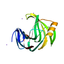
5ZO0
DownloadVisualize
BU of 5zo0 by Molmil
Neutron structure of xylanase at pD5.4
Descriptor: Endo-1,4-beta-xylanase 2
Authors:Wan, Q, Li, Z.H.
Deposit date:2018-04-12
Release date:2019-05-15
Last modified:2023-11-22
Method:NEUTRON DIFFRACTION (1.648 Å)
Cite:Neutron structure of xylanase at pD5.4
To be published
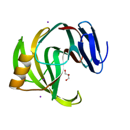
5ZKZ
DownloadVisualize
BU of 5zkz by Molmil
Crystal Structures of Mutant Endo-beta-1,4-xylanase II(Y77F) Complexed with Xylotriose
Descriptor: Endo-1,4-beta-xylanase 2, GLYCEROL, IODIDE ION, ...
Authors:Zhang, X, Wan, Q.
Deposit date:2018-03-26
Release date:2019-04-10
Last modified:2023-11-22
Method:X-RAY DIFFRACTION (1.3 Å)
Cite:Crystal Structures of Endo-beta-1,4-xylanase II
to be published
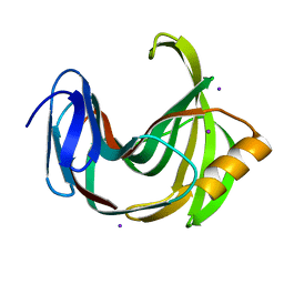
5ZIW
DownloadVisualize
BU of 5ziw by Molmil
Crystal Structures of Mutant Endo-beta-1,4-xylanase(Y77F)
Descriptor: Endo-1,4-beta-xylanase 2, IODIDE ION
Authors:Zhang, X, Wan, Q.
Deposit date:2018-03-17
Release date:2019-03-20
Last modified:2023-11-22
Method:X-RAY DIFFRACTION (1.3 Å)
Cite:Crystal Structures of Endo-beta-1,4-xylanase II
to be published
5ZII
DownloadVisualize
BU of 5zii by Molmil
Crystal Structures of Mutant Endo-beta-1,4-xylanase II (Y88F)Complexed with Xylotriose
Descriptor: Endo-1,4-beta-xylanase 2, IODIDE ION, beta-D-xylopyranose-(1-4)-beta-D-xylopyranose-(1-4)-beta-D-xylopyranose
Authors:Zhang, X, Wan, Q.
Deposit date:2018-03-15
Release date:2019-03-20
Last modified:2023-11-22
Method:X-RAY DIFFRACTION (1.3 Å)
Cite:Crystal Structures of Endo-beta-1,4-xylanase II
to be published
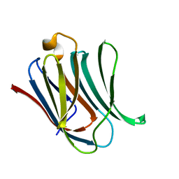
6EXY
DownloadVisualize
BU of 6exy by Molmil
Neutron crystal structure of perdeuterated galectin-3C in complex with glycerol
Descriptor: GLYCEROL, Galectin-3
Authors:Manzoni, F, Schrader, T.E, Ostermann, A, Oksanen, E, Logan, D.T.
Deposit date:2017-11-10
Release date:2018-09-12
Method:NEUTRON DIFFRACTION (1.1 Å), X-RAY DIFFRACTION
Cite:Elucidation of Hydrogen Bonding Patterns in Ligand-Free, Lactose- and Glycerol-Bound Galectin-3C by Neutron Crystallography to Guide Drug Design.
J. Med. Chem., 61, 2018
6EYM
DownloadVisualize
BU of 6eym by Molmil
Neutron crystal structure of perdeuterated galectin-3C in complex with lactose
Descriptor: Galectin-3, beta-D-galactopyranose-(1-4)-beta-D-glucopyranose
Authors:Manzoni, F, Coates, L, Blakeley, M.P, Oksanen, E, Logan, D.T.
Deposit date:2017-11-13
Release date:2018-09-12
Last modified:2020-07-29
Method:NEUTRON DIFFRACTION (1.7 Å), X-RAY DIFFRACTION
Cite:Elucidation of Hydrogen Bonding Patterns in Ligand-Free, Lactose- and Glycerol-Bound Galectin-3C by Neutron Crystallography to Guide Drug Design.
J. Med. Chem., 61, 2018
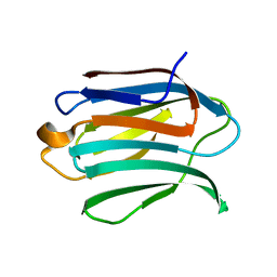
6F2Q
DownloadVisualize
BU of 6f2q by Molmil
Neutron crystal structure of perdeuterated galectin-3C in the ligand-free form
Descriptor: Galectin-3
Authors:Manzoni, F, Blakeley, M.P, Oksanen, E, Logan, D.T.
Deposit date:2017-11-27
Release date:2018-05-02
Last modified:2018-06-06
Method:NEUTRON DIFFRACTION (1.03 Å), X-RAY DIFFRACTION
Cite:Elucidation of Hydrogen Bonding Patterns in Ligand-Free, Lactose- and Glycerol-Bound Galectin-3C by Neutron Crystallography to Guide Drug Design.
J. Med. Chem., 61, 2018

218853

PDB entries from 2024-04-24

PDB statisticsPDBj update infoContact PDBjnumon