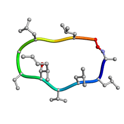
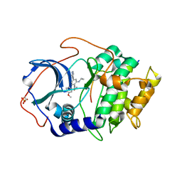
1DHM
 
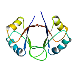 | | DNA-BINDING DOMAIN OF E2 FROM HUMAN PAPILLOMAVIRUS-31, NMR, MINIMIZED AVERAGE STRUCTURE | | 分子名称: | E2 PROTEIN | | 著者 | Liang, H, Petros, A.P, Meadows, R.P, Yoon, H.S, Egan, D.A, Walter, K, Holzman, T.F, Robins, T, Fesik, S.W. | | 登録日 | 1995-08-15 | | 公開日 | 1996-12-07 | | 最終更新日 | 2024-04-10 | | 実験手法 | SOLUTION NMR | | 主引用文献 | Solution structure of the DNA-binding domain of a human papillomavirus E2 protein: evidence for flexible DNA-binding regions.
Biochemistry, 35, 1996
|
|
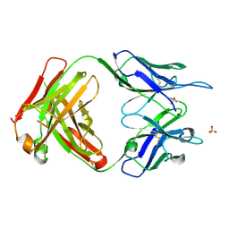
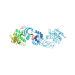
2JIX
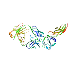 | | Crystal structure of ABT-007 FAB fragment with the soluble domain of EPO receptor | | 分子名称: | ABT-007 FAB FRAGMENT, ERYTHROPOIETIN RECEPTOR | | 著者 | Liu, Z, Stoll, V.S, DeVries, P, Jakob, C.G, Xie, N, Simmer, R.L, Lacy, S.E, Egan, D.A, Harlan, J.E, Lesniewski, R.R, Reilly, E.B. | | 登録日 | 2007-07-02 | | 公開日 | 2007-07-10 | | 最終更新日 | 2020-03-18 | | 実験手法 | X-RAY DIFFRACTION (3.2 Å) | | 主引用文献 | A Potent Erythropoietin-Mimicking Human Antibody Interacts Through a Novel Binding Site.
Blood, 110, 2007
|
|
1A7G
 | |
1CYB
 | |
1CYA
 | |
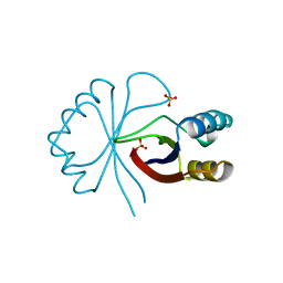
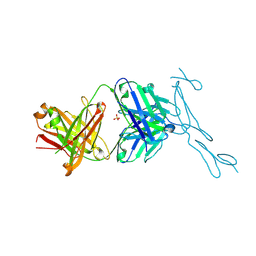
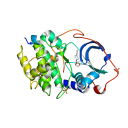
6PE7
 | | Crystal Structure of ABBV-323 FAB | | 分子名称: | FAB Heavy Chain, FAB Light chain, SULFATE ION | | 著者 | Argiriadi, M.A. | | 登録日 | 2019-06-20 | | 公開日 | 2019-08-14 | | 最終更新日 | 2023-10-11 | | 実験手法 | X-RAY DIFFRACTION (1.74 Å) | | 主引用文献 | CD40/anti-CD40 antibody complexes which illustrate agonist and antagonist structural switches.
BMC Mol Cell Biol, 20, 2019
|
|
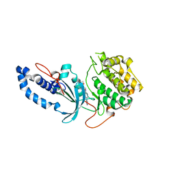
6PE9
 | | Crystal Structure of CD40 complexed to FAB516 | | 分子名称: | FAB Heavy chain, FAB Light chain, SULFATE ION, ... | | 著者 | Argiriadi, M.A. | | 登録日 | 2019-06-20 | | 公開日 | 2019-08-14 | | 最終更新日 | 2023-10-11 | | 実験手法 | X-RAY DIFFRACTION (3.13 Å) | | 主引用文献 | CD40/anti-CD40 antibody complexes which illustrate agonist and antagonist structural switches.
BMC Mol Cell Biol, 20, 2019
|
|
6PE8
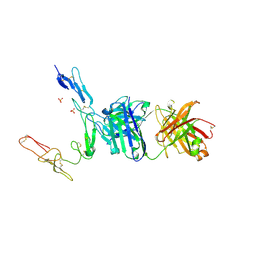 | | Crystal structure of CD40/ABBV-323 FAB complex | | 分子名称: | FAB Heavy chain, FAB Light chain, SULFATE ION, ... | | 著者 | Argiriadi, M.A. | | 登録日 | 2019-06-20 | | 公開日 | 2019-08-14 | | 最終更新日 | 2023-10-11 | | 実験手法 | X-RAY DIFFRACTION (2.84 Å) | | 主引用文献 | CD40/anti-CD40 antibody complexes which illustrate agonist and antagonist structural switches.
BMC Mol Cell Biol, 20, 2019
|
|
6E99
 | |
6E9W
 | |
6E9L
 | |
6ED6
 | |


