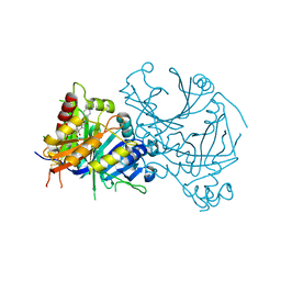
7L7E
 
 | |
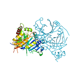
7L7D
 | |
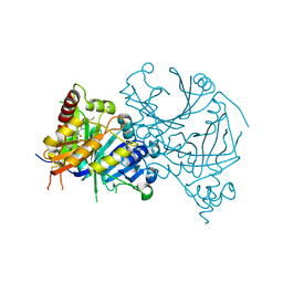
2AEF
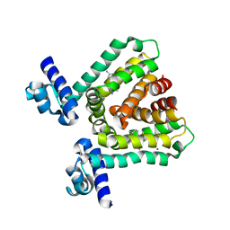
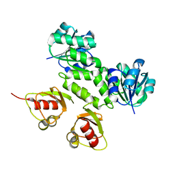 | | Crystal Structures of the MthK RCK Domain in Ca2+ bound form | | Descriptor: | CALCIUM ION, Calcium-gated potassium channel mthK | | Authors: | Dong, J, Shi, N, Berke, I, Chen, L, Jiang, Y. | | Deposit date: | 2005-07-22 | | Release date: | 2005-10-25 | | Last modified: | 2023-08-23 | | Method: | X-RAY DIFFRACTION (1.7 Å) | | Cite: | Structures of the MthK RCK Domain and the Effect of Ca2+ on Gating Ring Stability
J.Biol.Chem., 280, 2005
|
|
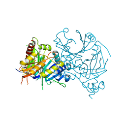
8GQT
 | |
3HTH
 | |
3HTJ
 | |
3HTA
 | |
3HTI
 | |
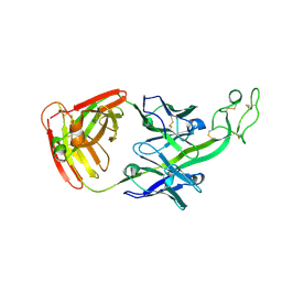
6E9I
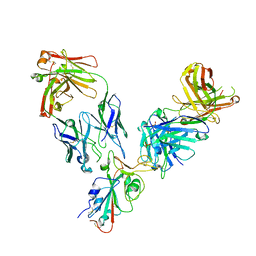
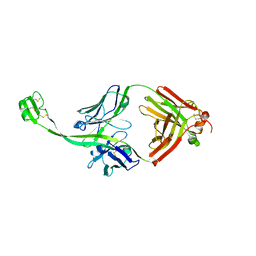 | | The crystal structure of bovine ultralong antibody BOV-4 | | Descriptor: | Bovine ultralong antibody BOV-4 heavy chain, Bovine ultralong antibody BOV-4 light chain | | Authors: | Dong, J, Crowe, J.E. | | Deposit date: | 2018-08-01 | | Release date: | 2019-05-01 | | Last modified: | 2019-05-08 | | Method: | X-RAY DIFFRACTION (2.5 Å) | | Cite: | Structural Diversity of Ultralong CDRH3s in Seven Bovine Antibody Heavy Chains.
Front Immunol, 10, 2019
|
|
6E9H
 | |
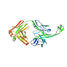
6E9G
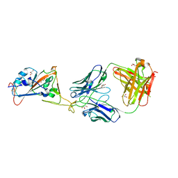
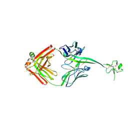
 | | The crystal structure of bovine ultralong antibody BOV-2 | | Descriptor: | Bovine ultralong antibody BOV-2 heavy chain, Bovine ultralong antibody BOV-2 light chain | | Authors: | Dong, J, Crowe, J.E. | | Deposit date: | 2018-08-01 | | Release date: | 2019-05-01 | | Method: | X-RAY DIFFRACTION (2.904 Å) | | Cite: | Structural Diversity of Ultralong CDRH3s in Seven Bovine Antibody Heavy Chains.
Front Immunol, 10, 2019
|
|
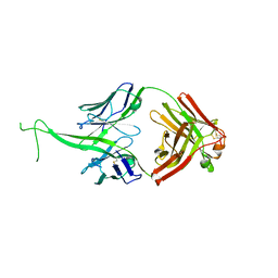
6E8V
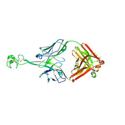
 | | The crystal structure of bovine ultralong antibody BOV-1 | | Descriptor: | Bovine ultralong antibody BOV-1 Heavy chain, Bovine ultralong antibody BOV-1 light chain | | Authors: | Dong, J, Crowe, J.E. | | Deposit date: | 2018-07-31 | | Release date: | 2019-09-04 | | Last modified: | 2020-10-21 | | Method: | X-RAY DIFFRACTION (3.79 Å) | | Cite: | Structural Diversity of Ultralong CDRH3s in Seven Bovine Antibody Heavy Chains.
Front Immunol, 10, 2019
|
|
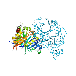
6E9U
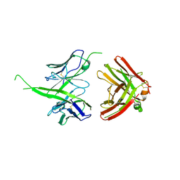
 | | The crystal structure of bovine ultralong antibody BOV-7 | | Descriptor: | Bovine ultralong antibody BOV-7 heavy chain, Bovine ultralong antibody BOV-7 light chain | | Authors: | Dong, J, Crowe, J.E. | | Deposit date: | 2018-08-01 | | Release date: | 2019-05-01 | | Method: | X-RAY DIFFRACTION (2.295 Å) | | Cite: | Structural Diversity of Ultralong CDRH3s in Seven Bovine Antibody Heavy Chains.
Front Immunol, 10, 2019
|
|
6E9K
 | |
6E9Q
 | |
8HZA
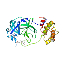
 | | Crystal structure of AtHPPD-YH20326 complex | | Descriptor: | 1,4-dimethyl-3-(oxan-4-ylmethyl)-5-(2-oxidanyl-6-oxidanylidene-cyclohexen-1-yl)carbonyl-benzimidazol-2-one, 4-hydroxyphenylpyruvate dioxygenase, COBALT (II) ION | | Authors: | Dong, J, Lin, H.-Y, Yang, G.-F. | | Deposit date: | 2023-01-08 | | Release date: | 2023-12-20 | | Method: | X-RAY DIFFRACTION (1.69 Å) | | Cite: | Crystal structure of AtHPPD-YH20326 complex
To Be Published
|
|
8I0Y
 | | Crystal structure of AtHPPD-Y191713 complex | | Descriptor: | 4-hydroxyphenylpyruvate dioxygenase, COBALT (II) ION, ethyl 5-methyl-6-[(2-methyl-3-oxidanylidene-1H-pyrazol-4-yl)carbonyl]-4-oxidanylidene-3-phenyl-quinazoline-2-carboxylate | | Authors: | Dong, J, Lin, H.-Y, Yang, G.-F. | | Deposit date: | 2023-01-11 | | Release date: | 2023-12-20 | | Method: | X-RAY DIFFRACTION (1.792 Å) | | Cite: | Crystal structure of AtHPPD-Y191713 complex
To Be Published
|
|
8I2S
 | | Crystal structure of AtHPPD-Y18979 complex | | Descriptor: | 1,5-dimethyl-3-(naphthalen-2-ylmethyl)-6-(2-oxidanyl-6-oxidanylidene-cyclohexen-1-yl)carbonyl-quinazoline-2,4-dione, 4-hydroxyphenylpyruvate dioxygenase, COBALT (II) ION | | Authors: | Dong, J, Lin, H.-Y, Yang, G.-F. | | Deposit date: | 2023-01-15 | | Release date: | 2023-12-20 | | Method: | X-RAY DIFFRACTION (1.592 Å) | | Cite: | Crystal structure of AtHPPD-Y18979 complex
To Be Published
|
|
8HXG
 | | Crystal structure of AtHPPD-Y14116 complex | | Descriptor: | (1S,2R,3R)-2-[(S)-(2-methoxy-8-methyl-quinolin-3-yl)-oxidanyl-methyl]-6,6-dimethyl-cyclohex-4-ene-1,3-diol, (4S)-2-METHYL-2,4-PENTANEDIOL, 4-hydroxyphenylpyruvate dioxygenase, ... | | Authors: | Dong, J, Lin, H.-Y, Yang, G.-F. | | Deposit date: | 2023-01-04 | | Release date: | 2023-12-20 | | Method: | X-RAY DIFFRACTION (1.702 Å) | | Cite: | Crystal structure of AtHPPD-Y14116 complex
To Be Published
|
|
8HZ7
 | | Crystal structure of AtHPPD-Y181135 complex | | Descriptor: | 3-butyl-5-methyl-6-[(2-methyl-3-oxidanylidene-1H-pyrazol-4-yl)carbonyl]-1,2,3-benzotriazin-4-one, 4-hydroxyphenylpyruvate dioxygenase, COBALT (II) ION | | Authors: | Dong, J, Lin, H.-Y, Yang, G.-F. | | Deposit date: | 2023-01-08 | | Release date: | 2023-12-20 | | Method: | X-RAY DIFFRACTION (1.802 Å) | | Cite: | Crystal structure of AtHPPD-Y181135 complex
To Be Published
|
|
8HYO
 | | Crystal structure of AtHPPD-Y18031 complex | | Descriptor: | 4-hydroxyphenylpyruvate dioxygenase, 6-[(R)-[(2R,6R)-2,6-bis(oxidanyl)cyclohexyl]-oxidanyl-methyl]-3-(furan-2-ylmethyl)-1,5-dimethyl-quinazoline-2,4-dione, COBALT (II) ION | | Authors: | Dong, J, Lin, H.-Y, Yang, G.-F. | | Deposit date: | 2023-01-07 | | Release date: | 2023-12-20 | | Method: | X-RAY DIFFRACTION (2 Å) | | Cite: | Crystal structure of AtHPPD-Y18031 complex
To Be Published
|
|
8HXF
 | | Crystal structure of AtHPPD-Y181188 complex | | Descriptor: | 4-hydroxyphenylpyruvate dioxygenase, 6-(2-oxidanyl-6-oxidanylidene-cyclohexen-1-yl)carbonyl-4-[[4-(trifluoromethyl)phenyl]methyl]-1,4-benzothiazin-3-one, COBALT (II) ION | | Authors: | Dong, J, Lin, H.-Y, Yang, G.-F. | | Deposit date: | 2023-01-04 | | Release date: | 2023-12-20 | | Method: | X-RAY DIFFRACTION (1.697 Å) | | Cite: | Crystal structure of AtHPPD-Y181188 complex
To Be Published
|
|
8HXH
 | | Crystal structure of AtHPPD-Y18024 complex | | Descriptor: | 1,5-dimethyl-6-(2-oxidanyl-6-oxidanylidene-cyclohexen-1-yl)carbonyl-3-propyl-quinazoline-2,4-dione, 4-hydroxyphenylpyruvate dioxygenase, COBALT (II) ION | | Authors: | Dong, J, Lin, H.-Y, Yang, G.-F. | | Deposit date: | 2023-01-04 | | Release date: | 2023-12-20 | | Method: | X-RAY DIFFRACTION (1.992 Å) | | Cite: | Crystal structure of AtHPPD-Y18024 complex
To Be Published
|
|
8HYS
 | | Crystal structure of AtHPPD-Y19802 complex | | Descriptor: | 1-methyl-3-[2-methyl-6-methylsulfonyl-3-(2-oxidanyl-6-oxidanylidene-cyclohexen-1-yl)carbonyl-phenyl]-6-(trifluoromethyl)pyrimidine-2,4-dione, 4-hydroxyphenylpyruvate dioxygenase, COBALT (II) ION | | Authors: | Dong, J, Lin, H.-Y, Yang, G.-F. | | Deposit date: | 2023-01-07 | | Release date: | 2023-12-20 | | Method: | X-RAY DIFFRACTION (1.7 Å) | | Cite: | Crystal structure of AtHPPD-Y19802 complex
To Be Published
|
|
8HYM
 | | Crystal structure of AtHPPD-Y18030 complex | | Descriptor: | 3-cyclopentyl-1,5-dimethyl-6-(2-oxidanyl-6-oxidanylidene-cyclohexen-1-yl)carbonyl-quinazoline-2,4-dione, 4-hydroxyphenylpyruvate dioxygenase, COBALT (II) ION | | Authors: | Dong, J, Lin, H.-Y, Yang, G.-F. | | Deposit date: | 2023-01-07 | | Release date: | 2023-12-20 | | Method: | X-RAY DIFFRACTION (1.794 Å) | | Cite: | Crystal structure of AtHPPD-Y18030 complex
To Be Published
|
|


