Loading
PDBj
MenuPDBj@FacebookPDBj@TwitterPDBj@YouTubewwPDB FoundationwwPDB
RCSB PDBPDBeBMRBAdv. SearchSearch help
Search by PDB author
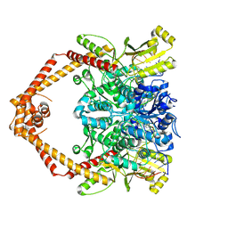
8GCC
DownloadVisualize
BU of 8gcc by Molmil
T. cruzi topoisomerase II alpha bound to dsDNA and the covalent inhibitor CT1
Descriptor: 2-{3-[(Z)-iminomethyl]-1H-1,2,4-triazol-1-yl}-1-{(3M)-3-[2-(trifluoromethyl)phenyl]-6H-pyrrolo[3,4-b]pyridin-6-yl}ethan-1-one, DNA (28-MER), DNA topoisomerase 2
Authors:Schenk, A, Deniston, C, Noeske, J.
Deposit date:2023-03-01
Release date:2023-07-12
Method:ELECTRON MICROSCOPY (2.94 Å)
Cite:Cyanotriazoles are selective topoisomerase II poisons that rapidly cure trypanosome infections.
Science, 380, 2023
6VNO
DownloadVisualize
BU of 6vno by Molmil
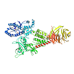
Cryo-EM structure of the C-terminal half of the Parkinson's Disease-linked protein Leucine Rich Repeat Kinase 2 (LRRK2)
Descriptor: GUANOSINE-5'-DIPHOSPHATE, Leucine-rich repeat serine/threonine-protein kinase 2, MAGNESIUM ION
Authors:Leschziner, A, Deniston, C, Lahiri, I.
Deposit date:2020-01-29
Release date:2020-08-26
Last modified:2020-12-23
Method:ELECTRON MICROSCOPY (3.5 Å)
Cite:Structure of LRRK2 in Parkinson's disease and model for microtubule interaction.
Nature, 588, 2020
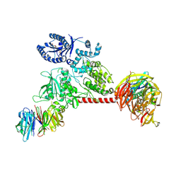
6VP8
DownloadVisualize
BU of 6vp8 by Molmil
Cryo-EM structure of the C-terminal half of the Parkinson's Disease-linked protein Leucine Rich Repeat Kinase 2 (LRRK2)
Descriptor: Leucine-rich repeat serine/threonine-protein kinase 2
Authors:Leschziner, A, Deniston, C, Lahiri, I.
Deposit date:2020-02-01
Release date:2020-08-26
Last modified:2022-03-16
Method:ELECTRON MICROSCOPY (3.5 Å)
Cite:Structure of LRRK2 in Parkinson's disease and model for microtubule interaction.
Nature, 588, 2020
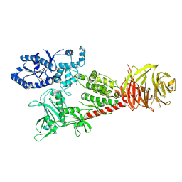
6VP6
DownloadVisualize
BU of 6vp6 by Molmil
Cryo-EM structure of the C-terminal half of the Parkinson's Disease-linked protein Leucine Rich Repeat Kinase 2 (LRRK2)
Descriptor: GUANOSINE-5'-DIPHOSPHATE, Leucine-rich repeat serine/threonine-protein kinase 2, MAGNESIUM ION
Authors:Leschziner, A, Deniston, C, Lahiri, I.
Deposit date:2020-02-01
Release date:2020-08-26
Last modified:2020-12-23
Method:ELECTRON MICROSCOPY (3.47 Å)
Cite:Structure of LRRK2 in Parkinson's disease and model for microtubule interaction.
Nature, 588, 2020
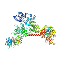
6VP7
DownloadVisualize
BU of 6vp7 by Molmil
Cryo-EM structure of the C-terminal half of the Parkinson's Disease-linked protein Leucine Rich Repeat Kinase 2 (LRRK2)
Descriptor: Leucine-rich repeat serine/threonine-protein kinase 2
Authors:Leschziner, A, Deniston, C, Lahiri, I.
Deposit date:2020-02-01
Release date:2020-08-26
Last modified:2020-12-23
Method:ELECTRON MICROSCOPY (3.5 Å)
Cite:Structure of LRRK2 in Parkinson's disease and model for microtubule interaction.
Nature, 588, 2020

221051

PDB entries from 2024-06-12

PDB statisticsPDBj update infoContact PDBjnumon