Loading
PDBj
MenuPDBj@FacebookPDBj@TwitterPDBj@YouTubewwPDB FoundationwwPDB
RCSB PDBPDBeBMRBAdv. SearchSearch help
Search by PDB author
6QQB
DownloadVisualize
BU of 6qqb by Molmil
Room temperature structure of the fluorescent protein Cerulean recorded after an accumulated dose of 147 kGy
Descriptor: Green fluorescent protein
Authors:Gotthard, G, Aumonier, S, Royant, A.
Deposit date:2019-02-18
Release date:2019-06-19
Last modified:2019-07-31
Method:X-RAY DIFFRACTION (2.45 Å)
Cite:Specific radiation damage is a lesser concern at room temperature.
Iucrj, 6, 2019
6QQH
DownloadVisualize
BU of 6qqh by Molmil
Cryogenic temperature structure of the ground state of AtPhot2LOV2 recorded after an accumulated dose of 2.68 MGy
Descriptor: FLAVIN MONONUCLEOTIDE, Phototropin-2
Authors:Aumonier, S, Gotthard, G, Royant, A.
Deposit date:2019-02-18
Release date:2019-06-19
Last modified:2024-01-24
Method:X-RAY DIFFRACTION (1.38 Å)
Cite:Specific radiation damage is a lesser concern at room temperature.
Iucrj, 6, 2019
6QQ9
DownloadVisualize
BU of 6qq9 by Molmil
Cryogenic temperature structure of the fluorescent protein Cerulean recorded after an accumulated dose of 5.8 MGy
Descriptor: CARBON DIOXIDE, Green fluorescent protein
Authors:Gotthard, G, Aumonier, S, Royant, A.
Deposit date:2019-02-18
Release date:2019-06-19
Last modified:2019-07-31
Method:X-RAY DIFFRACTION (1.82 Å)
Cite:Specific radiation damage is a lesser concern at room temperature.
Iucrj, 6, 2019
6QQE
DownloadVisualize
BU of 6qqe by Molmil
Room temperature structure of Hen Egg White Lysozyme recorded after an accumulated dose of 20 kGy
Descriptor: CHLORIDE ION, Lysozyme C
Authors:Gotthard, G, Aumonier, S, Royant, A.
Deposit date:2019-02-18
Release date:2019-06-19
Last modified:2019-07-31
Method:X-RAY DIFFRACTION (1.37 Å)
Cite:Specific radiation damage is a lesser concern at room temperature.
Iucrj, 6, 2019
6QQC
DownloadVisualize
BU of 6qqc by Molmil
Cryogenic temperature structure of Hen Egg White Lysozyme recorded after an accumulated dose of 110 kGy
Descriptor: CHLORIDE ION, Lysozyme C
Authors:Gotthard, G, Aumonier, S, Royant, A.
Deposit date:2019-02-18
Release date:2019-06-19
Last modified:2019-07-31
Method:X-RAY DIFFRACTION (1.42 Å)
Cite:Specific radiation damage is a lesser concern at room temperature.
Iucrj, 6, 2019
8BHD
DownloadVisualize
BU of 8bhd by Molmil
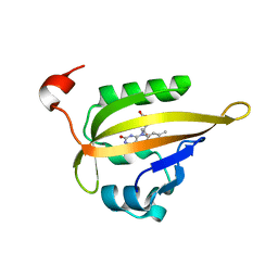
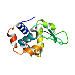
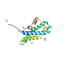
N-terminal domain of Plasmodium berghei glutamyl-tRNA synthetase (Tbxo4 derivative crystal structure)
Descriptor: GLYCEROL, Glutamate--tRNA ligase, SULFATE ION, ...
Authors:Benas, P, Jaramillo Ponce, J.R, Legrand, P, Frugier, M, Sauter, C.
Deposit date:2022-10-31
Release date:2023-01-25
Last modified:2024-06-19
Method:X-RAY DIFFRACTION (3.17 Å)
Cite:Solution X-ray scattering highlights discrepancies in Plasmodium multi-aminoacyl-tRNA synthetase complexes.
Protein Sci., 32, 2023
8S2X
DownloadVisualize
BU of 8s2x by Molmil
SSX structure of Arabidopsis thaliana Pdx1.3 grown in microfluidic droplets
Descriptor: CITRIC ACID, PHOSPHATE ION, Pyridoxal 5'-phosphate synthase subunit PDX1.3
Authors:Stubbs, J, Tews, I, Maly, M.
Deposit date:2024-02-19
Release date:2024-02-28
Last modified:2024-03-20
Method:X-RAY DIFFRACTION (2.5 Å)
Cite:Droplet microfluidics for time-resolved serial crystallography.
Iucrj, 11, 2024
8S2V
DownloadVisualize
BU of 8s2v by Molmil
SSX structure of Lysozyme grown in microfluidic droplets
Descriptor: CHLORIDE ION, Lysozyme C
Authors:Stubbs, J, Tews, I, Maly, M.
Deposit date:2024-02-19
Release date:2024-02-28
Last modified:2024-03-20
Method:X-RAY DIFFRACTION (1.8 Å)
Cite:Droplet microfluidics for time-resolved serial crystallography.
Iucrj, 11, 2024
8S2W
DownloadVisualize
BU of 8s2w by Molmil
SSX structure of Arabidopsis thaliana Pdx1.3 grown in seeded batch conditions
Descriptor: PHOSPHATE ION, Pyridoxal 5'-phosphate synthase subunit PDX1.3
Authors:Stubbs, J, Tews, I, Maly, M.
Deposit date:2024-02-19
Release date:2024-02-28
Last modified:2024-03-20
Method:X-RAY DIFFRACTION (2.5 Å)
Cite:Droplet microfluidics for time-resolved serial crystallography.
Iucrj, 11, 2024
8S2U
DownloadVisualize
BU of 8s2u by Molmil
SSX structure of Lysozyme grown in batch conditions
Descriptor: CHLORIDE ION, Lysozyme C
Authors:Stubbs, J, Tews, I, Maly, M.
Deposit date:2024-02-19
Release date:2024-02-28
Last modified:2024-03-20
Method:X-RAY DIFFRACTION (1.8 Å)
Cite:Droplet microfluidics for time-resolved serial crystallography.
Iucrj, 11, 2024
5II4
DownloadVisualize
BU of 5ii4 by Molmil
Crystal structure of red abalone VERL repeat 1 with linker at 2.0 A resolution
Descriptor: Maltose-binding periplasmic protein,Vitelline envelope sperm lysin receptor, TRIETHYLENE GLYCOL, alpha-D-glucopyranose-(1-4)-alpha-D-glucopyranose
Authors:Sadat Al-Hosseini, H, Raj, I, Nishimura, K, Jovine, L.
Deposit date:2016-03-01
Release date:2017-06-14
Last modified:2024-01-10
Method:X-RAY DIFFRACTION (2 Å)
Cite:Structural Basis of Egg Coat-Sperm Recognition at Fertilization.
Cell, 169, 2017
5II7
DownloadVisualize
BU of 5ii7 by Molmil
In-house sulfur-SAD structure of orthorhombic red abalone lysin at 1.66 A resolution
Descriptor: 2-(N-MORPHOLINO)-ETHANESULFONIC ACID, Egg-lysin, SULFATE ION
Authors:Sadat Al-Hosseini, H, Raj, I, Nishimura, K, Jovine, L.
Deposit date:2016-03-01
Release date:2017-06-14
Last modified:2024-01-10
Method:X-RAY DIFFRACTION (1.66 Å)
Cite:Structural Basis of Egg Coat-Sperm Recognition at Fertilization.
Cell, 169, 2017
5II5
DownloadVisualize
BU of 5ii5 by Molmil
Crystal structure of red abalone VERL repeat 1 at 1.8 A resolution
Descriptor: Maltose-binding periplasmic protein,Vitelline envelope sperm lysin receptor, TRIETHYLENE GLYCOL, alpha-D-glucopyranose-(1-4)-alpha-D-glucopyranose
Authors:Sadat Al-Hosseini, H, Raj, I, Nishimura, K, Jovine, L.
Deposit date:2016-03-01
Release date:2017-06-14
Last modified:2024-01-10
Method:X-RAY DIFFRACTION (1.8 Å)
Cite:Structural Basis of Egg Coat-Sperm Recognition at Fertilization.
Cell, 169, 2017
5II9
DownloadVisualize
BU of 5ii9 by Molmil
Monoclinic crystal structure of red abalone lysin at 2.11 A resolution
Descriptor: Egg-lysin
Authors:Sadat Al-Hosseini, H, Raj, I, Nishimura, K, Jovine, L.
Deposit date:2016-03-01
Release date:2017-06-14
Last modified:2024-01-10
Method:X-RAY DIFFRACTION (2.11 Å)
Cite:Structural Basis of Egg Coat-Sperm Recognition at Fertilization.
Cell, 169, 2017
5I05
DownloadVisualize
BU of 5i05 by Molmil
Crystal structure of human BMP9 at 1.87 A resolution
Descriptor: GLYCEROL, Growth/differentiation factor 2
Authors:Saito, T, Bokhove, M, Jovine, L.
Deposit date:2016-02-03
Release date:2017-06-07
Last modified:2024-01-10
Method:X-RAY DIFFRACTION (1.87 Å)
Cite:Structural Basis of the Human Endoglin-BMP9 Interaction: Insights into BMP Signaling and HHT1.
Cell Rep, 19, 2017
5IIC
DownloadVisualize
BU of 5iic by Molmil
Crystal structure of red abalone VERL repeat 3 at 2.9 A resolution
Descriptor: 2-acetamido-2-deoxy-beta-D-glucopyranose, Maltose-binding periplasmic protein,Vitelline envelope sperm lysin receptor, alpha-D-glucopyranose-(1-4)-alpha-D-glucopyranose
Authors:Sadat Al-Hosseini, H, Raj, I, Nishimura, K, Jovine, L.
Deposit date:2016-03-01
Release date:2017-06-14
Last modified:2024-01-10
Method:X-RAY DIFFRACTION (2.9 Å)
Cite:Structural Basis of Egg Coat-Sperm Recognition at Fertilization.
Cell, 169, 2017
4CW2
DownloadVisualize
BU of 4cw2 by Molmil
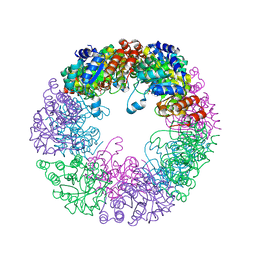
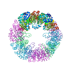
Crystal structure of cofactor-free urate oxidase in complex with the 5-peroxo derivative of 9-metyl uric acid (X-ray dose, 2.5 kGy)
Descriptor: (4S)-2-METHYL-2,4-PENTANEDIOL, (5S)-5-(dioxidanyl)-9-methyl-7H-purine-2,6,8-trione, URICASE
Authors:Bui, S, Steiner, R.A.
Deposit date:2014-04-01
Release date:2014-10-29
Last modified:2018-02-21
Method:X-RAY DIFFRACTION (1.32 Å)
Cite:Direct evidence for a peroxide intermediate and a reactive enzyme-substrate-dioxygen configuration in a cofactor-free oxidase.
Angew. Chem. Int. Ed. Engl., 53, 2014
8Q4S
DownloadVisualize
BU of 8q4s by Molmil
Crystal structure of phosphoserine phosphatase (SerB) from Brucella melitensis in complex with AP4 and magnesium.
Descriptor: (2S)-2-amino-4-phosphonobutanoic acid, GLYCEROL, MAGNESIUM ION, ...
Authors:Scaillet, T, Wouters, J.
Deposit date:2023-08-07
Release date:2024-08-21
Method:X-RAY DIFFRACTION (2.08 Å)
Cite:Crystal structure of phosphoserine phosphatase (SerB) from Brucella melitensis in complex with AP4 and magnesium.
To Be Published
6SAZ
DownloadVisualize
BU of 6saz by Molmil
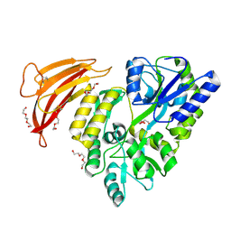
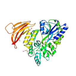
Cleaved human fetuin-b in complex with crayfish astacin
Descriptor: 2-acetamido-2-deoxy-beta-D-glucopyranose, 2-acetamido-2-deoxy-beta-D-glucopyranose-(1-4)-2-acetamido-2-deoxy-beta-D-glucopyranose, Astacin, ...
Authors:Gomis-Ruth, F.X, Guevara, T, Cuppari, A, Korschgen, H, Schmitz, C, Kuske, M, Yiallouros, I, Floehr, J, Jahnen-Dechent, W, Stocker, W.
Deposit date:2019-07-18
Release date:2019-10-23
Last modified:2024-01-24
Method:X-RAY DIFFRACTION (3 Å)
Cite:The C-terminal region of human plasma fetuin-B is dispensable for the raised-elephant-trunk mechanism of inhibition of astacin metallopeptidases.
Sci Rep, 9, 2019

225158

PDB entries from 2024-09-18

PDB statisticsPDBj update infoContact PDBjnumon