Loading
PDBj
MenuPDBj@FacebookPDBj@TwitterPDBj@YouTubewwPDB FoundationwwPDB
RCSB PDBPDBeBMRBAdv. SearchSearch help
Search by PDB author
8ERS
DownloadVisualize
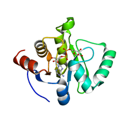
BU of 8ers by Molmil
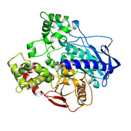
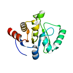
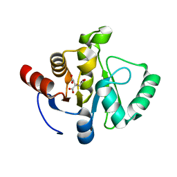
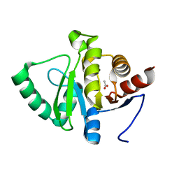
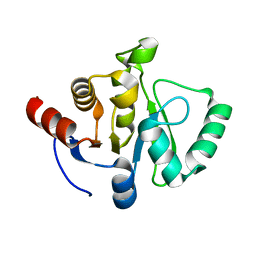
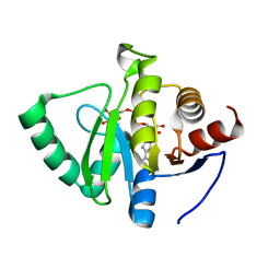
PanDDA analysis -- Crystal structure of SARS-CoV-2 NSP3 macrodomain in complex with Z4718398507 - (R,S) isomer
Descriptor: (1R,2S)-1-[(4-amino-2-hydroxybenzoyl)oxy]-2,3-dihydro-1H-indene-2-carboxylic acid, Non-structural protein 3
Authors:Correy, G.J, Fraser, J.S.
Deposit date:2022-10-12
Release date:2022-11-09
Last modified:2023-10-18
Method:X-RAY DIFFRACTION (1.05 Å)
Cite:Ligand screen against SARS-CoV-2 NSP3 macrodomain
To be published
5IVK
DownloadVisualize
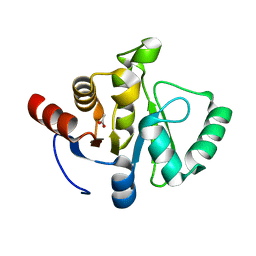
BU of 5ivk by Molmil
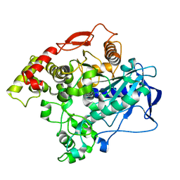
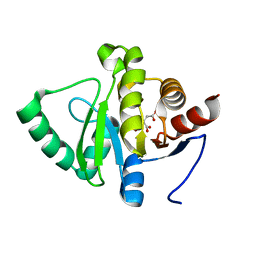
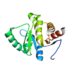
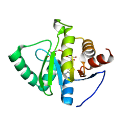
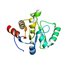
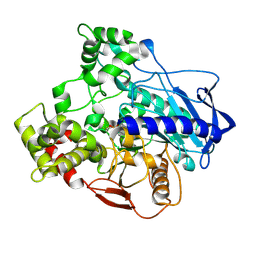
The alpha-esterase-7 carboxylesterase, E3, from the blowfly Lucilia cuprina: phosphorylated-enzyme ensemble refinement
Descriptor: Carboxylic ester hydrolase, DIETHYL HYDROGEN PHOSPHATE
Authors:Correy, G.J, Jackson, C.J.
Deposit date:2016-03-21
Release date:2016-06-22
Method:X-RAY DIFFRACTION (1.53 Å)
Cite:Mapping the Accessible Conformational Landscape of an Insect Carboxylesterase Using Conformational Ensemble Analysis and Kinetic Crystallography.
Structure, 24, 2016
5IVI
DownloadVisualize
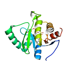
BU of 5ivi by Molmil
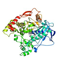
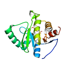
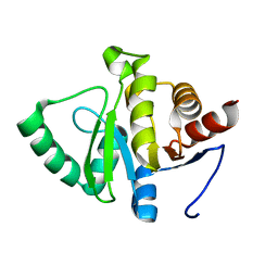
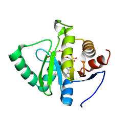
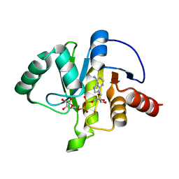
The alpha-esterase-7 carboxylesterase, E3, from the blowfly Lucilia cuprina: phosphorylated enzyme qFit multi-conformer model
Descriptor: Carboxylic ester hydrolase, DIETHYL HYDROGEN PHOSPHATE
Authors:Correy, G.J, Jackson, C.J.
Deposit date:2016-03-20
Release date:2016-06-15
Last modified:2023-09-27
Method:X-RAY DIFFRACTION (1.53 Å)
Cite:Mapping the Accessible Conformational Landscape of an Insect Carboxylesterase Using Conformational Ensemble Analysis and Kinetic Crystallography.
Structure, 24, 2016
5IVH
DownloadVisualize
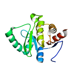
BU of 5ivh by Molmil
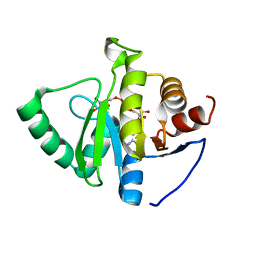
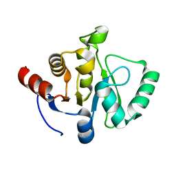
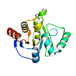
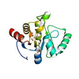
The alpha-esterase-7 carboxylesterase, E3, from the blowfly Lucilia cuprina: apo-enzyme ensemble refinement
Descriptor: Carboxylic ester hydrolase
Authors:Correy, G.J, Jackson, C.J.
Deposit date:2016-03-20
Release date:2016-06-22
Last modified:2023-09-27
Method:X-RAY DIFFRACTION (1.71 Å)
Cite:Mapping the Accessible Conformational Landscape of an Insect Carboxylesterase Using Conformational Ensemble Analysis and Kinetic Crystallography.
Structure, 24, 2016
5IVD
DownloadVisualize
BU of 5ivd by Molmil
The alpha-esterase-7 carboxylesterase, E3, from the blowfly Lucilia cuprina: apo-enzyme qFit multi-conformer model
Descriptor: Carboxylic ester hydrolase
Authors:Correy, G.J, Jackson, C.J.
Deposit date:2016-03-20
Release date:2016-06-15
Last modified:2023-09-27
Method:X-RAY DIFFRACTION (1.71 Å)
Cite:Mapping the Accessible Conformational Landscape of an Insect Carboxylesterase Using Conformational Ensemble Analysis and Kinetic Crystallography.
Structure, 24, 2016
7TWW
DownloadVisualize
BU of 7tww by Molmil
Crystal structure of SARS-CoV-2 NSP3 macrodomain in complex with ADP-ribose at pH 6 (P43 crystal form)
Descriptor: CITRIC ACID, Non-structural protein 3, [(2R,3S,4R,5R)-5-(6-AMINOPURIN-9-YL)-3,4-DIHYDROXY-OXOLAN-2-YL]METHYL [HYDROXY-[[(2R,3S,4R,5S)-3,4,5-TRIHYDROXYOXOLAN-2-YL]METHOXY]PHOSPHORYL] HYDROGEN PHOSPHATE
Authors:Correy, G.J, Fraser, J.S.
Deposit date:2022-02-07
Release date:2022-02-23
Last modified:2023-10-18
Method:X-RAY DIFFRACTION (0.9 Å)
Cite:Crystal structure of SARS-CoV-2 NSP3 macrodomain in complex with ADP-ribose at pH 6 (P43 crystal form)
To Be Published
7TWH
DownloadVisualize
BU of 7twh by Molmil
Crystal structure of SARS-CoV-2 NSP3 macrodomain at 293 K (P43 crystal form, 290 kGy)
Descriptor: Non-structural protein 3
Authors:Correy, G.J, Fraser, J.S.
Deposit date:2022-02-07
Release date:2022-02-23
Last modified:2023-10-18
Method:X-RAY DIFFRACTION (1.1 Å)
Cite:The mechanisms of catalysis and ligand binding for the SARS-CoV-2 NSP3 macrodomain from neutron and x-ray diffraction at room temperature.
Sci Adv, 8, 2022
7TWN
DownloadVisualize
BU of 7twn by Molmil
Crystal structure of SARS-CoV-2 NSP3 macrodomain at pH 5 (P43 crystal form)
Descriptor: 2-[N-CYCLOHEXYLAMINO]ETHANE SULFONIC ACID, CITRIC ACID, Non-structural protein 3
Authors:Correy, G.J, Fraser, J.S.
Deposit date:2022-02-07
Release date:2022-02-23
Last modified:2023-10-18
Method:X-RAY DIFFRACTION (0.9 Å)
Cite:The mechanisms of catalysis and ligand binding for the SARS-CoV-2 NSP3 macrodomain from neutron and x-ray diffraction at room temperature.
Sci Adv, 8, 2022
7TWJ
DownloadVisualize
BU of 7twj by Molmil
Crystal structure of SARS-CoV-2 NSP3 macrodomain at pH 4 (P43 crystal form)
Descriptor: CITRIC ACID, Non-structural protein 3
Authors:Correy, G.J, Fraser, J.S.
Deposit date:2022-02-07
Release date:2022-02-23
Last modified:2023-10-18
Method:X-RAY DIFFRACTION (0.9 Å)
Cite:The mechanisms of catalysis and ligand binding for the SARS-CoV-2 NSP3 macrodomain from neutron and x-ray diffraction at room temperature.
Sci Adv, 8, 2022
7TWX
DownloadVisualize
BU of 7twx by Molmil
Crystal structure of SARS-CoV-2 NSP3 macrodomain in complex with ADP-ribose at pH 7 (P43 crystal form)
Descriptor: Non-structural protein 3, [(2R,3S,4R,5R)-5-(6-AMINOPURIN-9-YL)-3,4-DIHYDROXY-OXOLAN-2-YL]METHYL [HYDROXY-[[(2R,3S,4R,5S)-3,4,5-TRIHYDROXYOXOLAN-2-YL]METHOXY]PHOSPHORYL] HYDROGEN PHOSPHATE
Authors:Correy, G.J, Fraser, J.S.
Deposit date:2022-02-07
Release date:2022-02-23
Last modified:2023-10-18
Method:X-RAY DIFFRACTION (0.9 Å)
Cite:Crystal structure of SARS-CoV-2 NSP3 macrodomain in complex with ADP-ribose at pH 7 (P43 crystal form)
To Be Published
7TWR
DownloadVisualize
BU of 7twr by Molmil
Crystal structure of SARS-CoV-2 NSP3 macrodomain at pH 8 (P43 crystal form)
Descriptor: ACETATE ION, Non-structural protein 3
Authors:Correy, G.J, Fraser, J.S.
Deposit date:2022-02-07
Release date:2022-02-23
Last modified:2023-10-18
Method:X-RAY DIFFRACTION (0.9 Å)
Cite:The mechanisms of catalysis and ligand binding for the SARS-CoV-2 NSP3 macrodomain from neutron and x-ray diffraction at room temperature.
Sci Adv, 8, 2022
7TWS
DownloadVisualize
BU of 7tws by Molmil
Crystal structure of SARS-CoV-2 NSP3 macrodomain at pH 10 (P43 crystal form)
Descriptor: Non-structural protein 3
Authors:Correy, G.J, Fraser, J.S.
Deposit date:2022-02-07
Release date:2022-02-23
Last modified:2023-10-18
Method:X-RAY DIFFRACTION (0.9 Å)
Cite:The mechanisms of catalysis and ligand binding for the SARS-CoV-2 NSP3 macrodomain from neutron and x-ray diffraction at room temperature.
Sci Adv, 8, 2022
7TWQ
DownloadVisualize
BU of 7twq by Molmil
Crystal structure of SARS-CoV-2 NSP3 macrodomain at pH 9 (P43 crystal form)
Descriptor: Non-structural protein 3
Authors:Correy, G.J, Fraser, J.S.
Deposit date:2022-02-07
Release date:2022-02-23
Last modified:2023-10-18
Method:X-RAY DIFFRACTION (0.9 Å)
Cite:The mechanisms of catalysis and ligand binding for the SARS-CoV-2 NSP3 macrodomain from neutron and x-ray diffraction at room temperature.
Sci Adv, 8, 2022
7TWI
DownloadVisualize
BU of 7twi by Molmil
Crystal structure of SARS-CoV-2 NSP3 macrodomain at 293 K (P43 crystal form, 539 kGy)
Descriptor: Non-structural protein 3
Authors:Correy, G.J, Fraser, J.S.
Deposit date:2022-02-07
Release date:2022-02-23
Last modified:2023-10-18
Method:X-RAY DIFFRACTION (1.1 Å)
Cite:The mechanisms of catalysis and ligand binding for the SARS-CoV-2 NSP3 macrodomain from neutron and x-ray diffraction at room temperature.
Sci Adv, 8, 2022
7TWG
DownloadVisualize
BU of 7twg by Molmil
Crystal structure of SARS-CoV-2 NSP3 macrodomain at 293 K (P43 crystal form, 153 kGy)
Descriptor: Non-structural protein 3
Authors:Correy, G.J, Fraser, J.S.
Deposit date:2022-02-07
Release date:2022-02-23
Last modified:2023-10-18
Method:X-RAY DIFFRACTION (1.1 Å)
Cite:The mechanisms of catalysis and ligand binding for the SARS-CoV-2 NSP3 macrodomain from neutron and x-ray diffraction at room temperature.
Sci Adv, 8, 2022
7TWY
DownloadVisualize
BU of 7twy by Molmil
Crystal structure of SARS-CoV-2 NSP3 macrodomain in complex with ADP-ribose at pH 8 (P43 crystal form)
Descriptor: Non-structural protein 3, [(2R,3S,4R,5R)-5-(6-AMINOPURIN-9-YL)-3,4-DIHYDROXY-OXOLAN-2-YL]METHYL [HYDROXY-[[(2R,3S,4R,5S)-3,4,5-TRIHYDROXYOXOLAN-2-YL]METHOXY]PHOSPHORYL] HYDROGEN PHOSPHATE
Authors:Correy, G.J, Fraser, J.S.
Deposit date:2022-02-07
Release date:2022-02-23
Last modified:2023-10-18
Method:X-RAY DIFFRACTION (0.9 Å)
Cite:Crystal structure of SARS-CoV-2 NSP3 macrodomain in complex with ADP-ribose at pH 8 (P43 crystal form)
To Be Published
7TWT
DownloadVisualize
BU of 7twt by Molmil
Crystal structure of SARS-CoV-2 NSP3 macrodomain in complex with ADP-ribose at pH 4 (P43 crystal form)
Descriptor: Non-structural protein 3, [(2R,3S,4R,5R)-5-(6-AMINOPURIN-9-YL)-3,4-DIHYDROXY-OXOLAN-2-YL]METHYL [HYDROXY-[[(2R,3S,4R,5S)-3,4,5-TRIHYDROXYOXOLAN-2-YL]METHOXY]PHOSPHORYL] HYDROGEN PHOSPHATE
Authors:Correy, G.J, Fraser, J.S.
Deposit date:2022-02-07
Release date:2022-02-23
Last modified:2023-10-18
Method:X-RAY DIFFRACTION (0.9 Å)
Cite:Crystal structure of SARS-CoV-2 NSP3 macrodomain in complex with ADP-ribose at pH 4 (P43 crystal form)
To Be Published
7TX4
DownloadVisualize
BU of 7tx4 by Molmil
Neutron crystal structure of SARS-CoV-2 NSP3 macrodomain at 293 K (P21 crystal form)
Descriptor: Papain-like protease nsp3
Authors:Correy, G.J, Fraser, J.S, Kovalevsky, A.
Deposit date:2022-02-07
Release date:2022-02-23
Last modified:2023-10-25
Method:NEUTRON DIFFRACTION (1.9 Å), X-RAY DIFFRACTION
Cite:The mechanisms of catalysis and ligand binding for the SARS-CoV-2 NSP3 macrodomain from neutron and x-ray diffraction at room temperature.
Sci Adv, 8, 2022
7TX1
DownloadVisualize
BU of 7tx1 by Molmil
Crystal structure of SARS-CoV-2 NSP3 macrodomain in complex with ADP-ribose at pH 10 (P43 crystal form)
Descriptor: Non-structural protein 3, [(2R,3S,4R,5R)-5-(6-AMINOPURIN-9-YL)-3,4-DIHYDROXY-OXOLAN-2-YL]METHYL [HYDROXY-[[(2R,3S,4R,5S)-3,4,5-TRIHYDROXYOXOLAN-2-YL]METHOXY]PHOSPHORYL] HYDROGEN PHOSPHATE
Authors:Correy, G.J, Fraser, J.S.
Deposit date:2022-02-07
Release date:2022-02-23
Last modified:2023-10-18
Method:X-RAY DIFFRACTION (0.9 Å)
Cite:Crystal structure of SARS-CoV-2 NSP3 macrodomain in complex with ADP-ribose at pH 10 (P43 crystal form)
To Be Published
7TWF
DownloadVisualize
BU of 7twf by Molmil
Crystal structure of SARS-CoV-2 NSP3 macrodomain at 293 K (P43 crystal form, 73 kGy)
Descriptor: Non-structural protein 3
Authors:Correy, G.J, Fraser, J.S.
Deposit date:2022-02-07
Release date:2022-02-23
Last modified:2023-10-18
Method:X-RAY DIFFRACTION (1.1 Å)
Cite:The mechanisms of catalysis and ligand binding for the SARS-CoV-2 NSP3 macrodomain from neutron and x-ray diffraction at room temperature.
Sci Adv, 8, 2022
7TX5
DownloadVisualize
BU of 7tx5 by Molmil
Neutron crystal structure of SARS-CoV-2 NSP3 macrodomain in complex with ADP-ribose at 293 K (C2 crystal form)
Descriptor: ADENOSINE-5-DIPHOSPHORIBOSE, Papain-like protease nsp3
Authors:Correy, G.J, Fraser, J.S, Kovalevsky, A.
Deposit date:2022-02-07
Release date:2022-02-23
Last modified:2023-10-25
Method:NEUTRON DIFFRACTION (1.95 Å), X-RAY DIFFRACTION
Cite:The mechanisms of catalysis and ligand binding for the SARS-CoV-2 NSP3 macrodomain from neutron and x-ray diffraction at room temperature.
Sci Adv, 8, 2022
7TWO
DownloadVisualize
BU of 7two by Molmil
Crystal structure of SARS-CoV-2 NSP3 macrodomain at pH 6 (P43 crystal form)
Descriptor: 2-[N-CYCLOHEXYLAMINO]ETHANE SULFONIC ACID, CITRIC ACID, Non-structural protein 3
Authors:Correy, G.J, Fraser, J.S.
Deposit date:2022-02-07
Release date:2022-02-23
Last modified:2023-10-18
Method:X-RAY DIFFRACTION (0.9 Å)
Cite:The mechanisms of catalysis and ligand binding for the SARS-CoV-2 NSP3 macrodomain from neutron and x-ray diffraction at room temperature.
Sci Adv, 8, 2022
7TX0
DownloadVisualize
BU of 7tx0 by Molmil
Crystal structure of SARS-CoV-2 NSP3 macrodomain in complex with ADP-ribose at pH 9 (P43 crystal form)
Descriptor: Non-structural protein 3, [(2R,3S,4R,5R)-5-(6-AMINOPURIN-9-YL)-3,4-DIHYDROXY-OXOLAN-2-YL]METHYL [HYDROXY-[[(2R,3S,4R,5S)-3,4,5-TRIHYDROXYOXOLAN-2-YL]METHOXY]PHOSPHORYL] HYDROGEN PHOSPHATE
Authors:Correy, G.J, Fraser, J.S.
Deposit date:2022-02-07
Release date:2022-02-23
Last modified:2023-10-18
Method:X-RAY DIFFRACTION (0.84 Å)
Cite:Crystal structure of SARS-CoV-2 NSP3 macrodomain in complex with ADP-ribose at pH 9 (P43 crystal form)
To Be Published
7TWP
DownloadVisualize
BU of 7twp by Molmil
Crystal structure of SARS-CoV-2 NSP3 macrodomain at pH 7 (P43 crystal form)
Descriptor: ACETATE ION, Non-structural protein 3
Authors:Correy, G.J, Fraser, J.S.
Deposit date:2022-02-07
Release date:2022-02-23
Last modified:2023-10-18
Method:X-RAY DIFFRACTION (0.9 Å)
Cite:The mechanisms of catalysis and ligand binding for the SARS-CoV-2 NSP3 macrodomain from neutron and x-ray diffraction at room temperature.
Sci Adv, 8, 2022
7TWV
DownloadVisualize
BU of 7twv by Molmil
Crystal structure of SARS-CoV-2 NSP3 macrodomain in complex with ADP-ribose at pH 5 (P43 crystal form)
Descriptor: CITRIC ACID, Non-structural protein 3, [(2R,3S,4R,5R)-5-(6-AMINOPURIN-9-YL)-3,4-DIHYDROXY-OXOLAN-2-YL]METHYL [HYDROXY-[[(2R,3S,4R,5S)-3,4,5-TRIHYDROXYOXOLAN-2-YL]METHOXY]PHOSPHORYL] HYDROGEN PHOSPHATE
Authors:Correy, G.J, Fraser, J.S.
Deposit date:2022-02-07
Release date:2022-02-23
Last modified:2023-10-18
Method:X-RAY DIFFRACTION (0.9 Å)
Cite:Crystal structure of SARS-CoV-2 NSP3 macrodomain in complex with ADP-ribose at pH 5 (P43 crystal form)
To Be Published

218853

PDB entries from 2024-04-24

PDB statisticsPDBj update infoContact PDBjnumon