Loading
PDBj
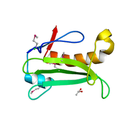
MenuPDBj@FacebookPDBj@TwitterPDBj@YouTubewwPDB FoundationwwPDB
RCSB PDBPDBeBMRBAdv. SearchSearch help
Search by PDB author
5CXT
DownloadVisualize
BU of 5cxt by Molmil
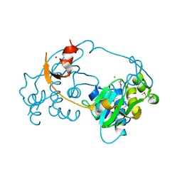
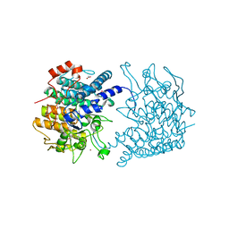
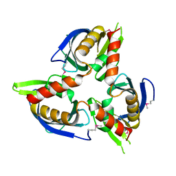
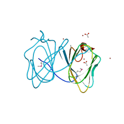
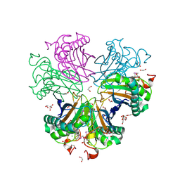
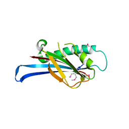
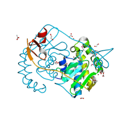
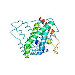
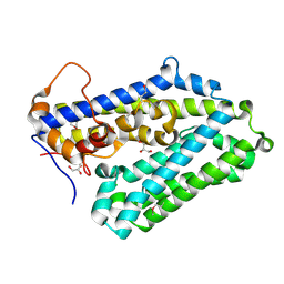
Crystal structure of a RNA-binding protein 39 (RBM39) in complex with fragment of splicing factor (U2AF) from Unknown at 2.20 A resolution
Descriptor: RNA-binding protein 39, Splicing factor U2AF 65 kDa subunit
Authors:Joint Center for Structural Genomics (JCSG), Partnership for T-Cell Biology (TCELL)
Deposit date:2015-07-29
Release date:2015-10-14
Last modified:2023-09-27
Method:X-RAY DIFFRACTION (2.2 Å)
Cite:UHM-ULM interactions in the RBM39-U2AF65 splicing-factor complex.
Acta Crystallogr D Struct Biol, 72, 2016
2IAY
DownloadVisualize
BU of 2iay by Molmil
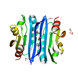
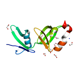
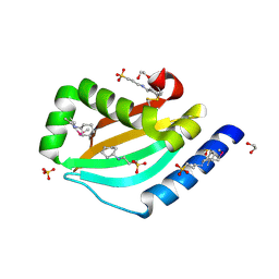
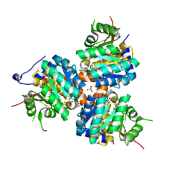
Crystal structure of a duf1831 family protein (lp2179) from lactobacillus plantarum at 1.20 A resolution
Descriptor: CHLORIDE ION, GLYCEROL, Hypothetical protein
Authors:Joint Center for Structural Genomics (JCSG)
Deposit date:2006-09-08
Release date:2006-10-10
Last modified:2023-01-25
Method:X-RAY DIFFRACTION (1.2 Å)
Cite:Structure of LP2179, the first representative of Pfam family PF08866, suggests a new fold with a role in amino-acid metabolism.
Acta Crystallogr.,Sect.F, 66, 2010
3D00
DownloadVisualize
BU of 3d00 by Molmil
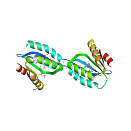
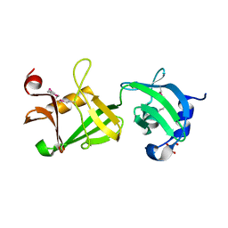
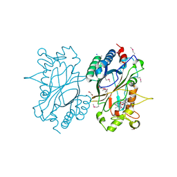
Crystal structure of a tungsten formylmethanofuran dehydrogenase subunit e (fmde)-like protein (syn_00638) from syntrophus aciditrophicus at 1.90 A resolution
Descriptor: CHLORIDE ION, Tungsten formylmethanofuran dehydrogenase subunit E, ZINC ION
Authors:Joint Center for Structural Genomics (JCSG)
Deposit date:2008-04-30
Release date:2008-05-20
Last modified:2023-02-01
Method:X-RAY DIFFRACTION (1.9 Å)
Cite:Structures of three members of Pfam PF02663 (FmdE) implicated in microbial methanogenesis reveal a conserved alpha+beta core domain and an auxiliary C-terminal treble-clef zinc finger.
Acta Crystallogr.,Sect.F, 66, 2010
3CGH
DownloadVisualize
BU of 3cgh by Molmil
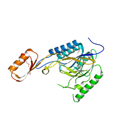
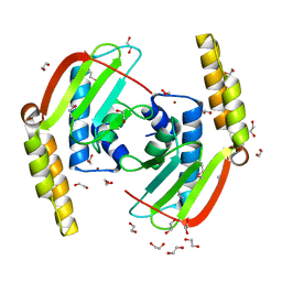
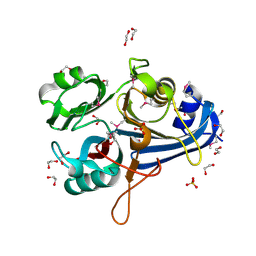
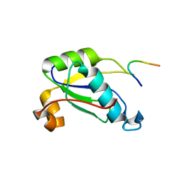
Crystal structure of a susd homolog (bt_3984) from bacteroides thetaiotaomicron vpi-5482 at 1.70 A resolution
Descriptor: ACETATE ION, SusD homolog, ZINC ION
Authors:Joint Center for Structural Genomics (JCSG)
Deposit date:2008-03-05
Release date:2008-03-18
Last modified:2023-02-01
Method:X-RAY DIFFRACTION (1.7 Å)
Cite:Structure of BT_3984, a member of the SusD/RagB family of nutrient-binding molecules.
Acta Crystallogr.,Sect.F, 66, 2010
3CM1
DownloadVisualize
BU of 3cm1 by Molmil
Crystal structure of SsgA-like sporulation-specific cell division protein (YP_290167.1) from Thermobifida fusca YX-ER1 at 2.60 A resolution
Descriptor: SsgA-like sporulation-specific cell division protein
Authors:Joint Center for Structural Genomics (JCSG), Chruszcz, M, Minor, W, Wang, S.
Deposit date:2008-03-20
Release date:2008-04-01
Last modified:2023-02-01
Method:X-RAY DIFFRACTION (2.6 Å)
Cite:Structural and functional characterizations of SsgB, a conserved activator of developmental cell division in morphologically complex actinomycetes.
J.Biol.Chem., 284, 2009
3H50
DownloadVisualize
BU of 3h50 by Molmil
CRYSTAL STRUCTURE OF A TETRACENOMYCIN POLYKETIDE SYNTHESIS PROTEIN (TCMJ) FROM XANTHOMONAS CAMPESTRIS PV. CAMPESTRIS AT 1.60 A RESOLUTION
Descriptor: ACETATE ION, Tetracenomycin polyketide synthesis protein, ZINC ION
Authors:Joint Center for Structural Genomics (JCSG)
Deposit date:2009-04-21
Release date:2009-05-05
Last modified:2023-02-01
Method:X-RAY DIFFRACTION (1.6 Å)
Cite:Conformational changes associated with the binding of zinc acetate at the putative active site of XcTcmJ, a cupin from Xanthomonas campestris pv. campestris.
Acta Crystallogr.,Sect.F, 66, 2010
3BYQ
DownloadVisualize
BU of 3byq by Molmil
Crystal structure of a duf1185 family protein (bb2672) from bordetella bronchiseptica rb50 at 1.70 A resolution
Descriptor: 1,2-ETHANEDIOL, CHLORIDE ION, TETRAETHYLENE GLYCOL, ...
Authors:Joint Center for Structural Genomics (JCSG)
Deposit date:2008-01-16
Release date:2008-01-29
Last modified:2023-02-01
Method:X-RAY DIFFRACTION (1.7 Å)
Cite:Structures of the first representatives of Pfam family PF06684 (DUF1185) reveal a novel variant of the Bacillus chorismate mutase fold and suggest a role in amino-acid metabolism.
Acta Crystallogr.,Sect.F, 66, 2010
2OOK
DownloadVisualize
BU of 2ook by Molmil
Crystal structure of a protein with unknown function (YP_749275.1) from Shewanella Frigidimarina NCIMB 400 at 1.80 A resolution
Descriptor: 1,2-ETHANEDIOL, Hypothetical protein
Authors:Joint Center for Structural Genomics (JCSG)
Deposit date:2007-01-25
Release date:2007-02-06
Last modified:2023-01-25
Method:X-RAY DIFFRACTION (1.8 Å)
Cite:Open and closed conformations of two SpoIIAA-like proteins (YP_749275.1 and YP_001095227.1) provide insights into membrane association and ligand binding.
Acta Crystallogr.,Sect.F, 66, 2010
2Q3L
DownloadVisualize
BU of 2q3l by Molmil
CRYSTAL STRUCTURE OF AN UNCHARACTERIZED PROTEIN FROM DUF3478 FAMILY WITH A SPOIIAA-LIKE FOLD (SHEW_3102) FROM SHEWANELLA LOIHICA PV-4 AT 2.25 A RESOLUTION
Descriptor: (4S)-2-METHYL-2,4-PENTANEDIOL, CHLORIDE ION, SODIUM ION, ...
Authors:Joint Center for Structural Genomics (JCSG)
Deposit date:2007-05-30
Release date:2007-06-12
Last modified:2023-01-25
Method:X-RAY DIFFRACTION (2.25 Å)
Cite:Open and closed conformations of two SpoIIAA-like proteins (YP_749275.1 and YP_001095227.1) provide insights into membrane association and ligand binding.
Acta Crystallogr.,Sect.F, 66, 2010
2Q8U
DownloadVisualize
BU of 2q8u by Molmil
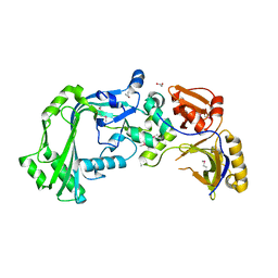
CRYSTAL STRUCTURE OF MRE11 FROM THERMOTOGA MARITIMA MSB8 (TM1635) AT 2.20 A RESOLUTION
Descriptor: Exonuclease, putative
Authors:Joint Center for Structural Genomics (JCSG)
Deposit date:2007-06-11
Release date:2007-06-26
Last modified:2023-01-25
Method:X-RAY DIFFRACTION (2.2 Å)
Cite:Crystal structure of the first eubacterial Mre11 nuclease reveals novel features that may discriminate substrates during DNA repair.
J.Mol.Biol., 397, 2010
2QTP
DownloadVisualize
BU of 2qtp by Molmil
Crystal structure of a duf1185 family protein (spo0826) from silicibacter pomeroyi dss-3 at 2.10 A resolution
Descriptor: Uncharacterized protein
Authors:Joint Center for Structural Genomics (JCSG)
Deposit date:2007-08-02
Release date:2007-08-21
Last modified:2023-01-25
Method:X-RAY DIFFRACTION (2.1 Å)
Cite:Structures of the first representatives of Pfam family PF06684 (DUF1185) reveal a novel variant of the Bacillus chorismate mutase fold and suggest a role in amino-acid metabolism.
Acta Crystallogr.,Sect.F, 66, 2010
2RA9
DownloadVisualize
BU of 2ra9 by Molmil
Crystal structure of a duf1285 family protein (sbal_2486) from shewanella baltica os155 at 1.40 A resolution
Descriptor: 1,2-ETHANEDIOL, SODIUM ION, Uncharacterized protein DUF1285
Authors:Joint Center for Structural Genomics (JCSG)
Deposit date:2007-09-14
Release date:2007-10-16
Last modified:2023-01-25
Method:X-RAY DIFFRACTION (1.4 Å)
Cite:Structures of the first representatives of Pfam family PF06938 (DUF1285) reveal a new fold with repeated structural motifs and possible involvement in signal transduction.
Acta Crystallogr.,Sect.F, 66, 2010
2RE3
DownloadVisualize
BU of 2re3 by Molmil
CRYSTAL STRUCTURE OF a DUF1285 family protein (SPO_0140) FROM SILICIBACTER POMEROYI DSS-3 AT 2.50 A RESOLUTION
Descriptor: GLYCEROL, Uncharacterized protein
Authors:Joint Center for Structural Genomics (JCSG)
Deposit date:2007-09-25
Release date:2007-10-09
Last modified:2023-01-25
Method:X-RAY DIFFRACTION (2.5 Å)
Cite:Structures of the first representatives of Pfam family PF06938 (DUF1285) reveal a new fold with repeated structural motifs and possible involvement in signal transduction.
Acta Crystallogr.,Sect.F, 66, 2010
2GLZ
DownloadVisualize
BU of 2glz by Molmil
Crystal structure of a formylmethanofuran dehydrogenase subunit e-like protein (dhaf_2992) from desulfitobacterium hafniense dcb-2 at 1.45 A resolution
Descriptor: 1,2-ETHANEDIOL, NICKEL (II) ION, ZINC ION, ...
Authors:Joint Center for Structural Genomics (JCSG)
Deposit date:2006-04-05
Release date:2006-04-18
Last modified:2023-01-25
Method:X-RAY DIFFRACTION (1.45 Å)
Cite:Structures of three members of Pfam PF02663 (FmdE) implicated in microbial methanogenesis reveal a conserved alpha+beta core domain and an auxiliary C-terminal treble-clef zinc finger.
Acta Crystallogr.,Sect.F, 66, 2010
2GVI
DownloadVisualize
BU of 2gvi by Molmil
Crystal structure of a putative formylmethanofuran dehydrogenase subunit e (ta1109) from thermoplasma acidophilum at 1.87 A resolution
Descriptor: 1,2-ETHANEDIOL, ACETIC ACID, UNKNOWN LIGAND, ...
Authors:Joint Center for Structural Genomics (JCSG)
Deposit date:2006-05-02
Release date:2006-06-13
Last modified:2023-01-25
Method:X-RAY DIFFRACTION (1.87 Å)
Cite:Structures of three members of Pfam PF02663 (FmdE) implicated in microbial methanogenesis reveal a conserved alpha+beta core domain and an auxiliary C-terminal treble-clef zinc finger.
Acta Crystallogr.,Sect.F, 66, 2010
3UFI
DownloadVisualize
BU of 3ufi by Molmil
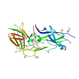
Crystal structure of a putative cell adhesion protein (BACOVA_04980) from Bacteroides ovatus ATCC 8483 at 2.18 A resolution
Descriptor: CHLORIDE ION, SULFATE ION, hypothetical protein BACOVA_04980
Authors:Joint Center for Structural Genomics (JCSG)
Deposit date:2011-11-01
Release date:2011-12-14
Last modified:2023-02-01
Method:X-RAY DIFFRACTION (2.18 Å)
Cite:A Distinct Type of Pilus from the Human Microbiome.
Cell, 165, 2016
3UP6
DownloadVisualize
BU of 3up6 by Molmil
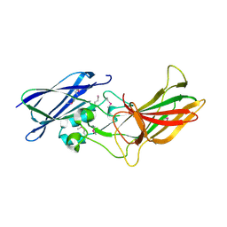
Crystal structure of a putative cell adhesion protein (BACOVA_04078) from Bacteroides ovatus ATCC 8483 at 2.80 A resolution
Descriptor: hypothetical protein BACOVA_04078
Authors:Joint Center for Structural Genomics (JCSG)
Deposit date:2011-11-17
Release date:2011-12-21
Last modified:2023-02-01
Method:X-RAY DIFFRACTION (2.8 Å)
Cite:A Distinct Type of Pilus from the Human Microbiome.
Cell, 165, 2016
3KK7
DownloadVisualize
BU of 3kk7 by Molmil
Crystal structure of Putative cell invasion protein with MAC/Perforin domain (NP_812351.1) from BACTERIODES THETAIOTAOMICRON VPI-5482 at 2.46 A resolution
Descriptor: (4S)-2-METHYL-2,4-PENTANEDIOL, 1,2-ETHANEDIOL, CHLORIDE ION, ...
Authors:Joint Center for Structural Genomics (JCSG)
Deposit date:2009-11-04
Release date:2009-11-24
Last modified:2023-02-01
Method:X-RAY DIFFRACTION (2.46 Å)
Cite:Structure of a membrane-attack complex/perforin (MACPF) family protein from the human gut symbiont Bacteroides thetaiotaomicron.
Acta Crystallogr.,Sect.F, 66, 2010
3NL9
DownloadVisualize
BU of 3nl9 by Molmil
Crystal structure of a putative NTP pyrophosphohydrolase (Exig_1061) from EXIGUOBACTERIUM SP. 255-15 at 1.78 A resolution
Descriptor: 1,2-ETHANEDIOL, putative NTP pyrophosphohydrolase
Authors:Joint Center for Structural Genomics (JCSG)
Deposit date:2010-06-21
Release date:2010-07-21
Last modified:2023-02-01
Method:X-RAY DIFFRACTION (1.78 Å)
Cite:Structure of a putative NTP pyrophosphohydrolase: YP_001813558.1 from Exiguobacterium sibiricum 255-15.
Acta Crystallogr.,Sect.F, 66, 2010
3NPD
DownloadVisualize
BU of 3npd by Molmil
Crystal structure of a putative secreted protein (PA3611) from PSEUDOMONAS AERUGINOSA at 1.60 A resolution
Descriptor: 1,2-ETHANEDIOL, 3-CYCLOHEXYL-1-PROPYLSULFONIC ACID, SULFATE ION, ...
Authors:Joint Center for Structural Genomics (JCSG)
Deposit date:2010-06-28
Release date:2010-07-28
Last modified:2023-02-01
Method:X-RAY DIFFRACTION (1.6 Å)
Cite:Crystal structure of a putative quorum sensing-regulated protein (PA3611) from the Pseudomonas-specific DUF4146 family.
Proteins, 82, 2014
2HUJ
DownloadVisualize
BU of 2huj by Molmil
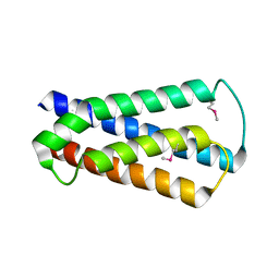
Crystal structure of a protein of uknown function (NP_471338.1) from Listeria innocua at 1.74 A resolution
Descriptor: Lin2004 protein
Authors:Joint Center for Structural Genomics (JCSG)
Deposit date:2006-07-26
Release date:2006-08-08
Last modified:2023-01-25
Method:X-RAY DIFFRACTION (1.74 Å)
Cite:Crystal structures of MW1337R and lin2004: representatives of a novel protein family that adopt a four-helical bundle fold.
Proteins, 71, 2008
3OHG
DownloadVisualize
BU of 3ohg by Molmil
Crystal structure of a protein with unknown function from DUF2233 family (BACOVA_00430) from Bacteroides ovatus at 1.80 A resolution
Descriptor: 1,2-ETHANEDIOL, CHLORIDE ION, SULFATE ION, ...
Authors:Joint Center for Structural Genomics (JCSG)
Deposit date:2010-08-17
Release date:2010-09-08
Last modified:2023-02-01
Method:X-RAY DIFFRACTION (1.8 Å)
Cite:Structure and function of the DUF2233 domain in bacteria and in the human mannose 6-phosphate uncovering enzyme.
J.Biol.Chem., 288, 2013
3OYV
DownloadVisualize
BU of 3oyv by Molmil
Crystal structure of an imelysin peptidase (BACOVA_03801) from Bacteroides ovatus ATCC 8483 at 1.25 A resolution
Descriptor: CHLORIDE ION, GLYCEROL, Imelysin, ...
Authors:Joint Center for Structural Genomics (JCSG)
Deposit date:2010-09-23
Release date:2010-10-20
Last modified:2023-02-01
Method:X-RAY DIFFRACTION (1.25 Å)
Cite:Structural and sequence analysis of imelysin-like proteins implicated in bacterial iron uptake.
Plos One, 6, 2011
1ESQ
DownloadVisualize
BU of 1esq by Molmil
CRYSTAL STRUCTURE OF THIAZOLE KINASE MUTANT (C198S) WITH ATP AND THIAZOLE PHOSPHATE.
Descriptor: 4-METHYL-5-HYDROXYETHYLTHIAZOLE PHOSPHATE, ADENOSINE-5'-TRIPHOSPHATE, HYDROXYETHYLTHIAZOLE KINASE, ...
Authors:Campobasso, N, Mathews, I.I, Begley, T.P, Ealick, S.E.
Deposit date:2000-04-10
Release date:2000-08-09
Last modified:2024-02-07
Method:X-RAY DIFFRACTION (2.5 Å)
Cite:Crystal structure of 4-methyl-5-beta-hydroxyethylthiazole kinase from Bacillus subtilis at 1.5 A resolution.
Biochemistry, 39, 2000
2IIZ
DownloadVisualize
BU of 2iiz by Molmil
Crystal structure of putative melanin biosynthesis protein TyrA with bound heme (NP_716371.1) from Shewanella Oneidensis at 2.30 A resolution
Descriptor: 1,2-ETHANEDIOL, ISOPROPYL ALCOHOL, Melanin biosynthesis protein TyrA, ...
Authors:Joint Center for Structural Genomics (JCSG)
Deposit date:2006-09-28
Release date:2006-11-14
Last modified:2023-11-15
Method:X-RAY DIFFRACTION (2.3 Å)
Cite:Identification and structural characterization of heme binding in a novel dye-decolorizing peroxidase, TyrA.
Proteins, 69, 2007

219869

數據於2024-05-15公開中

PDB statisticsPDBj update infoContact PDBjnumon