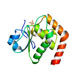
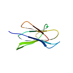
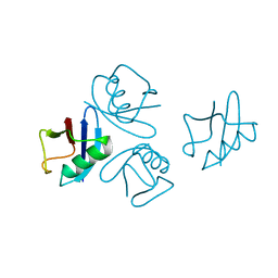
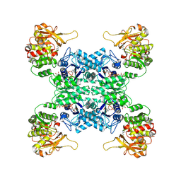
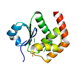
4UA1
 
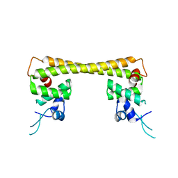 | | Crystal structure of dual function transcriptional regulator MerR form Bacillus megaterium MB1 in complex with mercury (II) ion | | 分子名称: | MERCURY (II) ION, Regulatory protein | | 著者 | Chang, C.C, Lin, L.Y, Zou, X.W, Huang, C.C, Chan, N.L. | | 登録日 | 2014-08-07 | | 公開日 | 2015-07-22 | | 最終更新日 | 2024-03-20 | | 実験手法 | X-RAY DIFFRACTION (2.56 Å) | | 主引用文献 | Structural basis of the mercury(II)-mediated conformational switching of the dual-function transcriptional regulator MerR
Nucleic Acids Res., 43, 2015
|
|
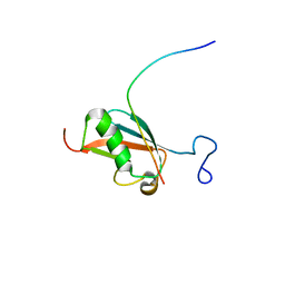
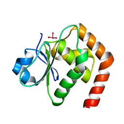
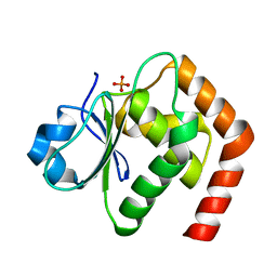
4UA2
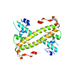 | | Crystal structure of dual function transcriptional regulator MerR from Bacillus megaterium MB1 | | 分子名称: | Regulatory protein | | 著者 | Lin, L.Y, Chang, C.C, Zou, X.W, Huang, C.C, Chan, N.L. | | 登録日 | 2014-08-07 | | 公開日 | 2015-07-22 | | 最終更新日 | 2020-01-29 | | 実験手法 | X-RAY DIFFRACTION (2.61 Å) | | 主引用文献 | Structural basis of the mercury(II)-mediated conformational switching of the dual-function transcriptional regulator MerR
Nucleic Acids Res., 43, 2015
|
|
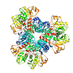
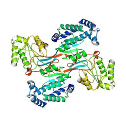
7DPW
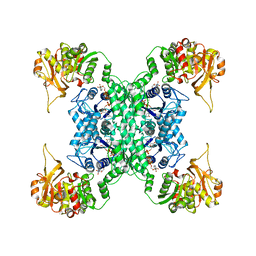 | | Structural basis for ligand binding modes of CTP synthase | | 分子名称: | CTP synthase, CYTIDINE-5'-TRIPHOSPHATE, MAGNESIUM ION | | 著者 | Liu, J.L, Zhou, X, Guo, C.J, Chang, C.C. | | 登録日 | 2020-12-21 | | 公開日 | 2021-09-15 | | 実験手法 | ELECTRON MICROSCOPY (2.65 Å) | | 主引用文献 | Structural basis for ligand binding modes of CTP synthase.
Proc.Natl.Acad.Sci.USA, 118, 2021
|
|
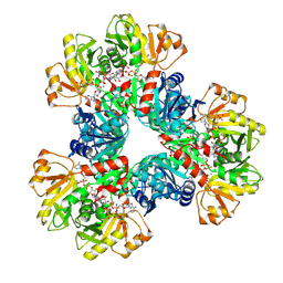
7DPT
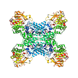 | | Structural basis for ligand binding modes of CTP synthase | | 分子名称: | 6-DIAZENYL-5-OXO-L-NORLEUCINE, ADENOSINE-5'-DIPHOSPHATE, CTP synthase, ... | | 著者 | Liu, J.L, Zhou, X, Guo, C.J, Chang, C.C. | | 登録日 | 2020-12-21 | | 公開日 | 2021-09-15 | | 実験手法 | ELECTRON MICROSCOPY (2.48 Å) | | 主引用文献 | Structural basis for ligand binding modes of CTP synthase.
Proc.Natl.Acad.Sci.USA, 118, 2021
|
|
6L1S
 | | Crystal structure of DUSP22 mutant_C88S | | 分子名称: | Dual specificity protein phosphatase 22, PHOSPHATE ION | | 著者 | Lai, C.H, Chang, C.C, Lyu, P.C. | | 登録日 | 2019-09-30 | | 公開日 | 2020-10-28 | | 最終更新日 | 2023-11-22 | | 実験手法 | X-RAY DIFFRACTION (1.3611 Å) | | 主引用文献 | Structural Insights into the Active Site Formation of DUSP22 in N-loop-containing Protein Tyrosine Phosphatases.
Int J Mol Sci, 21, 2020
|
|
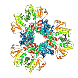
7XMV
 | | E.coli phosphoribosylpyrophosphate (PRPP) synthetase type A(AMP/ADP) filament bound with ADP, AMP and R5P | | 分子名称: | 5-O-phosphono-alpha-D-ribofuranose, ADENOSINE MONOPHOSPHATE, ADENOSINE-5'-DIPHOSPHATE, ... | | 著者 | Hu, H.H, Lu, G.M, Chang, C.C, Liu, J.L. | | 登録日 | 2022-04-27 | | 公開日 | 2022-06-29 | | 最終更新日 | 2022-07-06 | | 実験手法 | ELECTRON MICROSCOPY (2.6 Å) | | 主引用文献 | Filamentation modulates allosteric regulation of PRPS.
Elife, 11, 2022
|
|
7XMU
 | | E.coli phosphoribosylpyrophosphate (PRPP) synthetase type A filament bound with ADP, Pi and R5P | | 分子名称: | 5-O-phosphono-alpha-D-ribofuranose, ADENOSINE-5'-DIPHOSPHATE, MAGNESIUM ION, ... | | 著者 | Hu, H.H, Lu, G.M, Chang, C.C, Liu, J.L. | | 登録日 | 2022-04-26 | | 公開日 | 2022-06-29 | | 最終更新日 | 2022-07-06 | | 実験手法 | ELECTRON MICROSCOPY (2.3 Å) | | 主引用文献 | Filamentation modulates allosteric regulation of PRPS.
Elife, 11, 2022
|
|
7XN3
 | | E.coli phosphoribosylpyrophosphate (PRPP) synthetase type B filament bound with Pi | | 分子名称: | PHOSPHATE ION, Ribose-phosphate pyrophosphokinase | | 著者 | Hu, H.H, Lu, G.M, Chang, C.C, Liu, J.L. | | 登録日 | 2022-04-27 | | 公開日 | 2022-06-29 | | 最終更新日 | 2022-07-06 | | 実験手法 | ELECTRON MICROSCOPY (2.9 Å) | | 主引用文献 | Filamentation modulates allosteric regulation of PRPS.
Elife, 11, 2022
|
|
4M9O
 | |
4M8V
 | |
2KQS
 | |
7C8S
 | | Crystal structure of DUSP22 mutant_N128A | | 分子名称: | Dual specificity protein phosphatase 22, SULFATE ION | | 著者 | Lai, C.H, Lyu, P.C. | | 登録日 | 2020-06-03 | | 公開日 | 2020-10-28 | | 最終更新日 | 2023-11-29 | | 実験手法 | X-RAY DIFFRACTION (1.31 Å) | | 主引用文献 | Structural Insights into the Active Site Formation of DUSP22 in N-loop-containing Protein Tyrosine Phosphatases.
Int J Mol Sci, 21, 2020
|
|
6AHO
 | |
4NPN
 | | Crystal structure of human tetra-SUMO-2 | | 分子名称: | Small ubiquitin-related modifier 2 | | 著者 | Kung, C.C.-H, Naik, M.T, Chen, C.L, Ma, C, Huang, T.H. | | 登録日 | 2013-11-22 | | 公開日 | 2014-10-15 | | 最終更新日 | 2024-03-20 | | 実験手法 | X-RAY DIFFRACTION (1.633 Å) | | 主引用文献 | Structural analysis of poly-SUMO chain recognition by the RNF4-SIMs domain.
Biochem.J., 462, 2014
|
|
7DQ8
 | |
7DQ0
 | |
7WJ4
 | |
7WIZ
 | |
1TXB
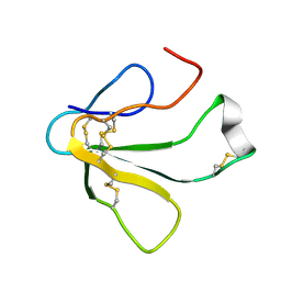 | | SOLUTION NMR STRUCTURE OF TOXIN B, A LONG NEUROTOXIN FROM THE VENOM OF THE KING COBRA, 10 STRUCTURES | | 分子名称: | TOXIN B | | 著者 | Peng, S.-S, Kumar, T.K.S, Jayaraman, G, Chang, C.-C, Yu, C. | | 登録日 | 1996-07-20 | | 公開日 | 1997-10-15 | | 最終更新日 | 2017-11-29 | | 実験手法 | SOLUTION NMR | | 主引用文献 | Solution structure of toxin b, a long neurotoxin from the venom of the king cobra (Ophiophagus hannah).
J.Biol.Chem., 272, 1997
|
|
1TXA
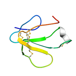 | | SOLUTION NMR STRUCTURE OF TOXIN B, A LONG NEUROTOXIN FROM THE VENOM OF THE KING COBRA, MINIMIZED AVERAGE STRUCTURE | | 分子名称: | TOXIN B | | 著者 | Peng, S.-S, Kumar, T.K.S, Jayaraman, G, Chang, C.-C, Yu, C. | | 登録日 | 1996-07-20 | | 公開日 | 1997-10-15 | | 最終更新日 | 2017-11-29 | | 実験手法 | SOLUTION NMR | | 主引用文献 | Solution structure of toxin b, a long neurotoxin from the venom of the king cobra (Ophiophagus hannah).
J.Biol.Chem., 272, 1997
|
|
6LMY
 | | Crystal structure of DUSP22 mutant_C88S/S93A | | 分子名称: | Dual specificity protein phosphatase 22, PHOSPHATE ION | | 著者 | Lai, C.H, Lyu, P.C. | | 登録日 | 2019-12-27 | | 公開日 | 2020-10-28 | | 最終更新日 | 2023-11-22 | | 実験手法 | X-RAY DIFFRACTION (1.5 Å) | | 主引用文献 | Structural Insights into the Active Site Formation of DUSP22 in N-loop-containing Protein Tyrosine Phosphatases.
Int J Mol Sci, 21, 2020
|
|
6LOT
 | | Crystal structure of DUSP22 mutant_N128D | | 分子名称: | Dual specificity protein phosphatase 22, SULFATE ION | | 著者 | Lai, C.H, Lyu, P.C. | | 登録日 | 2020-01-07 | | 公開日 | 2020-10-28 | | 最終更新日 | 2023-11-29 | | 実験手法 | X-RAY DIFFRACTION (1.69 Å) | | 主引用文献 | Structural Insights into the Active Site Formation of DUSP22 in N-loop-containing Protein Tyrosine Phosphatases.
Int J Mol Sci, 21, 2020
|
|
6LVQ
 | | Crystal structure of DUSP22_VO4 | | 分子名称: | Dual specificity protein phosphatase 22, VANADATE ION | | 著者 | Lai, C.H, Lyu, P.C. | | 登録日 | 2020-02-04 | | 公開日 | 2020-10-28 | | 最終更新日 | 2023-11-29 | | 実験手法 | X-RAY DIFFRACTION (1.38 Å) | | 主引用文献 | Structural Insights into the Active Site Formation of DUSP22 in N-loop-containing Protein Tyrosine Phosphatases.
Int J Mol Sci, 21, 2020
|
|
6LOU
 | | Crystal structure of DUSP22 mutant_C88S/S93N | | 分子名称: | Dual specificity protein phosphatase 22, PHOSPHATE ION | | 著者 | Lai, C.H, Lyu, P.C. | | 登録日 | 2020-01-07 | | 公開日 | 2020-10-28 | | 最終更新日 | 2023-11-29 | | 実験手法 | X-RAY DIFFRACTION (1.5301 Å) | | 主引用文献 | Structural Insights into the Active Site Formation of DUSP22 in N-loop-containing Protein Tyrosine Phosphatases.
Int J Mol Sci, 21, 2020
|
|
7WXH
 | | GPR domain open form of Drosophila P5CS filament with glutamate, ATP, and NADPH | | 分子名称: | Delta-1-pyrroline-5-carboxylate synthase | | 著者 | Liu, J.L, Zhong, J, Guo, C.J, Zhou, X. | | 登録日 | 2022-02-14 | | 公開日 | 2022-03-30 | | 最終更新日 | 2022-04-06 | | 実験手法 | ELECTRON MICROSCOPY (4.3 Å) | | 主引用文献 | Structural basis of dynamic P5CS filaments.
Elife, 11, 2022
|
|


