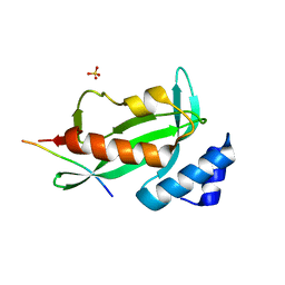
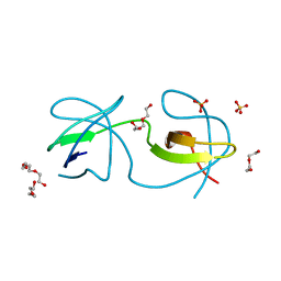
1M3X
 
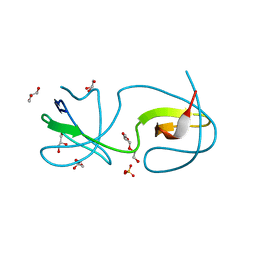
 | | Photosynthetic Reaction Center From Rhodobacter Sphaeroides | | Descriptor: | 1,2-DIACYL-SN-GLYCERO-3-PHOSPHOCHOLINE, BACTERIOCHLOROPHYLL A, BACTERIOPHEOPHYTIN A, ... | | Authors: | Camara-Artigas, A, Brune, D, Allen, J.P. | | Deposit date: | 2002-07-01 | | Release date: | 2002-08-28 | | Last modified: | 2024-02-14 | | Method: | X-RAY DIFFRACTION (2.55 Å) | | Cite: | Interactions between lipids and bacterial reaction centers determined by protein crystallography.
Proc.Natl.Acad.Sci.USA, 99, 2002
|
|
4EJE
 | |
6TG7
 | |
5ECA
 | |
4ZNX
 | |
4ZNY
 | |
4Y92
 | |
4YC1
 | |
5EC7
 | |
5DK8
 | |
2O88
 | |
4RTX
 | |
4RTU
 | |
4RTZ
 | |
4RTW
 | |
6QJJ
 | |
6QJD
 | |
6QJN
 | |
6QJI
 | |
3I9Q
 | |
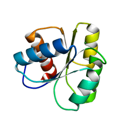
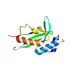
3I4W
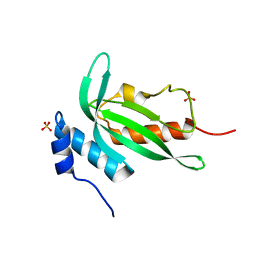
 | | Crystal Structure of the third PDZ domain of PSD-95 | | Descriptor: | ACETATE ION, Disks large homolog 4 | | Authors: | Camara-Artigas, A, Gavira, J.A. | | Deposit date: | 2009-07-03 | | Release date: | 2010-04-07 | | Last modified: | 2023-11-15 | | Method: | X-RAY DIFFRACTION (1.35 Å) | | Cite: | Novel conformational aspects of the third PDZ domain of the neuronal post-synaptic density-95 protein revealed from two 1.4A X-ray structures
J.Struct.Biol., 170, 2010
|
|
4J9E
 | |
4J9B
 | |
4J9D
 | |
7ZLX
 | |


