Loading
PDBj
MenuPDBj@FacebookPDBj@TwitterPDBj@YouTubewwPDB FoundationwwPDB
RCSB PDBPDBeBMRBAdv. SearchSearch help
Search by PDB author
4XZP
DownloadVisualize
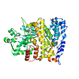
BU of 4xzp by Molmil
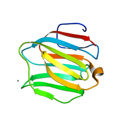
Crystal structure of the N-terminal domain of human galectin-4
Descriptor: CALCIUM ION, Galectin-4
Authors:Rustiguel, J.K, Nonato, M.C.
Deposit date:2015-02-04
Release date:2016-03-02
Last modified:2023-09-27
Method:X-RAY DIFFRACTION (1.48 Å)
Cite:Full-length model of the human galectin-4 and insights into dynamics of inter-domain communication.
Sci Rep, 6, 2016
4DJD
DownloadVisualize
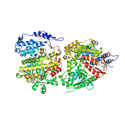
BU of 4djd by Molmil
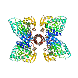
Crystal structure of folate-free corrinoid iron-sulfur protein (CFeSP) in complex with its methyltransferase (MeTr)
Descriptor: 5-methyltetrahydrofolate corrinoid/iron sulfur protein methyltransferase, CALCIUM ION, COBALAMIN, ...
Authors:Kung, Y, Doukov, T.I, Blasiak, L.C, Drennan, C.L.
Deposit date:2012-02-01
Release date:2012-03-14
Last modified:2024-02-28
Method:X-RAY DIFFRACTION (2.38 Å)
Cite:Visualizing molecular juggling within a B12-dependent methyltransferase complex.
Nature, 484, 2012
4DJF
DownloadVisualize
BU of 4djf by Molmil
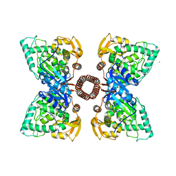
Crystal structure of folate-bound corrinoid iron-sulfur protein (CFeSP) in complex with its methyltransferase (MeTr), co-crystallized with folate and Ti(III) citrate reductant
Descriptor: 5-METHYL-5,6,7,8-TETRAHYDROFOLIC ACID, 5-methyltetrahydrofolate corrinoid/iron sulfur protein methyltransferase, CALCIUM ION, ...
Authors:Kung, Y, Drennan, C.L.
Deposit date:2012-02-01
Release date:2012-03-14
Last modified:2024-02-28
Method:X-RAY DIFFRACTION (3.03 Å)
Cite:Visualizing molecular juggling within a B12-dependent methyltransferase complex.
Nature, 484, 2012
4DJE
DownloadVisualize
BU of 4dje by Molmil
Crystal structure of folate-bound corrinoid iron-sulfur protein (CFeSP) in complex with its methyltransferase (MeTr), co-crystallized with folate
Descriptor: 5-METHYL-5,6,7,8-TETRAHYDROFOLIC ACID, 5-methyltetrahydrofolate corrinoid/iron sulfur protein methyltransferase, CALCIUM ION, ...
Authors:Kung, Y, Drennan, C.L.
Deposit date:2012-02-01
Release date:2012-03-14
Last modified:2024-02-28
Method:X-RAY DIFFRACTION (3.504 Å)
Cite:Visualizing molecular juggling within a B12-dependent methyltransferase complex.
Nature, 484, 2012
5EGQ
DownloadVisualize
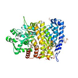
BU of 5egq by Molmil
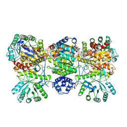
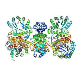
Structure of tetrameric rat phenylalanine hydroxylase mutant R270K, residues 25-453
Descriptor: Phenylalanine-4-hydroxylase, SULFATE ION
Authors:Taylor, A.B, Khan, C.A, Fitzpatrick, P.F.
Deposit date:2015-10-27
Release date:2016-05-18
Last modified:2023-09-27
Method:X-RAY DIFFRACTION (2.5 Å)
Cite:Domain Movements upon Activation of Phenylalanine Hydroxylase Characterized by Crystallography and Chromatography-Coupled Small-Angle X-ray Scattering.
J.Am.Chem.Soc., 138, 2016
5FGJ
DownloadVisualize
BU of 5fgj by Molmil
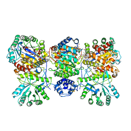
Structure of tetrameric rat phenylalanine hydroxylase, residues 1-453
Descriptor: FE (III) ION, MAGNESIUM ION, Phenylalanine-4-hydroxylase
Authors:Taylor, A.B, Roberts, K.M, Fitzpatrick, P.F.
Deposit date:2015-12-20
Release date:2016-05-18
Last modified:2023-09-27
Method:X-RAY DIFFRACTION (3.6 Å)
Cite:Domain Movements upon Activation of Phenylalanine Hydroxylase Characterized by Crystallography and Chromatography-Coupled Small-Angle X-ray Scattering.
J.Am.Chem.Soc., 138, 2016
6CGM
DownloadVisualize
BU of 6cgm by Molmil
X-ray crystal structure of Bacillus subtilis ribonucleotide reductase NrdE alpha subunit (nucleotide free)
Descriptor: 1,2-ETHANEDIOL, Ribonucleoside-diphosphate reductase
Authors:Maggiolo, A.O, Boal, A.K.
Deposit date:2018-02-20
Release date:2018-05-09
Last modified:2024-03-13
Method:X-RAY DIFFRACTION (2 Å)
Cite:An endogenous dAMP ligand inBacillus subtilisclass Ib RNR promotes assembly of a noncanonical dimer for regulation by dATP.
Proc. Natl. Acad. Sci. U.S.A., 115, 2018
6CGL
DownloadVisualize
BU of 6cgl by Molmil
X-ray crystal structure of Bacillus subtilis ribonucleotide reductase NrdE alpha subunit dAMP-bound as-isolated (pH 4)
Descriptor: 2'-DEOXYADENOSINE-5'-MONOPHOSPHATE, Ribonucleoside-diphosphate reductase, SULFATE ION
Authors:Maggiolo, A.O, Boal, A.K.
Deposit date:2018-02-20
Release date:2018-05-09
Last modified:2023-10-04
Method:X-RAY DIFFRACTION (3.2 Å)
Cite:An endogenous dAMP ligand inBacillus subtilisclass Ib RNR promotes assembly of a noncanonical dimer for regulation by dATP.
Proc. Natl. Acad. Sci. U.S.A., 115, 2018
6CGN
DownloadVisualize
BU of 6cgn by Molmil
X-ray crystal structure of Bacillus subtilis ribonucleotide reductase NrdE alpha subunit dAMP-bound (pH 7)
Descriptor: 1,2-ETHANEDIOL, 2'-DEOXYADENOSINE-5'-MONOPHOSPHATE, PHOSPHATE ION, ...
Authors:Maggiolo, A.O, Boal, A.K.
Deposit date:2018-02-20
Release date:2018-05-09
Last modified:2024-03-13
Method:X-RAY DIFFRACTION (2.26 Å)
Cite:An endogenous dAMP ligand inBacillus subtilisclass Ib RNR promotes assembly of a noncanonical dimer for regulation by dATP.
Proc. Natl. Acad. Sci. U.S.A., 115, 2018
3DQ1
DownloadVisualize
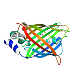
BU of 3dq1 by Molmil
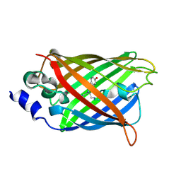
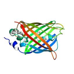
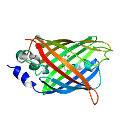
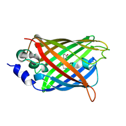
Structure of the Yellow Fluorescent Protein Citrine Frozen at 4000 Atmospheres Number 2: Structure 24 in a Series of 26 High Pressure Structures
Descriptor: Green fluorescent protein
Authors:Barstow, B, Kim, C.U.
Deposit date:2008-07-09
Release date:2008-09-23
Last modified:2023-11-15
Method:X-RAY DIFFRACTION (1.7 Å)
Cite:Alteration of citrine structure by hydrostatic pressure explains the accompanying spectral shift.
Proc.Natl.Acad.Sci.Usa, 105, 2008
3DQA
DownloadVisualize
BU of 3dqa by Molmil
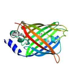
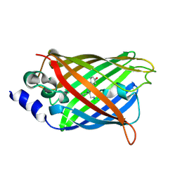
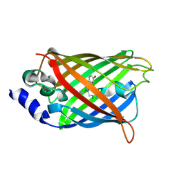
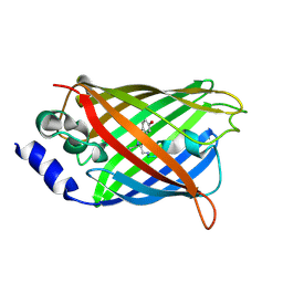
Structure of the Yellow Fluorescent Protein Citrine Frozen at 1250 Atmospheres Number 4: Structure 14 in a Series of 26 High Pressure Structures
Descriptor: Green fluorescent protein
Authors:Barstow, B, Kim, C.U.
Deposit date:2008-07-09
Release date:2008-09-23
Last modified:2023-11-15
Method:X-RAY DIFFRACTION (1.44 Å)
Cite:Alteration of citrine structure by hydrostatic pressure explains the accompanying spectral shift.
Proc.Natl.Acad.Sci.Usa, 105, 2008
3DQH
DownloadVisualize
BU of 3dqh by Molmil
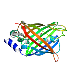
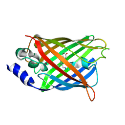
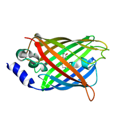
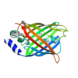
Structure of the Yellow Fluorescent Protein Citrine Frozen at 1000 Atmospheres Number 5: Structure 9 in a Series of 26 High Pressure Structures
Descriptor: Green fluorescent protein
Authors:Barstow, B, Kim, C.U.
Deposit date:2008-07-09
Release date:2008-09-23
Last modified:2023-11-15
Method:X-RAY DIFFRACTION (1.45 Å)
Cite:Alteration of citrine structure by hydrostatic pressure explains the accompanying spectral shift.
Proc.Natl.Acad.Sci.Usa, 105, 2008
3DQ9
DownloadVisualize
BU of 3dq9 by Molmil
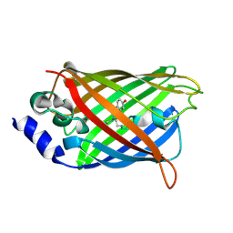
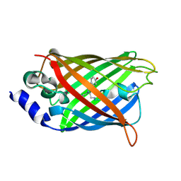
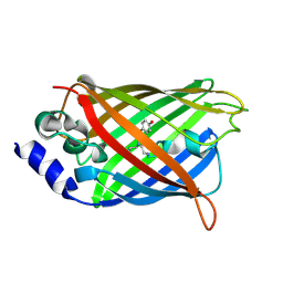
Structure of the Yellow Fluorescent Protein Citrine Frozen at 1500 Atmospheres Number 1: Structure 15 in a Series of 26 High Pressure Structures
Descriptor: Green fluorescent protein
Authors:Barstow, B, Kim, C.U.
Deposit date:2008-07-09
Release date:2008-09-23
Last modified:2023-11-15
Method:X-RAY DIFFRACTION (1.4 Å)
Cite:Alteration of citrine structure by hydrostatic pressure explains the accompanying spectral shift.
Proc.Natl.Acad.Sci.Usa, 105, 2008
3DQM
DownloadVisualize
BU of 3dqm by Molmil
Structure of the Yellow Fluorescent Protein Citrine Frozen at 750 Atmospheres: Structure 4 in a Series of 26 High Pressure Structures
Descriptor: Green fluorescent protein
Authors:Barstow, B, Kim, C.U.
Deposit date:2008-07-09
Release date:2008-09-23
Last modified:2023-11-15
Method:X-RAY DIFFRACTION (1.44 Å)
Cite:Alteration of citrine structure by hydrostatic pressure explains the accompanying spectral shift.
Proc.Natl.Acad.Sci.Usa, 105, 2008
3DQ2
DownloadVisualize
BU of 3dq2 by Molmil
Structure of the Yellow Fluorescent Protein Citrine Frozen at 4000 Atmospheres Number 1: Structure 23 in a Series of 26 High Pressure Structures
Descriptor: Green fluorescent protein
Authors:Barstow, B, Kim, C.U.
Deposit date:2008-07-09
Release date:2008-09-23
Last modified:2023-11-15
Method:X-RAY DIFFRACTION (1.6 Å)
Cite:Alteration of citrine structure by hydrostatic pressure explains the accompanying spectral shift.
Proc.Natl.Acad.Sci.Usa, 105, 2008
3DQC
DownloadVisualize
BU of 3dqc by Molmil
Structure of the Yellow Fluorescent Protein Citrine Frozen at 1250 Atmospheres Number 3: Structure 13 in a Series of 26 High Pressure Structures
Descriptor: Green fluorescent protein
Authors:Barstow, B, Kim, C.U.
Deposit date:2008-07-09
Release date:2008-09-23
Last modified:2023-11-15
Method:X-RAY DIFFRACTION (1.49 Å)
Cite:Alteration of citrine structure by hydrostatic pressure explains the accompanying spectral shift.
Proc.Natl.Acad.Sci.Usa, 105, 2008
3DQ6
DownloadVisualize
BU of 3dq6 by Molmil
Structure of the Yellow Fluorescent Protein Citrine Frozen at 1920 Atmospheres Number 2: Structure 18 in a Series of 26 High Pressure Structures
Descriptor: Green fluorescent protein
Authors:Barstow, B, Kim, C.U.
Deposit date:2008-07-09
Release date:2008-09-23
Last modified:2023-11-15
Method:X-RAY DIFFRACTION (1.6 Å)
Cite:Alteration of citrine structure by hydrostatic pressure explains the accompanying spectral shift.
Proc.Natl.Acad.Sci.Usa, 105, 2008
3DQL
DownloadVisualize
BU of 3dql by Molmil
Structure of the Yellow Fluorescent Protein Citrine Frozen at 1000 Atmospheres Number 1: Structure 5 in a Series of 26 High Pressure Structures
Descriptor: Green fluorescent protein
Authors:Barstow, B, Kim, C.U.
Deposit date:2008-07-09
Release date:2008-09-23
Last modified:2023-11-15
Method:X-RAY DIFFRACTION (1.47 Å)
Cite:Alteration of citrine structure by hydrostatic pressure explains the accompanying spectral shift.
Proc.Natl.Acad.Sci.Usa, 105, 2008
3DQ3
DownloadVisualize
BU of 3dq3 by Molmil
Structure of the Yellow Fluorescent Protein Citrine Frozen at 2500 Atmospheres: Structure 22 in a Series of 26 High Pressure Structures
Descriptor: Green fluorescent protein
Authors:Barstow, B, Kim, C.U.
Deposit date:2008-07-09
Release date:2008-09-23
Last modified:2023-11-15
Method:X-RAY DIFFRACTION (1.7 Å)
Cite:Alteration of citrine structure by hydrostatic pressure explains the accompanying spectral shift.
Proc.Natl.Acad.Sci.Usa, 105, 2008
3DQD
DownloadVisualize
BU of 3dqd by Molmil
Structure of the Yellow Fluorescent Protein Citrine Frozen at 1250 Atmospheres Number 2: Structure 12 in a Series of 26 High Pressure Structures
Descriptor: Green fluorescent protein
Authors:Barstow, B, Kim, C.U.
Deposit date:2008-07-09
Release date:2008-09-23
Last modified:2023-11-15
Method:X-RAY DIFFRACTION (1.4 Å)
Cite:Alteration of citrine structure by hydrostatic pressure explains the accompanying spectral shift.
Proc.Natl.Acad.Sci.Usa, 105, 2008
3DQN
DownloadVisualize
BU of 3dqn by Molmil
Structure of the Yellow Fluorescent Protein Citrine Frozen at 500 Atmospheres: Structure 3 in a Series of 26 High Pressure Structures
Descriptor: Green fluorescent protein
Authors:Barstow, B, Kim, C.U.
Deposit date:2008-07-09
Release date:2008-09-23
Last modified:2023-11-15
Method:X-RAY DIFFRACTION (1.44 Å)
Cite:Alteration of citrine structure by hydrostatic pressure explains the accompanying spectral shift.
Proc.Natl.Acad.Sci.Usa, 105, 2008
3DPW
DownloadVisualize
BU of 3dpw by Molmil
Structure of the Yellow Fluorescent Protein Citrine Frozen at 1 Atmosphere Number 1: Structure 1 in a Series of 26 High Pressure Structures
Descriptor: Green fluorescent protein
Authors:Barstow, B, Kim, C.U.
Deposit date:2008-07-09
Release date:2008-09-23
Last modified:2023-11-15
Method:X-RAY DIFFRACTION (1.59 Å)
Cite:Alteration of citrine structure by hydrostatic pressure explains the accompanying spectral shift.
Proc.Natl.Acad.Sci.Usa, 105, 2008
3DQ4
DownloadVisualize
BU of 3dq4 by Molmil
Structure of the Yellow Fluorescent Protein Citrine Frozen at 2000 Atmospheres Number 2: Structure 20 in a Series of 26 High Pressure Structures
Descriptor: Green fluorescent protein
Authors:Barstow, B, Kim, C.U.
Deposit date:2008-07-09
Release date:2008-09-23
Last modified:2023-11-15
Method:X-RAY DIFFRACTION (1.47 Å)
Cite:Alteration of citrine structure by hydrostatic pressure explains the accompanying spectral shift.
Proc.Natl.Acad.Sci.Usa, 105, 2008
3DQF
DownloadVisualize
BU of 3dqf by Molmil
Structure of the Yellow Fluorescent Protein Citrine Frozen at 1000 Atmospheres Number 6: Structure 10 in a Series of 26 High Pressure Structures
Descriptor: Green fluorescent protein
Authors:Barstow, B, Kim, C.U.
Deposit date:2008-07-09
Release date:2008-09-23
Last modified:2023-11-15
Method:X-RAY DIFFRACTION (1.46 Å)
Cite:Alteration of citrine structure by hydrostatic pressure explains the accompanying spectral shift.
Proc.Natl.Acad.Sci.Usa, 105, 2008
3DQO
DownloadVisualize
BU of 3dqo by Molmil
Structure of the Yellow Fluorescent Protein Citrine Frozen at 1 Atmosphere Number 2: Structure 2 in a Series of 26 High Pressure Structures
Descriptor: Green fluorescent protein
Authors:Barstow, B, Kim, C.U.
Deposit date:2008-07-09
Release date:2008-09-23
Last modified:2023-11-15
Method:X-RAY DIFFRACTION (1.5 Å)
Cite:Alteration of citrine structure by hydrostatic pressure explains the accompanying spectral shift.
Proc.Natl.Acad.Sci.Usa, 105, 2008

219140

PDB entries from 2024-05-01

PDB statisticsPDBj update infoContact PDBjnumon