Loading
PDBj
MenuPDBj@FacebookPDBj@TwitterPDBj@YouTubewwPDB FoundationwwPDB
RCSB PDBPDBeBMRBAdv. SearchSearch help
Search by PDB author
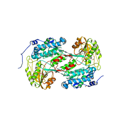
4O5O
DownloadVisualize
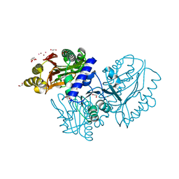
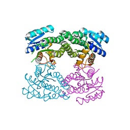
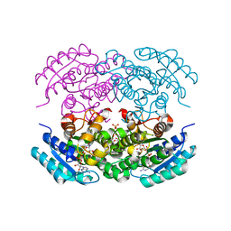
BU of 4o5o by Molmil
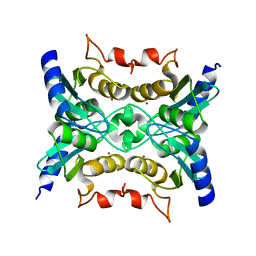
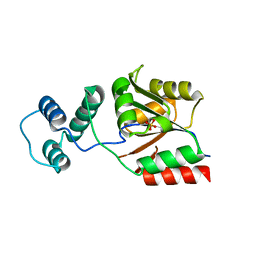
X-ray Crystal Structure of a 3-hydroxyacyl-CoA dehydrogenase from Brucella suis
Descriptor: 1,2-ETHANEDIOL, 2-AMINO-2-HYDROXYMETHYL-PROPANE-1,3-DIOL, 3-hydroxyacyl-CoA dehydrogenase, ...
Authors:Seattle Structural Genomics Center for Infectious Disease (SSGCID)
Deposit date:2013-12-19
Release date:2014-01-01
Last modified:2024-02-28
Method:X-RAY DIFFRACTION (1.4 Å)
Cite:X-ray Crystal Structure of a 3-hydroxyacyl-CoA dehydrogenase from Brucella suis
TO BE PUBLISHED
4O5H
DownloadVisualize
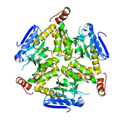
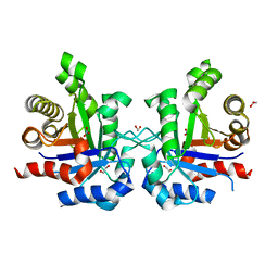
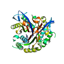
BU of 4o5h by Molmil
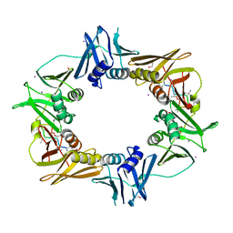
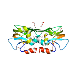
X-ray crystal structure of a putative phenylacetaldehyde dehydrogenase from Burkholderia cenocepacia
Descriptor: 1,2-ETHANEDIOL, GLYCEROL, Phenylacetaldehyde dehydrogenase, ...
Authors:Seattle Structural Genomics Center for Infectious Disease (SSGCID)
Deposit date:2013-12-19
Release date:2014-02-19
Last modified:2023-09-20
Method:X-RAY DIFFRACTION (2 Å)
Cite:X-ray crystal structure of a putative phenylacetaldehyde dehydrogenase from Burkholderia cenocepacia
TO BE PUBLISHED
4F3W
DownloadVisualize
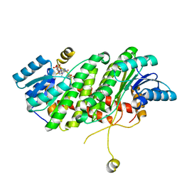
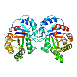
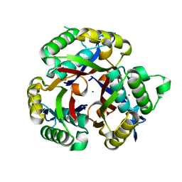
BU of 4f3w by Molmil
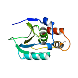
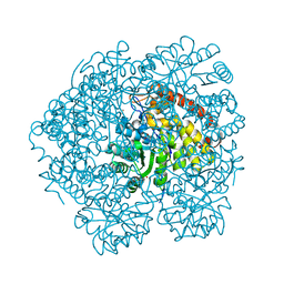
Crystal structure of cytidine deaminase Cdd from Mycobacterium marinum
Descriptor: Cytidine deaminase Cdd, ZINC ION
Authors:Seattle Structural Genomics Center for Infectious Disease (SSGCID)
Deposit date:2012-05-09
Release date:2012-05-23
Last modified:2023-09-13
Method:X-RAY DIFFRACTION (1.8 Å)
Cite:Increasing the structural coverage of tuberculosis drug targets.
Tuberculosis (Edinb), 95, 2015
6PTR
DownloadVisualize
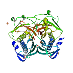
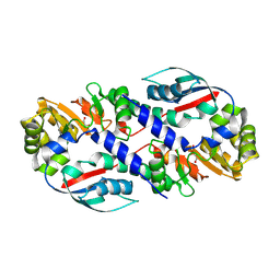
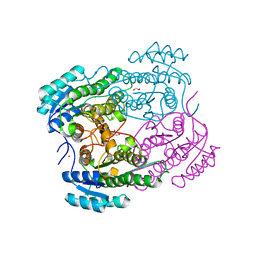
BU of 6ptr by Molmil
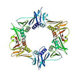
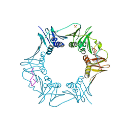
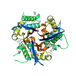
Crystal structure of a DnaN sliding clamp (DNA polymerase III subunit beta) from Bartonella birtlesii bound to griselimycin
Descriptor: 1,2-ETHANEDIOL, ACE-MVA-MP8-NZC-LEU-MP8-LEU-MVA-PRO-MLU-GLY, Beta sliding clamp, ...
Authors:Seattle Structural Genomics Center for Infectious Disease (SSGCID)
Deposit date:2019-07-16
Release date:2019-08-07
Last modified:2023-11-15
Method:X-RAY DIFFRACTION (1.75 Å)
Cite:Crystal structure of a DnaN sliding clamp (DNA polymerase III subunit beta) from Bartonella birtlesii bound to griselimycin
To Be Published
6Q10
DownloadVisualize
BU of 6q10 by Molmil
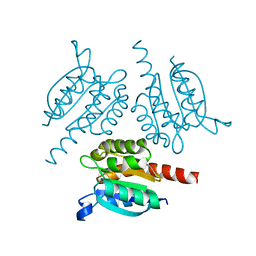
Crystal structure of the soluble domain (residues 71-217) of a conserved hypothetical secreted protein (Rv2700 ortholog) from Mycobacterium marinum
Descriptor: 1,2-ETHANEDIOL, MymaA.19257.a.B3
Authors:Seattle Structural Genomics Center for Infectious Disease (SSGCID)
Deposit date:2019-08-02
Release date:2019-08-14
Method:X-RAY DIFFRACTION (1.6 Å)
Cite:SSGCID, Mycobacerium marinum, Structural Genomics, Seattle Structural Genomics Center for Infectious Disease, secreted protein, Rv2700 ortholog
to be published
6PTV
DownloadVisualize
BU of 6ptv by Molmil
Crystal structure of a DnaN sliding clamp (DNA polymerase III subunit beta) from Rickettsia rickettsii bound to griselimycin
Descriptor: (4S)-2-METHYL-2,4-PENTANEDIOL, ACE-MVA-MP8-NZC-LEU-MP8-LEU-MVA-PRO-MLU-GLY, Beta sliding clamp
Authors:Seattle Structural Genomics Center for Infectious Disease (SSGCID)
Deposit date:2019-07-16
Release date:2019-08-07
Last modified:2023-11-15
Method:X-RAY DIFFRACTION (1.85 Å)
Cite:Crystal structure of a DnaN sliding clamp (DNA polymerase III subunit beta) from Rickettsia rickettsii bound to griselimycin
To Be Published
6PTH
DownloadVisualize
BU of 6pth by Molmil
Crystal structure of a DnaN sliding clamp (DNA polymerase III subunit beta) from Pseudomonas aeruginosa bound to griselimycin
Descriptor: Beta sliding clamp, Griselimycin, SULFATE ION
Authors:Seattle Structural Genomics Center for Infectious Disease (SSGCID)
Deposit date:2019-07-15
Release date:2019-08-07
Last modified:2023-11-15
Method:X-RAY DIFFRACTION (3.05 Å)
Cite:Crystal structure of a DnaN sliding clamp (DNA polymerase III subunit beta) from Pseudomonas aeruginosa bound to griselimycin
To Be Published
3TCR
DownloadVisualize
BU of 3tcr by Molmil
Crystal structure of a molybdopterin biosynthesis protein from Mycobacterium abscessus
Descriptor: Molybdopterin biosynthesis protein
Authors:Seattle Structural Genomics Center for Infectious Disease (SSGCID)
Deposit date:2011-08-09
Release date:2011-08-24
Last modified:2023-09-13
Method:X-RAY DIFFRACTION (2.3 Å)
Cite:Increasing the structural coverage of tuberculosis drug targets.
Tuberculosis (Edinb), 95, 2015
3TAV
DownloadVisualize
BU of 3tav by Molmil
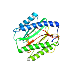
Crystal structure of a Methionine Aminopeptidase from Mycobacterium abscessus
Descriptor: (2S)-2-hydroxybutanedioic acid, CHLORIDE ION, D-MALATE, ...
Authors:Seattle Structural Genomics Center for Infectious Disease (SSGCID)
Deposit date:2011-08-04
Release date:2011-08-17
Last modified:2024-02-28
Method:X-RAY DIFFRACTION (2.15 Å)
Cite:Increasing the structural coverage of tuberculosis drug targets.
Tuberculosis (Edinb), 95, 2015
4EMD
DownloadVisualize
BU of 4emd by Molmil
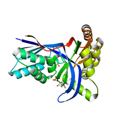
Crystal structure of IspE (4-diphosphocytidyl-2-C-methyl-D-erythritol kinase) from Mycobacterium abcessus, bound to CMP and SO4
Descriptor: 4-diphosphocytidyl-2-C-methyl-D-erythritol kinase, CYTIDINE-5'-MONOPHOSPHATE, SULFATE ION
Authors:Seattle Structural Genomics Center for Infectious Disease (SSGCID)
Deposit date:2012-04-11
Release date:2012-04-25
Last modified:2023-09-13
Method:X-RAY DIFFRACTION (1.75 Å)
Cite:Increasing the structural coverage of tuberculosis drug targets.
Tuberculosis (Edinb), 95, 2015
4EGE
DownloadVisualize
BU of 4ege by Molmil
Crystal Structure of Dipeptidase PepE from Mycobacterium ulcerans
Descriptor: 1,2-ETHANEDIOL, Dipeptidase PepE, UNKNOWN ATOM OR ION, ...
Authors:Seattle Structural Genomics Center for Infectious Disease (SSGCID)
Deposit date:2012-03-30
Release date:2012-04-25
Last modified:2023-09-13
Method:X-RAY DIFFRACTION (2.2 Å)
Cite:Increasing the structural coverage of tuberculosis drug targets.
Tuberculosis (Edinb), 95, 2015
3TLF
DownloadVisualize
BU of 3tlf by Molmil
Crystal structure of an enoyl-CoA hydratase/isomerase from Mycobacterium paratuberculosis
Descriptor: 1,2-ETHANEDIOL, enoyl-CoA hydratase/isomerase
Authors:Seattle Structural Genomics Center for Infectious Disease (SSGCID)
Deposit date:2011-08-29
Release date:2011-10-05
Last modified:2023-09-13
Method:X-RAY DIFFRACTION (2.15 Å)
Cite:Increasing the structural coverage of tuberculosis drug targets.
Tuberculosis (Edinb), 95, 2015
3TJR
DownloadVisualize
BU of 3tjr by Molmil
Crystal structure of a Rv0851c ortholog short chain dehydrogenase from Mycobacterium paratuberculosis
Descriptor: UNKNOWN LIGAND, short chain dehydrogenase
Authors:Seattle Structural Genomics Center for Infectious Disease (SSGCID)
Deposit date:2011-08-24
Release date:2011-08-31
Last modified:2023-09-13
Method:X-RAY DIFFRACTION (1.6 Å)
Cite:Increasing the structural coverage of tuberculosis drug targets.
Tuberculosis (Edinb), 95, 2015
3U0A
DownloadVisualize
BU of 3u0a by Molmil
Crystal structure of an Acyl-CoA thioesterase II TesB2 from Mycobacterium marinum
Descriptor: Acyl-CoA thioesterase II TesB2, GLYCEROL, SODIUM ION, ...
Authors:Arakaki, T.L, Staker, B.L, Clifton, M.C, Sankaran, B, Seattle Structural Genomics Center for Infectious Disease (SSGCID)
Deposit date:2011-09-28
Release date:2011-10-05
Last modified:2023-09-13
Method:X-RAY DIFFRACTION (2.5 Å)
Cite:Increasing the structural coverage of tuberculosis drug targets.
Tuberculosis (Edinb), 95, 2015
3TL3
DownloadVisualize
BU of 3tl3 by Molmil
Structure of a short-chain type dehydrogenase/reductase from Mycobacterium ulcerans
Descriptor: SODIUM ION, Short-chain type dehydrogenase/reductase
Authors:Seattle Structural Genomics Center for Infectious Disease (SSGCID)
Deposit date:2011-08-29
Release date:2011-09-07
Last modified:2023-09-13
Method:X-RAY DIFFRACTION (1.85 Å)
Cite:Increasing the structural coverage of tuberculosis drug targets.
Tuberculosis (Edinb), 95, 2015
3KXQ
DownloadVisualize
BU of 3kxq by Molmil
Crystal structure of triosephosphate isomerase from bartonella henselae at 1.6A resolution
Descriptor: 1,2-ETHANEDIOL, NITRATE ION, Triosephosphate isomerase
Authors:Seattle Structural Genomics Center for Infectious Disease (SSGCID)
Deposit date:2009-12-03
Release date:2009-12-22
Last modified:2023-09-06
Method:X-RAY DIFFRACTION (1.6 Å)
Cite:Crystal structure of triosephosphate isomerase from bartonella henselae at 1.6A resolution
To be Published
3KRS
DownloadVisualize
BU of 3krs by Molmil
Structure of Triosephosphate Isomerase from Cryptosporidium Parvum at 1.55A Resolution
Descriptor: SODIUM ION, Triosephosphate isomerase, UNKNOWN ATOM OR ION
Authors:Seattle Structural Genomics Center for Infectious Disease (SSGCID)
Deposit date:2009-11-19
Release date:2009-12-01
Last modified:2023-09-06
Method:X-RAY DIFFRACTION (1.55 Å)
Cite:Structure of triosephosphate isomerase from Cryptosporidium parvum.
Acta Crystallogr.,Sect.F, 67, 2011
3L0G
DownloadVisualize
BU of 3l0g by Molmil
Crystal structure of Nicotinate-nucleotide pyrophosphorylase from Ehrlichia chaffeensis at 2.05A resolution
Descriptor: 1,2-ETHANEDIOL, FORMIC ACID, Nicotinate-nucleotide pyrophosphorylase
Authors:Seattle Structural Genomics Center for Infectious Disease (SSGCID)
Deposit date:2009-12-09
Release date:2009-12-22
Last modified:2023-09-06
Method:X-RAY DIFFRACTION (2.05 Å)
Cite:Crystal structure of Nicotinate-nucleotide pyrophosphorylase from Ehrlichia chaffeensis at 2.05A resolution
To be Published
3KZX
DownloadVisualize
BU of 3kzx by Molmil
Crystal structure of a Had-superfamily hydrolase from Ehrlichia chaffeensis at 1.9A resolution
Descriptor: HAD-superfamily hydrolase, subfamily IA, variant 1, ...
Authors:Seattle Structural Genomics Center for Infectious Disease (SSGCID)
Deposit date:2009-12-08
Release date:2009-12-22
Last modified:2023-09-06
Method:X-RAY DIFFRACTION (1.9 Å)
Cite:Crystal structure of a Had-superfamily hydrolase from Ehrlichia chaffeensis at 1.9A resolution
To be Published
3TZU
DownloadVisualize
BU of 3tzu by Molmil
Crystal structure of a glycine cleavage system H protein (GCVH) from Mycobacterium marinum
Descriptor: 1,2-ETHANEDIOL, ACETATE ION, DI(HYDROXYETHYL)ETHER, ...
Authors:Seattle Structural Genomics Center for Infectious Disease (SSGCID)
Deposit date:2011-09-27
Release date:2011-10-05
Last modified:2023-09-13
Method:X-RAY DIFFRACTION (2.3 Å)
Cite:Increasing the structural coverage of tuberculosis drug targets.
Tuberculosis (Edinb), 95, 2015
4O0K
DownloadVisualize
BU of 4o0k by Molmil
Crystal structure of 1-pyrroline-4-hydroxy-2-carboxylate deaminase from Brucella melitensis with covalently bound substrate
Descriptor: 1,2-ETHANEDIOL, Dihydrodipicolinate synthetase
Authors:Seattle Structural Genomics Center for Infectious Disease (SSGCID)
Deposit date:2013-12-13
Release date:2014-01-15
Last modified:2023-11-29
Method:X-RAY DIFFRACTION (1.5 Å)
Cite:Crystal structure of 1-pyrroline-4-hydroxy-2-carboxylate deaminase from Brucella melitensis with covalently bound substrate
To be Published
3F0G
DownloadVisualize
BU of 3f0g by Molmil
Co-crystal structure of 2C-methyl-D-erythritol 2,4-cyclodiphosphate synthase with CMP
Descriptor: 2-C-methyl-D-erythritol 2,4-cyclodiphosphate synthase, CYTIDINE-5'-MONOPHOSPHATE, ZINC ION
Authors:Seattle Structural Genomics Center for Infectious Disease (SSGCID)
Deposit date:2008-10-24
Release date:2008-11-04
Last modified:2023-09-06
Method:X-RAY DIFFRACTION (2.08 Å)
Cite:Leveraging structure determination with fragment screening for infectious disease drug targets: MECP synthase from Burkholderia pseudomallei.
J Struct Funct Genomics, 12, 2011
5EJ2
DownloadVisualize
BU of 5ej2 by Molmil
Crystal structure of Carveol dehydrogenase from Mycobacterium avium in complex with NAD
Descriptor: (4S)-2-METHYL-2,4-PENTANEDIOL, Carveol dehydrogenase, NICOTINAMIDE-ADENINE-DINUCLEOTIDE, ...
Authors:Seattle Structural Genomics Center for Infectious Disease (SSGCID)
Deposit date:2015-10-31
Release date:2015-12-02
Last modified:2023-09-27
Method:X-RAY DIFFRACTION (2.15 Å)
Cite:Mycofactocin-associated mycobacterial dehydrogenases with non-exchangeable NAD cofactors.
Sci Rep, 7, 2017
3F0D
DownloadVisualize
BU of 3f0d by Molmil
High resolution crystal structure of 2C-methyl-D-erythritol 2,4-cyclodiphosphatase synthase from Burkholderia pseudomallei
Descriptor: 2-C-methyl-D-erythritol 2,4-cyclodiphosphate synthase, ZINC ION
Authors:Seattle Structural Genomics Center for Infectious Disease (SSGCID)
Deposit date:2008-10-24
Release date:2008-11-04
Last modified:2023-12-27
Method:X-RAY DIFFRACTION (1.2 Å)
Cite:Leveraging structure determination with fragment screening for infectious disease drug targets: MECP synthase from Burkholderia pseudomallei.
J Struct Funct Genomics, 12, 2011
3F0E
DownloadVisualize
BU of 3f0e by Molmil
Crystal structure of 2C-methyl-D-erythritol 2,4-cyclodiphosphate synthase from Burkholderia pseudomallei
Descriptor: 2-C-methyl-D-erythritol 2,4-cyclodiphosphate synthase, MAGNESIUM ION, ZINC ION
Authors:Seattle Structural Genomics Center for Infectious Disease (SSGCID)
Deposit date:2008-10-24
Release date:2008-11-04
Last modified:2023-12-27
Method:X-RAY DIFFRACTION (2.05 Å)
Cite:Leveraging structure determination with fragment screening for infectious disease drug targets: MECP synthase from Burkholderia pseudomallei.
J Struct Funct Genomics, 12, 2011

219140

PDB entries from 2024-05-01

PDB statisticsPDBj update infoContact PDBjnumon