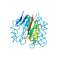
Loading
PDBj
MenuPDBj@FacebookPDBj@TwitterPDBj@YouTubewwPDB FoundationwwPDB
RCSB PDBPDBeBMRBAdv. SearchSearch help
Search by PDB author
2RHE
DownloadVisualize
BU of 2rhe by Molmil
STRUCTURE OF A NOVEL BENCE-JONES PROTEIN (RHE) FRAGMENT AT 1.6 ANGSTROMS RESOLUTION
Descriptor: BENCE-JONES PROTEIN RHE (LIGHT CHAIN)
Authors:Fureyjunior, W, Wang, B.C, Yoo, C.S, Sax, M.
Deposit date:1983-06-13
Release date:1983-09-15
Last modified:2017-11-29
Method:X-RAY DIFFRACTION (1.6 Å)
Cite:Structure of a novel Bence-Jones protein (Rhe) fragment at 1.6 A resolution.
J.Mol.Biol., 167, 1983
1SL8
DownloadVisualize
BU of 1sl8 by Molmil
Calcium-loaded apo-aequorin from Aequorea victoria
Descriptor: Aequorin 1, CALCIUM ION
Authors:Deng, L, Markova, S.V, Vysotski, E.S, Liu, Z.J, Lee, J, Rose, J, Wang, B.C, Southeast Collaboratory for Structural Genomics (SECSG)
Deposit date:2004-03-05
Release date:2004-12-28
Last modified:2023-11-29
Method:X-RAY DIFFRACTION (1.7 Å)
Cite:All three Ca2+-binding loops of photoproteins bind calcium ions: The crystal structures of calcium-loaded apo-aequorin and apo-obelin.
Protein Sci., 14, 2005
1SL7
DownloadVisualize
BU of 1sl7 by Molmil
Crystal structure of calcium-loaded apo-obelin from Obelia longissima
Descriptor: CALCIUM ION, Obelin
Authors:Deng, L, Markova, S.V, Vysotski, E.S, Liu, Z.J, Lee, J, Rose, J, Wang, B.C, Southeast Collaboratory for Structural Genomics (SECSG)
Deposit date:2004-03-05
Release date:2004-12-28
Last modified:2023-08-23
Method:X-RAY DIFFRACTION (2.2 Å)
Cite:All three Ca2+-binding loops of photoproteins bind calcium ions: The crystal structures of calcium-loaded apo-aequorin and apo-obelin.
Protein Sci., 14, 2005
4WBX
DownloadVisualize
BU of 4wbx by Molmil
Conserved hypothetical protein PF1771 from Pyrococcus furiosus solved by sulfur SAD using Swiss Light Source data
Descriptor: 2-keto acid:ferredoxin oxidoreductase subunit alpha
Authors:Weinert, T, Waltersperger, S, Olieric, V, Panepucci, E, Chen, L, Rose, J.P, Wang, M, Wang, B.C, Southeast Collaboratory for Structural Genomics (SECSG)
Deposit date:2014-09-04
Release date:2014-12-10
Last modified:2023-12-27
Method:X-RAY DIFFRACTION (2.301 Å)
Cite:Fast native-SAD phasing for routine macromolecular structure determination.
Nat.Methods, 12, 2015
1SGW
DownloadVisualize
BU of 1sgw by Molmil
Putative ABC transporter (ATP-binding protein) from Pyrococcus furiosus Pfu-867808-001
Descriptor: CHLORIDE ION, SODIUM ION, putative ABC transporter
Authors:Liu, Z.J, Tempel, W, Shah, A, Chen, L, Lee, D, Kelley, L.-L.C, Dillard, B.D, Rose, J.P, Eneh, J.C, Hopkins, R.C, Jenney Jr, F.E, Lee, H.S, Poole II, F.L, Shah, C, Sugar, F.J, Adams, M.W.W, Richardson, D.C, Richardson, J.S, Wang, B.C, Southeast Collaboratory for Structural Genomics (SECSG)
Deposit date:2004-02-24
Release date:2004-08-10
Last modified:2024-02-14
Method:X-RAY DIFFRACTION (1.7 Å)
Cite:Putative ABC transporter (ATP-binding protein) from Pyrococcus furiosus Pfu-867808-001
To be Published
2F8P
DownloadVisualize
BU of 2f8p by Molmil
Crystal structure of obelin following Ca2+ triggered bioluminescence suggests neutral coelenteramide as the primary excited state
Descriptor: CALCIUM ION, N-[3-BENZYL-5-(4-HYDROXYPHENYL)PYRAZIN-2-YL]-2-(4-HYDROXYPHENYL)ACETAMIDE, Obelin
Authors:Liu, Z.J, Stepanyuk, G.A, Vysotski, E.S, Lee, J, Wang, B.C, Southeast Collaboratory for Structural Genomics (SECSG)
Deposit date:2005-12-03
Release date:2006-02-14
Last modified:2023-08-30
Method:X-RAY DIFFRACTION (1.93 Å)
Cite:Crystal structure of obelin after Ca2+-triggered bioluminescence suggests neutral coelenteramide as the primary excited state.
Proc.Natl.Acad.Sci.Usa, 103, 2006
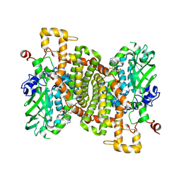
2BN2
DownloadVisualize
BU of 2bn2 by Molmil
CRYSTAL STRUCTURE OF BOVINE NEUROPHYSIN II COMPLEXED WITH THE VASOPRESSIN ANALOGUE PHE-TYR AMIDE
Descriptor: NEUROPHYSIN II, PHENYLALANINE, TYROSINE
Authors:Rose, J.P, Wang, B.C.
Deposit date:1998-12-18
Release date:1999-02-16
Last modified:2024-06-05
Method:X-RAY DIFFRACTION (2.8 Å)
Cite:Crystal structure of a bovine neurophysin II dipeptide complex at 2.8 A determined from the single-wavelength anomalous scattering signal of an incorporated iodine atom.
Proc.Natl.Acad.Sci.USA, 88, 1991
2I0X
DownloadVisualize
BU of 2i0x by Molmil
Hypothetical protein PF1117 from Pyrococcus furiosus
Descriptor: Hypothetical protein PF1117
Authors:Chen, L.Q, Fu, Z.-Q, Liu, Z.-J, Rose, J.P, Wang, B.C, Southeast Collaboratory for Structural Genomics (SECSG)
Deposit date:2006-08-11
Release date:2006-10-10
Last modified:2024-02-21
Method:X-RAY DIFFRACTION (2.7 Å)
Cite:Crystal Structure of Hypothetical Protein Pf1117 from Pyrococcus furiosus
To be Published
3R7C
DownloadVisualize
BU of 3r7c by Molmil
The structure of a hexahestidine-tagged form of augmenter of liver regeneration reveals a novel Cd(2)Cl(4)O(6) cluster that aids in crystal packing
Descriptor: CADMIUM ION, CHLORIDE ION, FAD-linked sulfhydryl oxidase ALR, ...
Authors:Florence, Q.J, Wu, C.-K, Swindell II, J.T, Wang, B.C, Rose, J.P.
Deposit date:2011-03-22
Release date:2012-03-28
Last modified:2017-11-08
Method:X-RAY DIFFRACTION (2.4 Å)
Cite:The structure of a hexahestidine-tagged form of augmenter of liver regeneration reveals a novel Cd(2)Cl(4)O(6) cluster that aids in crystal packing
To be Published
1EL4
DownloadVisualize
BU of 1el4 by Molmil
STRUCTURE OF THE CALCIUM-REGULATED PHOTOPROTEIN OBELIN DETERMINED BY SULFUR SAS
Descriptor: C2-HYDROXY-COELENTERAZINE, CHLORIDE ION, OBELIN
Authors:Liu, Z.J, Vysotski, E.S, Rose, J, Lee, J, Wang, B.C.
Deposit date:2000-03-13
Release date:2001-03-13
Last modified:2024-02-07
Method:X-RAY DIFFRACTION (1.73 Å)
Cite:Structure of the Ca2+-regulated photoprotein obelin at 1.7 A resolution determined directly from its sulfur substructure.
Protein Sci., 9, 2000
1VK1
DownloadVisualize
BU of 1vk1 by Molmil
Conserved hypothetical protein from Pyrococcus furiosus Pfu-392566-001
Descriptor: Conserved hypothetical protein, DI(HYDROXYETHYL)ETHER, PHOSPHATE ION, ...
Authors:Shah, A, Liu, Z.J, Tempel, W, Chen, L, Lee, D, Yang, H, Chang, J, Zhao, M, Ng, J, Rose, J, Brereton, P.S, Izumi, M, Jenney Jr, F.E, Poole II, F.L, Shah, C, Sugar, F.J, Adams, M.W.W, Richardson, D.C, Richardson, J.S, Wang, B.C, Southeast Collaboratory for Structural Genomics (SECSG)
Deposit date:2004-04-13
Release date:2004-08-10
Last modified:2023-12-27
Method:X-RAY DIFFRACTION (1.2 Å)
Cite:(NZ)CH...O contacts assist crystallization of a ParB-like nuclease.
Bmc Struct.Biol., 7, 2007
9BJF
DownloadVisualize
BU of 9bjf by Molmil
Tungstate binding protein (Tungbindin) from Eubacterium limosum in apo form
Descriptor: Molybdenum-pterin binding domain-containing protein, SULFATE ION
Authors:Zhou, D, Rose, J.P, Chen, L, Wang, B.C.
Deposit date:2024-04-25
Release date:2024-09-04
Method:X-RAY DIFFRACTION (2.89 Å)
Cite:Tungstate binding protein (Tungbindin) from Eubacterium limosum in apo form
To Be Published
9BKA
DownloadVisualize
BU of 9bka by Molmil
Crystal structure of Rid family protein PSPTO3006
Descriptor: Rid family protein PSPTO3006
Authors:Zhou, D, Chen, L, Rose, J.P, Wang, B.C.
Deposit date:2024-04-26
Release date:2024-09-04
Method:X-RAY DIFFRACTION (1.86 Å)
Cite:Crystal structure of Rid family protein PSPTO3006
To Be Published
9BKB
DownloadVisualize
BU of 9bkb by Molmil
Crystal structure of Rid family protein ACIAD3089 from Acinetobacter baylyi
Descriptor: Rid family protein ACIAD3089
Authors:Zhou, D, Chen, L, Rose, J.P, Wang, B.C.
Deposit date:2024-04-26
Release date:2024-09-04
Method:X-RAY DIFFRACTION (2.1 Å)
Cite:Crystal structure of Rid family protein ACIAD3089 from Acinetobacter baylyi
To Be Published
9BKH
DownloadVisualize
BU of 9bkh by Molmil
Crystal structure of RidA family protein PA5083 from Pseudomonas aeruginosa with Acetaldehyde bound
Descriptor: ACETYL GROUP, RidA family protein PA5083
Authors:Zhou, D, Chen, L, Rose, J.P, Wang, B.C.
Deposit date:2024-04-28
Release date:2024-09-04
Method:X-RAY DIFFRACTION (1.67 Å)
Cite:Crystal structure of RidA family protein PA5083 from Pseudomonas aeruginosa with Acetaldehyde bound
To Be Published
9BKE
DownloadVisualize
BU of 9bke by Molmil
STRUCTURE OF 4-HYDROXYPHENYLACETATE 3-MONOOXYGENASE (HPAB), OXYGENASE COMPONENT FROM ESCHERICHIA COLI MUTANT XS6 WITH AMP BOUND
Descriptor: 4-hydroxyphenylacetate 3-monooxygenase oxygenase component, ADENOSINE MONOPHOSPHATE
Authors:Zhou, D, Chen, L, Rose, J.P, Wang, B.C.
Deposit date:2024-04-27
Release date:2024-09-04
Method:X-RAY DIFFRACTION (1.95 Å)
Cite:STRUCTURE OF 4-HYDROXYPHENYLACETATE 3-MONOOXYGENASE (HPAB), OXYGENASE COMPONENT FROM ESCHERICHIA COLI MUTANT XS6 WITH AMP BOUND
To Be Published
9BJV
DownloadVisualize
BU of 9bjv by Molmil
Crystal structure of hypothetical protein PA5083 from Pseudomonas aeruginosa Sulfur SAD phased
Descriptor: RidA family protein PA5083
Authors:Zhou, D, Chen, L, Rose, J.P, Wang, B.C.
Deposit date:2024-04-25
Release date:2024-09-04
Method:X-RAY DIFFRACTION (1.8 Å)
Cite:Structure of the Rid protein PA5083 from Pseudomonas aeruginosa (Apo form) solved by Sulfur SAD
To Be Published
9BKG
DownloadVisualize
BU of 9bkg by Molmil
Crystal structure of selenomethionine labeled bovine trypsin mutant - S195A solved by Sulphur-SAD at 1.54A wavelength
Descriptor: Serine protease 1
Authors:Zhou, D, Chen, L, Rose, J.P, Wang, B.C.
Deposit date:2024-04-27
Release date:2024-06-05
Method:X-RAY DIFFRACTION (1.57 Å)
Cite:Crystal structure of selenomethionine labeled bovine trypsin mutant - S195A solved by Sulphur-SAD at 1.54A wavelength
To Be Published
9BK4
DownloadVisualize
BU of 9bk4 by Molmil
Crystal structure of RidA family protein PA5083 from Pseudomonas aeruginosa with pyruvic acid bound
Descriptor: PYRUVIC ACID, RidA family protein PA5083, SULFATE ION
Authors:Zhou, D, Chen, L, Rose, J.P, Wang, B.C.
Deposit date:2024-04-26
Release date:2024-09-04
Method:X-RAY DIFFRACTION (2.08 Å)
Cite:Crystal structure of RidA family protein PA5083 from Pseudomonas aeruginosa with pyruvic acid bound
To Be Published
9BED
DownloadVisualize
BU of 9bed by Molmil
Tungstate binding protein (Tungbindin) from Eubacterium limosum with eight molybdates bound
Descriptor: MOLYBDATE ION, Molybdenum-pterin-binding protein
Authors:Zhou, D, Rose, J.P, Chen, L, Wang, B.C.
Deposit date:2024-04-15
Release date:2024-09-04
Method:X-RAY DIFFRACTION (2.02 Å)
Cite:Tungstate binding protein (Tungbindin) from Eubacterium limosum with eight molybdates bound
To Be Published
9D2C
DownloadVisualize
BU of 9d2c by Molmil
Crystal Structure of Tungbindin Treated with Proteinase K
Descriptor: MOLYBDATE ION, Molybdenum-pterin binding domain-containing protein
Authors:Zhou, D, Chen, L, Rose, J.P, Wang, B.C.
Deposit date:2024-08-08
Release date:2024-09-04
Method:X-RAY DIFFRACTION (2.38 Å)
Cite:Crystal structure of Tungbindin treated with proteinase K
To Be Published
9BJW
DownloadVisualize
BU of 9bjw by Molmil
Crystal structure of RidA family protein PA5083 from Pseudomonas aeruginosa
Descriptor: RidA family protein PA5083
Authors:Zhou, D, Chen, L, Rose, J.P, Wang, B.C.
Deposit date:2024-04-25
Release date:2024-09-04
Method:X-RAY DIFFRACTION (1.54 Å)
Cite:Structure of the Rid protein PA5083 from Pseudomonas aeruginosa (Apo form)
To Be Published
9BK1
DownloadVisualize
BU of 9bk1 by Molmil
Crystal structure of Endoribonuclease L-PSP family protein PSPTO0102 Sulfur SAD phased
Descriptor: L-PSP family protein PSPTO_0102
Authors:Zhou, D, Chen, L, Rose, J.P, Wang, B.C.
Deposit date:2024-04-26
Release date:2024-09-04
Method:X-RAY DIFFRACTION (1.57 Å)
Cite:Crystal structure of Endoribonuclease L-PSP family protein PSPTO0102 Sulfur SAD phased
To Be Published
9BK8
DownloadVisualize
BU of 9bk8 by Molmil
Crystal structure of RidA family protein PA5083 from Pseudomonas aeruginosa with 2-ketobutyric acid bound
Descriptor: 2-KETOBUTYRIC ACID, RidA family protein PA5083
Authors:Zhou, D, Chen, L, Rose, J.P, Wang, B.C.
Deposit date:2024-04-26
Release date:2024-09-04
Method:X-RAY DIFFRACTION (1.67 Å)
Cite:Crystal structure of RidA family protein PA5083 from Pseudomonas aeruginosa with 2-ketobutyric acid bound
To Be Published
9BJY
DownloadVisualize
BU of 9bjy by Molmil
Crystal structure of RidA family protein PA5083 from Pseudomonas aeruginosa with TMAO bound
Descriptor: RidA family protein, SULFATE ION, trimethylamine oxide
Authors:Zhou, D, Chen, L, Rose, J.P, Wang, B.C.
Deposit date:2024-04-26
Release date:2024-09-04
Method:X-RAY DIFFRACTION (1.58 Å)
Cite:Structure of the RidA protein PA5083 from Pseudomonas aeruginosa with TMAO bound
To Be Published

226262

PDB entries from 2024-10-16

PDB statisticsPDBj update infoContact PDBjnumon