Loading
PDBj
MenuPDBj@FacebookPDBj@TwitterPDBj@YouTubewwPDB FoundationwwPDB
RCSB PDBPDBeBMRBAdv. SearchSearch help
Search by PDB author
1LS1
DownloadVisualize
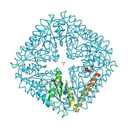
BU of 1ls1 by Molmil
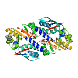
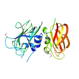
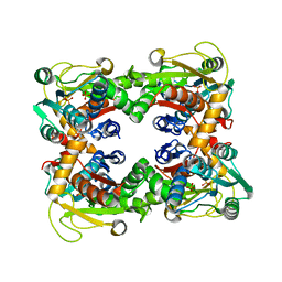
T. aquaticus Ffh NG Domain at 1.1A Resolution
Descriptor: MAGNESIUM ION, OXYGEN MOLECULE, SIGNAL RECOGNITION PARTICLE PROTEIN
Authors:Ramirez, U.D, Minasov, G, Freymann, D.M.
Deposit date:2002-05-16
Release date:2002-11-16
Last modified:2024-02-14
Method:X-RAY DIFFRACTION (1.1 Å)
Cite:Structural Basis for Mobility in the 1.1 A Crystal Structure of the NG Domain of Thermus aquaticus Ffh
J.Mol.Biol., 320, 2002
1O5U
DownloadVisualize
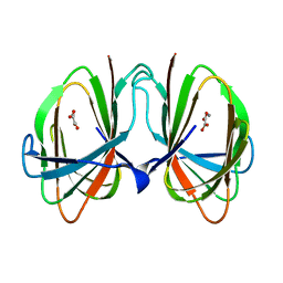
BU of 1o5u by Molmil
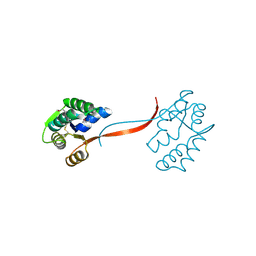
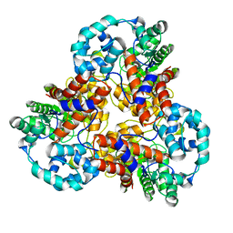
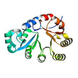
Crystal structure of a duf861 family protein (tm1112) from thermotoga maritima at 1.83 A resolution
Descriptor: UNKNOWN LIGAND, novel Thermotoga maritima enzyme TM1112
Authors:Joint Center for Structural Genomics (JCSG)
Deposit date:2003-10-06
Release date:2003-11-11
Last modified:2023-09-20
Method:X-RAY DIFFRACTION (1.83 Å)
Cite:Crystal structure of a novel Thermotoga maritima enzyme (TM1112) from the cupin family at 1.83 A resolution
Proteins, 56, 2004
1O4U
DownloadVisualize
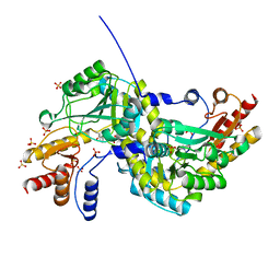
BU of 1o4u by Molmil
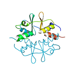
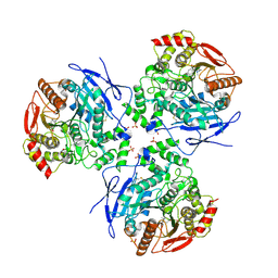
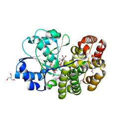
Crystal structure of a nicotinate nucleotide pyrophosphorylase (tm1645) from thermotoga maritima at 2.50 A resolution
Descriptor: Type II quinolic acid phosphoribosyltransferase
Authors:Joint Center for Structural Genomics (JCSG)
Deposit date:2003-07-09
Release date:2003-07-22
Last modified:2023-09-20
Method:X-RAY DIFFRACTION (2.5 Å)
Cite:Crystal structure of a type II quinolic acid phosphoribosyltransferase (TM1645) from Thermotoga maritima at 2.50 A resolution
Proteins, 55, 2004
1O4W
DownloadVisualize
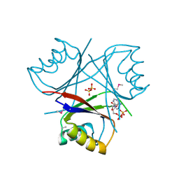
BU of 1o4w by Molmil
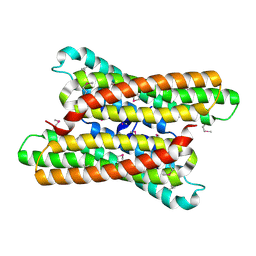
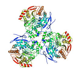
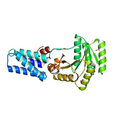
CRYSTAL STRUCTURE OF a PIN (PILT N-TERMINUS) DOMAIN CONTAINING PROTEIN (AF0591) FROM ARCHAEOGLOBUS FULGIDUS AT 1.90 A RESOLUTION
Descriptor: PIN (PilT N-terminus) domain
Authors:Joint Center for Structural Genomics (JCSG)
Deposit date:2003-07-16
Release date:2003-07-29
Last modified:2023-01-25
Method:X-RAY DIFFRACTION (1.9 Å)
Cite:Crystal structure of a PIN (PilT N-terminus) domain (AF0591) from Archaeoglobus fulgidus at 1.90 A resolution
Proteins, 56, 2004
1O50
DownloadVisualize
BU of 1o50 by Molmil
Crystal structure of a cbs domain-containing protein (tm0935) from thermotoga maritima at 1.87 A resolution
Descriptor: CBS domain-containing predicted protein TM0935
Authors:Joint Center for Structural Genomics (JCSG)
Deposit date:2003-07-31
Release date:2003-08-12
Last modified:2024-05-22
Method:X-RAY DIFFRACTION (1.87 Å)
Cite:Crystal structure of a tandem cystathionine-beta-synthase (CBS) domain protein (TM0935) from Thermotoga maritima at 1.87 A resolution
Proteins, 57, 2004
1O5H
DownloadVisualize
BU of 1o5h by Molmil
Crystal structure of formiminotetrahydrofolate cyclodeaminase (TM1560) from Thermotoga maritima at 2.80 A resolution
Descriptor: Formiminotetrahydrofolate cyclodeaminase
Authors:Joint Center for Structural Genomics (JCSG)
Deposit date:2003-09-17
Release date:2003-09-30
Last modified:2023-01-25
Method:X-RAY DIFFRACTION (2.8 Å)
Cite:Crystal structure of a formiminotetrahydrofolate cyclodeaminase (TM1560) from Thermotoga maritima at 2.80 A resolution reveals a new fold
Proteins, 58, 2005
1O4V
DownloadVisualize
BU of 1o4v by Molmil
Crystal structure of the catalytic subunit of a phosphoribosylaminoimidazole mutase (tm0446) from thermotoga maritima at 1.77 A resolution
Descriptor: SULFATE ION, phosphoribosylaminoimidazole mutase PurE
Authors:Joint Center for Structural Genomics (JCSG)
Deposit date:2003-07-11
Release date:2003-07-22
Last modified:2023-09-20
Method:X-RAY DIFFRACTION (1.77 Å)
Cite:Crystal structure of a phosphoribosylaminoimidazole mutase PurE (TM0446) from Thermotoga maritima at 1.77 A resolution
Proteins, 55, 2004
1O4T
DownloadVisualize
BU of 1o4t by Molmil
Crystal structure of a predicted oxalate decarboxylase (tm1287) from thermotoga maritima at 1.95 A resolution
Descriptor: MANGANESE (II) ION, OXALATE ION, putative oxalate decarboxylase
Authors:Joint Center for Structural Genomics (JCSG)
Deposit date:2003-07-03
Release date:2003-07-15
Last modified:2023-09-20
Method:X-RAY DIFFRACTION (1.95 Å)
Cite:Crystal structure of a putative oxalate decarboxylase (TM1287) from Thermotoga maritima at 1.95 A resolution
Proteins, 56, 2004
1O4S
DownloadVisualize
BU of 1o4s by Molmil
Crystal structure of Aspartate aminotransferase (TM1255) from Thermotoga maritima at 1.90 A resolution
Descriptor: Aspartate aminotransferase, PYRIDOXAL-5'-PHOSPHATE, SULFATE ION
Authors:Joint Center for Structural Genomics (JCSG)
Deposit date:2003-06-26
Release date:2003-07-08
Last modified:2023-09-20
Method:X-RAY DIFFRACTION (1.9 Å)
Cite:Crystal structure of an aspartate aminotransferase (TM1255) from Thermotoga maritima at 1.90 A resolution
Proteins, 55, 2004
1O51
DownloadVisualize
BU of 1o51 by Molmil
Crystal structure of a putative PII-like signaling protein (TM0021) from Thermotoga maritima at 2.50 A resolution
Descriptor: ADENOSINE-5'-DIPHOSPHATE, Hypothetical protein TM0021, SULFATE ION
Authors:Joint Center for Structural Genomics (JCSG)
Deposit date:2003-08-01
Release date:2003-08-19
Last modified:2023-01-25
Method:X-RAY DIFFRACTION (2.5 Å)
Cite:Crystal structure of a putative PII-like signaling protein (TM0021) from Thermotoga maritima at 2.5 A resolution
Proteins, 54, 2004
1O59
DownloadVisualize
BU of 1o59 by Molmil
Crystal structure of Allantoicase (yir029w) from Saccharomyces cerevisiae at 2.40 A resolution
Descriptor: Allantoicase
Authors:Joint Center for Structural Genomics (JCSG)
Deposit date:2003-08-22
Release date:2003-09-02
Last modified:2023-01-25
Method:X-RAY DIFFRACTION (2.4 Å)
Cite:Crystal structure of an allantoicase (YIR029W) from Saccharomyces cerevisiae at 2.4 A resolution
Proteins, 56, 2004
1J5S
DownloadVisualize
BU of 1j5s by Molmil
Crystal structure of uronate isomerase (TM0064) from Thermotoga maritima at 2.85 A resolution
Descriptor: URONATE ISOMERASE
Authors:Joint Center for Structural Genomics (JCSG)
Deposit date:2002-07-02
Release date:2002-07-17
Last modified:2024-05-22
Method:X-RAY DIFFRACTION (2.85 Å)
Cite:Crystal structure of uronate isomerase (TM0064) from Thermotoga maritima at 2.85 A resolution.
Proteins, 53, 2003
2DQZ
DownloadVisualize
BU of 2dqz by Molmil
Crystal structure of human carboxylesterase in complex with homatropine, coenzyme A, and palmitate
Descriptor: 2-acetamido-2-deoxy-beta-D-glucopyranose, COENZYME A, FLUORIDE ION, ...
Authors:Bencharit, S, Redinbo, M.R.
Deposit date:2006-06-02
Release date:2006-08-29
Last modified:2023-10-25
Method:X-RAY DIFFRACTION (2.8 Å)
Cite:Multisite promiscuity in the processing of endogenous substrates by human carboxylesterase 1
J.Mol.Biol., 363, 2006
2DQY
DownloadVisualize
BU of 2dqy by Molmil
Crystal structure of human carboxylesterase in complex with cholate and palmitate
Descriptor: 2-acetamido-2-deoxy-beta-D-glucopyranose, CHOLIC ACID, Liver carboxylesterase 1, ...
Authors:Bencharit, S, Edwards, C.C, Morton, C.L, Howard-Williams, E.L, Potter, P.M, Redinbo, M.R.
Deposit date:2006-06-02
Release date:2006-08-29
Last modified:2023-10-25
Method:X-RAY DIFFRACTION (3 Å)
Cite:Multisite promiscuity in the processing of endogenous substrates by human carboxylesterase 1
J.Mol.Biol., 363, 2006
2DR0
DownloadVisualize
BU of 2dr0 by Molmil
Crystal structure of human carboxylesterase in complex with taurocholate
Descriptor: 2-acetamido-2-deoxy-beta-D-glucopyranose, Liver carboxylesterase 1, N-acetyl-alpha-neuraminic acid, ...
Authors:Bencharit, S, Redinbo, M.R.
Deposit date:2006-06-02
Release date:2006-08-29
Last modified:2020-07-29
Method:X-RAY DIFFRACTION (3.2 Å)
Cite:Multisite promiscuity in the processing of endogenous substrates by human carboxylesterase 1
J.Mol.Biol., 363, 2006
2H1J
DownloadVisualize
BU of 2h1j by Molmil
3.1 A X-ray structure of putative Oligoendopeptidase F: Crystals grown by microfluidic seeding
Descriptor: Oligoendopeptidase F, ZINC ION
Authors:Gerdts, C.J, Tereshko, V, Dementieva, I, Collart, F, Joachimiak, A, Kossiakoff, A, Ismagilov, R.F, Accelerated Technologies Center for Gene to 3D Structure (ATCG3D)
Deposit date:2006-05-16
Release date:2006-06-13
Last modified:2011-07-13
Method:X-RAY DIFFRACTION (3.1 Å)
Cite:Time-Controlled Microfluidic Seeding in nL-Volume Droplets To Separate Nucleation and Growth Stages of Protein Crystallization.
Angew.Chem.Int.Ed.Engl., 45, 2006
2H7C
DownloadVisualize
BU of 2h7c by Molmil
Crystal structure of human carboxylesterase in complex with Coenzyme A
Descriptor: 2-acetamido-2-deoxy-beta-D-glucopyranose, 2-acetamido-2-deoxy-beta-D-glucopyranose-(1-4)-2-acetamido-2-deoxy-beta-D-glucopyranose, COENZYME A, ...
Authors:Bencharit, S, Edwards, C.C, Morton, C.L, Howard-Williams, E.L, Potter, P.M, Redinbo, M.R.
Deposit date:2006-06-02
Release date:2006-08-29
Last modified:2023-10-25
Method:X-RAY DIFFRACTION (2 Å)
Cite:Multisite promiscuity in the processing of endogenous substrates by human carboxylesterase 1
J.Mol.Biol., 363, 2006
2H1N
DownloadVisualize
BU of 2h1n by Molmil
3.0 A X-ray structure of putative oligoendopeptidase F: crystals grown by vapor diffusion technique
Descriptor: Oligoendopeptidase F, UNKNOWN LIGAND, ZINC ION
Authors:Gerdts, C.J, Tereshko, V, Dementieva, I, Collart, F, Joachimiak, A, Kossiakoff, A, Ismagilov, R.F, Midwest Center for Structural Genomics (MCSG)
Deposit date:2006-05-16
Release date:2006-06-13
Last modified:2011-07-13
Method:X-RAY DIFFRACTION (3 Å)
Cite:Time-Controlled Microfluidic Seeding in nL-Volume Droplets To Separate Nucleation and Growth Stages of Protein Crystallization.
Angew.Chem.Int.Ed.Engl., 45, 2006
1IJC
DownloadVisualize
BU of 1ijc by Molmil
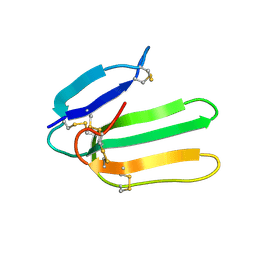
Solution Structure of Bucandin, a Neurotoxin from the Venom of the Malayan Krait
Descriptor: bucandin
Authors:Torres, A.M, Kini, R.M, Nirthanan, S, Kuchel, P.W.
Deposit date:2001-04-25
Release date:2001-12-21
Last modified:2022-02-23
Method:SOLUTION NMR
Cite:NMR structure of bucandin, a neurotoxin from the venom of the Malayan krait (Bungarus candidus).
Biochem.J., 360, 2001
1VJ2
DownloadVisualize
BU of 1vj2 by Molmil
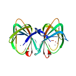
Crystal structure of a novel family of manganese-containing cupin (tm1459) from thermotoga maritima at 1.65 A resolution
Descriptor: MANGANESE (II) ION, UNKNOWN LIGAND, novel manganese-containing cupin TM1459
Authors:Joint Center for Structural Genomics (JCSG)
Deposit date:2003-12-03
Release date:2003-12-09
Last modified:2023-09-20
Method:X-RAY DIFFRACTION (1.65 Å)
Cite:Crystal structure of a novel manganese-containing cupin (TM1459) from Thermotoga maritima at 1.65 A resolution.
Proteins, 56, 2004
1KQ3
DownloadVisualize
BU of 1kq3 by Molmil
CRYSTAL STRUCTURE OF A GLYCEROL DEHYDROGENASE (TM0423) FROM THERMOTOGA MARITIMA AT 1.5 A RESOLUTION
Descriptor: 2-AMINO-2-HYDROXYMETHYL-PROPANE-1,3-DIOL, CHLORIDE ION, ZINC ION, ...
Authors:Wilson, I.A, Miller, M.D, Joint Center for Structural Genomics (JCSG)
Deposit date:2002-01-03
Release date:2002-02-27
Last modified:2024-02-14
Method:X-RAY DIFFRACTION (1.5 Å)
Cite:Structural genomics of the Thermotoga maritima proteome implemented in a high-throughput structure determination pipeline
Proc.Natl.Acad.Sci.USA, 99, 2002
1KQ4
DownloadVisualize
BU of 1kq4 by Molmil
CRYSTAL STRUCTURE OF A THY1-COMPLEMENTING PROTEIN (TM0449) FROM THERMOTOGA MARITIMA AT 2.25 A RESOLUTION
Descriptor: FLAVIN-ADENINE DINUCLEOTIDE, HYPOTHETICAL PROTEIN TM0449
Authors:Wilson, I.A, Miller, M.D, Joint Center for Structural Genomics (JCSG)
Deposit date:2002-01-03
Release date:2002-02-27
Last modified:2017-10-11
Method:X-RAY DIFFRACTION (2.25 Å)
Cite:Structural genomics of the Thermotoga maritima proteome implemented in a high-throughput structure determination pipeline
Proc.Natl.Acad.Sci.USA, 99, 2002
1O58
DownloadVisualize
BU of 1o58 by Molmil
Crystal structure of O-acetylserine sulfhydrylase (TM0665) from Thermotoga maritima at 1.80 A resolution
Descriptor: O-acetylserine sulfhydrylase, PHOSPHATE ION
Authors:Joint Center for Structural Genomics (JCSG)
Deposit date:2003-08-20
Release date:2003-09-02
Last modified:2024-05-22
Method:X-RAY DIFFRACTION (1.8 Å)
Cite:Crystal structure of O-acetylserine sulfhydrylase (TM0665) from Thermotoga maritima at 1.8 A resolution
Proteins, 56, 2004
1O1Z
DownloadVisualize
BU of 1o1z by Molmil
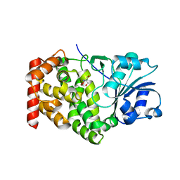
Crystal structure of glycerophosphodiester phosphodiesterase (GDPD) (TM1621) from Thermotoga maritima at 1.60 A resolution
Descriptor: SODIUM ION, glycerophosphodiester phosphodiesterase
Authors:Joint Center for Structural Genomics (JCSG)
Deposit date:2003-02-12
Release date:2003-04-01
Last modified:2024-05-22
Method:X-RAY DIFFRACTION (1.6 Å)
Cite:Crystal structure of a glycerophosphodiester phosphodiesterase (GDPD) from Thermotoga maritima (TM1621) at 1.60 A resolution.
Proteins, 56, 2004
1O2D
DownloadVisualize
BU of 1o2d by Molmil
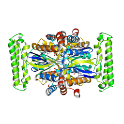
Crystal structure of Alcohol dehydrogenase, iron-containing (TM0920) from Thermotoga maritima at 1.30 A resolution
Descriptor: 2-AMINO-2-HYDROXYMETHYL-PROPANE-1,3-DIOL, Alcohol dehydrogenase, iron-containing, ...
Authors:Joint Center for Structural Genomics (JCSG)
Deposit date:2003-02-27
Release date:2003-06-10
Last modified:2023-01-25
Method:X-RAY DIFFRACTION (1.3 Å)
Cite:Crystal structure of an iron-containing 1,3-propanediol dehydrogenase (TM0920) from Thermotoga maritima at 1.3 A resolution
Proteins, 54, 2004

220113

PDB entries from 2024-05-22

PDB statisticsPDBj update infoContact PDBjnumon