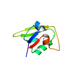
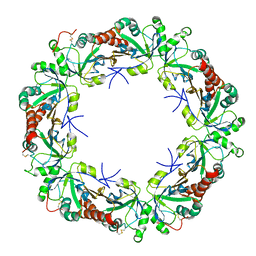
2HI7
 
 | | Crystal structure of DsbA-DsbB-ubiquinone complex | | 分子名称: | Disulfide bond formation protein B, Thiol:disulfide interchange protein dsbA, UBIQUINONE-1, ... | | 著者 | Inaba, K, Murakami, S, Suzuki, M, Nakagawa, A, Yamashita, E, Okada, K, Ito, K. | | 登録日 | 2006-06-29 | | 公開日 | 2006-12-05 | | 最終更新日 | 2021-11-10 | | 実験手法 | X-RAY DIFFRACTION (3.7 Å) | | 主引用文献 | Crystal Structure of the DsbB-DsbA Complex Reveals a Mechanism of Disulfide Bond Generation
Cell(Cambridge,Mass.), 127, 2006
|
|
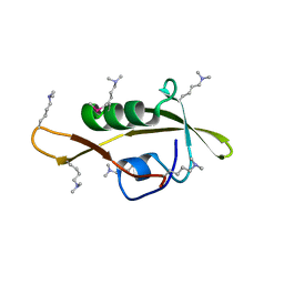
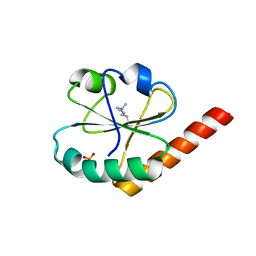
7EBW
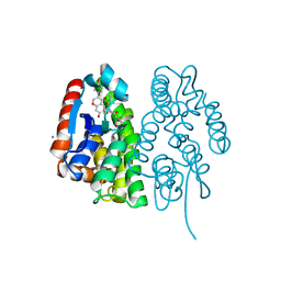 | | Crystal structure of Aedes aegypti Noppera-bo, glutathione S-transferase epsilon 8, in desmethylglycitein and glutathione-bound form | | 分子名称: | 6,7-dihydroxy-3-(4-hydroxyphenyl)-4H-chromen-4-one, CALCIUM ION, GLUTATHIONE, ... | | 著者 | Inaba, K, Koiwai, K, Senda, M, Senda, T, Niwa, R. | | 登録日 | 2021-03-11 | | 公開日 | 2022-01-26 | | 最終更新日 | 2023-11-29 | | 実験手法 | X-RAY DIFFRACTION (1.94 Å) | | 主引用文献 | Molecular action of larvicidal flavonoids on ecdysteroidogenic glutathione S-transferase Noppera-bo in Aedes aegypti.
Bmc Biol., 20, 2022
|
|
7EBV
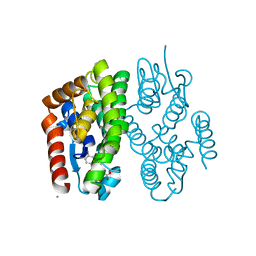 | | Crystal structure of Aedes aegypti Noppera-bo, glutathione S-transferase epsilon 8, in luteolin- and glutathione-bound form | | 分子名称: | 2-(3,4-dihydroxyphenyl)-5,7-dihydroxy-4H-chromen-4-one, CALCIUM ION, GLUTATHIONE, ... | | 著者 | Inaba, K, Koiwai, K, Senda, M, Senda, T, Niwa, R. | | 登録日 | 2021-03-11 | | 公開日 | 2022-01-26 | | 最終更新日 | 2023-11-29 | | 実験手法 | X-RAY DIFFRACTION (1.5 Å) | | 主引用文献 | Molecular action of larvicidal flavonoids on ecdysteroidogenic glutathione S-transferase Noppera-bo in Aedes aegypti.
Bmc Biol., 20, 2022
|
|
7EBT
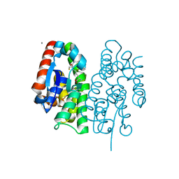 | | Crystal structure of Aedes aegypti Noppera-bo, glutathione S-transferase epsilon 8, in glutathione-bound form | | 分子名称: | CALCIUM ION, GLUTATHIONE, Glutathione transferase | | 著者 | Inaba, K, Koiwai, K, Senda, M, Senda, T, Niwa, R. | | 登録日 | 2021-03-11 | | 公開日 | 2022-01-26 | | 最終更新日 | 2023-11-29 | | 実験手法 | X-RAY DIFFRACTION (1.51 Å) | | 主引用文献 | Molecular action of larvicidal flavonoids on ecdysteroidogenic glutathione S-transferase Noppera-bo in Aedes aegypti.
Bmc Biol., 20, 2022
|
|
7EBU
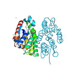 | | Crystal structure of Aedes aegypti Noppera-bo, glutathione S-transferase epsilon 8, in Daidzein- and glutathione-bound form | | 分子名称: | 7-hydroxy-3-(4-hydroxyphenyl)-4H-chromen-4-one, CALCIUM ION, GLUTATHIONE, ... | | 著者 | Inaba, K, Koiwai, K, Senda, M, Senda, T, Niwa, R. | | 登録日 | 2021-03-11 | | 公開日 | 2022-01-26 | | 最終更新日 | 2023-11-29 | | 実験手法 | X-RAY DIFFRACTION (1.95 Å) | | 主引用文献 | Molecular action of larvicidal flavonoids on ecdysteroidogenic glutathione S-transferase Noppera-bo in Aedes aegypti.
Bmc Biol., 20, 2022
|
|
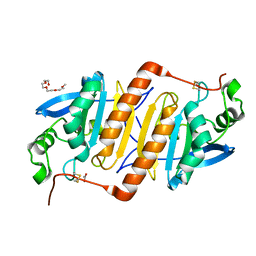
2ZUP
 | | Updated crystal structure of DsbB-DsbA complex from E. coli | | 分子名称: | Disulfide bond formation protein B, Thiol:disulfide interchange protein dsbA, UBIQUINONE-1, ... | | 著者 | Inaba, K, Suzuki, M, Murakami, S, Nakagawa, A. | | 登録日 | 2008-10-28 | | 公開日 | 2009-04-14 | | 最終更新日 | 2023-11-01 | | 実験手法 | X-RAY DIFFRACTION (3.7 Å) | | 主引用文献 | Dynamic nature of disulphide bond formation catalysts revealed by crystal structures of DsbB
Embo J., 28, 2009
|
|
2ZPL
 | |
2ZUQ
 | | Crystal structure of DsbB-Fab complex | | 分子名称: | Disulfide bond formation protein B, Fab fragment heavy chain, Fab fragment light chain, ... | | 著者 | Inaba, K, Suzuki, M, Murakami, S. | | 登録日 | 2008-10-28 | | 公開日 | 2009-04-14 | | 最終更新日 | 2023-11-01 | | 実験手法 | X-RAY DIFFRACTION (3.3 Å) | | 主引用文献 | Dynamic nature of disulphide bond formation catalysts revealed by crystal structures of DsbB
Embo J., 28, 2009
|
|
2ZPM
 | |
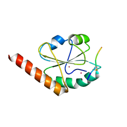
3AHR
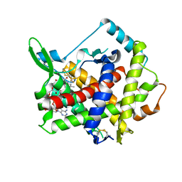 | | Inactive human Ero1 | | 分子名称: | ERO1-like protein alpha, FLAVIN-ADENINE DINUCLEOTIDE | | 著者 | Inaba, K, Sitia, R, Suzuki, M. | | 登録日 | 2010-04-26 | | 公開日 | 2010-12-22 | | 最終更新日 | 2023-11-01 | | 実験手法 | X-RAY DIFFRACTION (3.07 Å) | | 主引用文献 | Crystal structures of human Ero1-alpha reveal the mechanisms of regulated and targeted oxidation of PDI
Embo J., 29, 2010
|
|
3AHQ
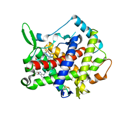 | | hyperactive human Ero1 | | 分子名称: | ERO1-like protein alpha, FLAVIN-ADENINE DINUCLEOTIDE | | 著者 | Inaba, K, Sitia, R, Suzuki, M. | | 登録日 | 2010-04-26 | | 公開日 | 2010-12-22 | | 最終更新日 | 2023-11-01 | | 実験手法 | X-RAY DIFFRACTION (2.35 Å) | | 主引用文献 | Crystal structures of human Ero1-alpha reveal the mechanisms of regulated and targeted oxidation of PDI
Embo J., 29, 2010
|
|
3APQ
 | |
3APO
 | |
3APS
 | |
3VWU
 | |
3VWW
 | |
3VWV
 | |
3W8J
 | | Crystal structure of P5 a0 in a complex with Prx4 c-term | | 分子名称: | C-terminal peptide from Peroxiredoxin-4, GLYCEROL, POTASSIUM ION, ... | | 著者 | Inaba, K, Suzuki, M, Kojima, R. | | 登録日 | 2013-03-13 | | 公開日 | 2013-09-04 | | 実験手法 | X-RAY DIFFRACTION (2.1 Å) | | 主引用文献 | Synergistic cooperation of PDI family members in peroxiredoxin 4-driven oxidative protein folding
Sci Rep, 3, 2013
|
|
3WGE
 | | Crystal structure of ERp46 Trx2 | | 分子名称: | GLYCEROL, Thioredoxin domain-containing protein 5 | | 著者 | Inaba, K, Suzuki, M, Kojima, R. | | 登録日 | 2013-08-04 | | 公開日 | 2014-06-25 | | 実験手法 | X-RAY DIFFRACTION (0.95 Å) | | 主引用文献 | Radically different thioredoxin domain arrangement of ERp46, an efficient disulfide bond introducer of the mammalian PDI family
Structure, 22, 2014
|
|
3WGD
 | | Crystal structure of ERp46 Trx1 | | 分子名称: | GLYCEROL, PHOSPHATE ION, POTASSIUM ION, ... | | 著者 | Inaba, K, Suzuki, M, Kojima, R. | | 登録日 | 2013-08-04 | | 公開日 | 2014-06-25 | | 実験手法 | X-RAY DIFFRACTION (2.5 Å) | | 主引用文献 | Radically different thioredoxin domain arrangement of ERp46, an efficient disulfide bond introducer of the mammalian PDI family
Structure, 22, 2014
|
|
3WGX
 | | Crystal structure of ERp46 Trx2 in a complex with Prx4 C-term | | 分子名称: | GLYCEROL, Peroxiredoxin-4, Thioredoxin domain-containing protein 5 | | 著者 | Inaba, K, Suzuki, M, Kojima, R. | | 登録日 | 2013-08-13 | | 公開日 | 2014-06-25 | | 実験手法 | X-RAY DIFFRACTION (0.92 Å) | | 主引用文献 | Radically different thioredoxin domain arrangement of ERp46, an efficient disulfide bond introducer of the mammalian PDI family
Structure, 22, 2014
|
|
1CH4
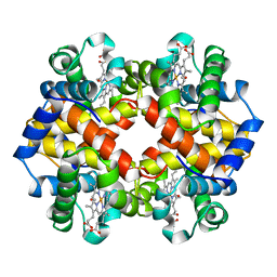 | | MODULE-SUBSTITUTED CHIMERA HEMOGLOBIN BETA-ALPHA (F133V) | | 分子名称: | CARBON MONOXIDE, MODULE-SUBSTITUTED CHIMERA HEMOGLOBIN BETA-ALPHA, PROTOPORPHYRIN IX CONTAINING FE | | 著者 | Shirai, T, Fujikake, M, Yamane, T, Inaba, K, Ishimori, K, Morishima, I. | | 登録日 | 1998-06-11 | | 公開日 | 1999-04-27 | | 最終更新日 | 2024-04-03 | | 実験手法 | X-RAY DIFFRACTION (2.5 Å) | | 主引用文献 | Crystal structure of a protein with an artificial exon-shuffling, module M4-substituted chimera hemoglobin beta alpha, at 2.5 A resolution.
J.Mol.Biol., 287, 1999
|
|
8J7Y
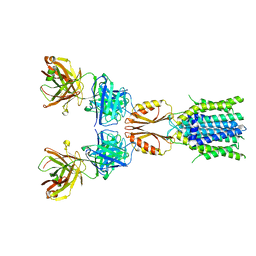 | | Cryo-EM structure of hZnT7DeltaHis-loop-Fab complex in zinc-bound state, determined in outward-facing conformation | | 分子名称: | Heavy chain of YN7114-08 Fab, Light chain of YN7114-08 Fab, ZINC ION, ... | | 著者 | Han, B.B, Inaba, K, Watanabe, S. | | 登録日 | 2023-04-28 | | 公開日 | 2023-09-20 | | 実験手法 | ELECTRON MICROSCOPY (3.4 Å) | | 主引用文献 | Cryo-EM structures of human zinc transporter ZnT7 reveal the mechanism of Zn 2+ uptake into the Golgi apparatus.
Nat Commun, 14, 2023
|
|
8J7U
 | | Cryo-EM structure of hZnT7-Fab complex in zinc-bound state, determined in outward-facing conformation | | 分子名称: | Heavy chain of YN7114-08 Fab, Light chain of YN7114-08 Fab, ZINC ION, ... | | 著者 | Han, B.B, Inaba, K, Watanabe, S. | | 登録日 | 2023-04-28 | | 公開日 | 2023-09-20 | | 実験手法 | ELECTRON MICROSCOPY (3.12 Å) | | 主引用文献 | Cryo-EM structures of human zinc transporter ZnT7 reveal the mechanism of Zn 2+ uptake into the Golgi apparatus.
Nat Commun, 14, 2023
|
|
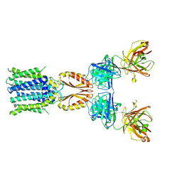
8J7V
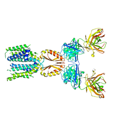 | | Cryo-EM structure of hZnT7-Fab complex in zinc-unbound state, determined in heterogeneous conformations- one subunit in an inward-facing and the other in an outward-facing conformation | | 分子名称: | Heavy chain of YN7114-08 Fab, Light chain of YN7114-08 Fab, Zinc transporter 7 | | 著者 | Han, B.B, Inaba, K, Watanabe, S. | | 登録日 | 2023-04-28 | | 公開日 | 2023-09-20 | | 実験手法 | ELECTRON MICROSCOPY (2.79 Å) | | 主引用文献 | Cryo-EM structures of human zinc transporter ZnT7 reveal the mechanism of Zn 2+ uptake into the Golgi apparatus.
Nat Commun, 14, 2023
|
|


