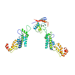
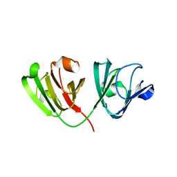
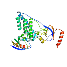
Loading
PDBj
MenuPDBj@FacebookPDBj@TwitterPDBj@YouTubewwPDB FoundationwwPDB
RCSB PDBPDBeBMRBAdv. SearchSearch help
Search by PDB author
7SD4
DownloadVisualize
BU of 7sd4 by Molmil
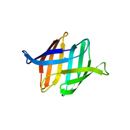
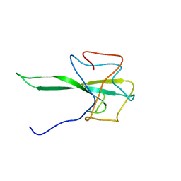
SARS-CoV-2 Nucleocapsid N-terminal domain (N-NTD) protein
Descriptor: Nucleoprotein
Authors:Sarkar, S, Runge, B, Russell, R.W, Calero, D, Zeinalilathori, S, Quinn, C.M, Lu, M, Calero, G, Gronenborn, A.M, Polenova, T.
Deposit date:2021-09-29
Release date:2022-06-08
Last modified:2023-06-14
Method:SOLID-STATE NMR
Cite:Atomic-Resolution Structure of SARS-CoV-2 Nucleocapsid Protein N-Terminal Domain.
J.Am.Chem.Soc., 144, 2022
1BBN
DownloadVisualize
BU of 1bbn by Molmil
THREE-DIMENSIONAL SOLUTION STRUCTURE OF HUMAN INTERLEUKIN-4 BY MULTI-DIMENSIONAL HETERONUCLEAR MAGNETIC RESONANCE SPECTROSCOPY
Descriptor: INTERLEUKIN-4
Authors:Clore, G.M, Powers, B, Garrett, D.S, Gronenborn, A.M.
Deposit date:1992-05-01
Release date:1993-10-31
Last modified:2012-09-05
Method:SOLUTION NMR
Cite:Three-dimensional solution structure of human interleukin-4 by multidimensional heteronuclear magnetic resonance spectroscopy.
Science, 256, 1992
3S60
DownloadVisualize
BU of 3s60 by Molmil
Structure of the cyanobacterial Oscillatoria Agardhii Agglutinin (OAA) in free state obtained at 25 degree Celsius
Descriptor: Lectin
Authors:Koharudin, L.M.I, Gronenborn, A.M.
Deposit date:2011-05-23
Release date:2011-06-15
Last modified:2023-09-13
Method:X-RAY DIFFRACTION (1.6 Å)
Cite:Structural basis of the anti-HIV activity of the cyanobacterial Oscillatoria Agardhii agglutinin.
Structure, 19, 2011
3S5X
DownloadVisualize
BU of 3s5x by Molmil
Structure of the cyanobacterial Oscillatoria Agardhii Agglutinin (OAA) in complex with a3,a6 mannopentaose
Descriptor: Lectin, alpha-D-mannopyranose, alpha-D-mannopyranose-(1-3)-alpha-D-mannopyranose-(1-6)-[alpha-D-mannopyranose-(1-3)]beta-D-mannopyranose
Authors:Koharudin, L.M.I, Gronenborn, A.M.
Deposit date:2011-05-23
Release date:2011-06-15
Last modified:2023-09-13
Method:X-RAY DIFFRACTION (1.65 Å)
Cite:Structural basis of the anti-HIV activity of the cyanobacterial Oscillatoria Agardhii agglutinin.
Structure, 19, 2011
5FJB
DownloadVisualize
BU of 5fjb by Molmil
Cyclophilin A Stabilize HIV-1 Capsid through a Novel Non- canonical Binding Site
Descriptor: GAG POLYPROTEIN, PEPTIDYL-PROLYL CIS-TRANS ISOMERASE A
Authors:Liu, C, Perilla, J.R, Ning, J, Lu, M, Hou, G, Ramalhu, R, Bedwell, G.J, Ahn, J, Shi, J, Gronenborn, A.M, Prevelige Jr, P.E, Rousso, I, Aiken, C, Polenova, T, Schulten, K, Zhang, P.
Deposit date:2015-10-07
Release date:2016-03-16
Last modified:2017-08-23
Method:ELECTRON MICROSCOPY (9 Å)
Cite:Cyclophilin a Stabilizes the HIV-1 Capsid Through a Novel Non-Canonical Binding Site.
Nat.Commun., 7, 2016
5GAT
DownloadVisualize
BU of 5gat by Molmil
SOLUTION NMR STRUCTURE OF THE WILD TYPE DNA BINDING DOMAIN OF AREA COMPLEXED TO A 13BP DNA CONTAINING A CGATA SITE, 35 STRUCTURES
Descriptor: DNA (5'-D(*CP*AP*GP*CP*GP*AP*TP*AP*GP*AP*GP*AP*C)-3'), DNA (5'-D(*GP*TP*CP*TP*CP*TP*AP*TP*CP*GP*CP*TP*G)-3'), NITROGEN REGULATORY PROTEIN AREA, ...
Authors:Clore, G.M, Starich, M, Wikstrom, M, Gronenborn, A.M.
Deposit date:1997-11-07
Release date:1998-01-28
Last modified:2022-03-16
Method:SOLUTION NMR
Cite:The solution structure of a fungal AREA protein-DNA complex: an alternative binding mode for the basic carboxyl tail of GATA factors.
J.Mol.Biol., 277, 1998
5HDW
DownloadVisualize
BU of 5hdw by Molmil
ApaG Domain of FBxo3
Descriptor: F-box only protein 3
Authors:Krzysiak, T.C, Chen, B.B, Mallampalli, R.K, Gronenborn, A.M.
Deposit date:2016-01-05
Release date:2016-04-06
Last modified:2023-09-27
Method:X-RAY DIFFRACTION (2 Å)
Cite:Crystal structure and interaction studies of the human FBxo3 ApaG domain.
Febs J., 283, 2016
3OBL
DownloadVisualize
BU of 3obl by Molmil
Crystal structure of the potent anti-HIV cyanobacterial lectin from Oscillatoria Agardhii
Descriptor: 3-CYCLOHEXYL-1-PROPYLSULFONIC ACID, Lectin
Authors:Koharudin, L.M.I, Furey, W, Gronenborn, A.M.
Deposit date:2010-08-06
Release date:2010-10-20
Last modified:2024-02-21
Method:X-RAY DIFFRACTION (1.2 Å)
Cite:Novel fold and carbohydrate specificity of the potent anti-HIV cyanobacterial lectin from Oscillatoria agardhii.
J.Biol.Chem., 286, 2011
1ATA
DownloadVisualize
BU of 1ata by Molmil
HIGH-RESOLUTION STRUCTURE OF ASCARIS TRYPSIN INHIBITOR IN SOLUTION: DIRECT EVIDENCE FOR A PH INDUCED CONFORMATIONAL TRANSITION IN THE REACTIVE SITE
Descriptor: ASCARIS TRYPSIN INHIBITOR
Authors:Clore, G.M, Grasberger, B.L, Gronenborn, A.M.
Deposit date:1994-05-20
Release date:1994-08-31
Last modified:2022-02-16
Method:SOLUTION NMR
Cite:High-resolution structure of Ascaris trypsin inhibitor in solution: direct evidence for a pH-induced conformational transition in the reactive site.
Structure, 2, 1994
1ATB
DownloadVisualize
BU of 1atb by Molmil
HIGH-RESOLUTION STRUCTURE OF ASCARIS TRYPSIN INHIBITOR IN SOLUTION: DIRECT EVIDENCE FOR A PH INDUCED CONFORMATIONAL TRANSITION IN THE REACTIVE SITE
Descriptor: ASCARIS TRYPSIN INHIBITOR
Authors:Clore, G.M, Grasberger, B.L, Gronenborn, A.M.
Deposit date:1994-05-20
Release date:1994-08-31
Last modified:2022-02-16
Method:SOLUTION NMR
Cite:High-resolution structure of Ascaris trypsin inhibitor in solution: direct evidence for a pH-induced conformational transition in the reactive site.
Structure, 2, 1994
1ATE
DownloadVisualize
BU of 1ate by Molmil
HIGH-RESOLUTION STRUCTURE OF ASCARIS TRYPSIN INHIBITOR IN SOLUTION: DIRECT EVIDENCE FOR A PH INDUCED CONFORMATIONAL TRANSITION IN THE REACTIVE SITE
Descriptor: ASCARIS TRYPSIN INHIBITOR
Authors:Clore, G.M, Grasberger, B.L, Gronenborn, A.M.
Deposit date:1994-05-20
Release date:1994-08-31
Last modified:2022-02-16
Method:SOLUTION NMR
Cite:High-resolution structure of Ascaris trypsin inhibitor in solution: direct evidence for a pH-induced conformational transition in the reactive site.
Structure, 2, 1994
6W5B
DownloadVisualize
BU of 6w5b by Molmil
N124D Deamidation Mutant of Human gammaD-Crystallin
Descriptor: Gamma-crystallin D
Authors:Whitley, M.J, Rathi, N, Ambarian, M, Gronenborn, A.M.
Deposit date:2020-03-12
Release date:2021-01-20
Last modified:2023-10-18
Method:X-RAY DIFFRACTION (1.15 Å)
Cite:Assessing the Structures and Interactions of gamma D-Crystallin Deamidation Variants.
Structure, 29, 2021
1ATD
DownloadVisualize
BU of 1atd by Molmil
HIGH-RESOLUTION STRUCTURE OF ASCARIS TRYPSIN INHIBITOR IN SOLUTION: DIRECT EVIDENCE FOR A PH INDUCED CONFORMATIONAL TRANSITION IN THE REACTIVE SITE
Descriptor: ASCARIS TRYPSIN INHIBITOR
Authors:Clore, G.M, Grasberger, B.L, Gronenborn, A.M.
Deposit date:1994-05-20
Release date:1994-08-31
Last modified:2022-02-16
Method:SOLUTION NMR
Cite:High-resolution structure of Ascaris trypsin inhibitor in solution: direct evidence for a pH-induced conformational transition in the reactive site.
Structure, 2, 1994
6WAP
DownloadVisualize
BU of 6wap by Molmil
Atomic-Resolution Structure of HIV-1 Capsid Tubes by Magic Angle Spinning NMR
Descriptor: HIV-1 capsid protein
Authors:Lu, M, Russell, R.W, Bryer, A, Quinn, C.M, Hou, G, Zhang, H, Schwieters, C.D, Perilla, J.R, Gronenborn, A.M, Polenova, T.
Deposit date:2020-03-25
Release date:2020-09-02
Last modified:2023-06-14
Method:SOLID-STATE NMR
Cite:Atomic-resolution structure of HIV-1 capsid tubes by magic-angle spinning NMR.
Nat.Struct.Mol.Biol., 27, 2020
6WCY
DownloadVisualize
BU of 6wcy by Molmil
N160D Deamidation Mutant of Human gammaD-Crystallin
Descriptor: Gamma-crystallin D, SULFATE ION
Authors:Whitley, M.J, Rathi, N, Ambarian, M, Gronenborn, A.M.
Deposit date:2020-03-31
Release date:2021-01-20
Last modified:2023-10-18
Method:X-RAY DIFFRACTION (1.204 Å)
Cite:Assessing the Structures and Interactions of gamma D-Crystallin Deamidation Variants.
Structure, 29, 2021
1WJF
DownloadVisualize
BU of 1wjf by Molmil
SOLUTION STRUCTURE OF H12C MUTANT OF THE N-TERMINAL ZN BINDING DOMAIN OF HIV-1 INTEGRASE COMPLEXED TO CADMIUM, NMR, 40 STRUCTURES
Descriptor: CADMIUM ION, HIV-1 INTEGRASE
Authors:Cai, M, Gronenborn, A.M, Clore, G.M.
Deposit date:1998-06-11
Release date:1998-12-16
Last modified:2024-05-01
Method:SOLUTION NMR
Cite:Solution structure of the His12 --> Cys mutant of the N-terminal zinc binding domain of HIV-1 integrase complexed to cadmium.
Protein Sci., 7, 1998
4FBV
DownloadVisualize
BU of 4fbv by Molmil
Crystal structure of the Myxococcus Xanthus hemagglutinin in complex with a3,a6-mannopentaose
Descriptor: 1,2-ETHANEDIOL, Myxobacterial hemagglutinin, alpha-D-mannopyranose, ...
Authors:Koharudin, L.M.I, Gronenborn, A.M.
Deposit date:2012-05-23
Release date:2012-08-15
Last modified:2023-09-13
Method:X-RAY DIFFRACTION (1.76 Å)
Cite:Structural Insights into the Anti-HIV Activity of the Oscillatoria agardhii Agglutinin Homolog Lectin Family.
J.Biol.Chem., 287, 2012
4FBR
DownloadVisualize
BU of 4fbr by Molmil
Crystal structure of the Myxococcus Xanthus hemagglutinin (MBHA)
Descriptor: Myxobacterial hemagglutinin
Authors:Koharudin, L.M.I, Gronenborn, A.M.
Deposit date:2012-05-23
Release date:2012-08-15
Last modified:2023-09-13
Method:X-RAY DIFFRACTION (1.6 Å)
Cite:Structural Insights into the Anti-HIV Activity of the Oscillatoria agardhii Agglutinin Homolog Lectin Family.
J.Biol.Chem., 287, 2012
4FBO
DownloadVisualize
BU of 4fbo by Molmil
Crystal structure of the Pseudomonas fluorescens agglutinin (PFA)
Descriptor: Pseudomonas fluorescens agglutinin
Authors:Koharudin, L.M.I, Gronenborn, A.M.
Deposit date:2012-05-23
Release date:2012-08-15
Last modified:2023-09-13
Method:X-RAY DIFFRACTION (1.7 Å)
Cite:Structural Insights into the Anti-HIV Activity of the Oscillatoria agardhii Agglutinin Homolog Lectin Family.
J.Biol.Chem., 287, 2012
1SAL
DownloadVisualize
BU of 1sal by Molmil
HIGH RESOLUTION SOLUTION NMR STRUCTURE OF THE OLIGOMERIZATION DOMAIN OF P53 BY MULTI-DIMENSIONAL NMR (SAD STRUCTURES)
Descriptor: TUMOR SUPPRESSOR P53
Authors:Clore, G.M, Omichinski, J.G, Gronenborn, A.M.
Deposit date:1995-03-12
Release date:1995-10-15
Last modified:2022-03-02
Method:SOLUTION NMR
Cite:Refined solution structure of the oligomerization domain of the tumour suppressor p53.
Nat.Struct.Biol., 2, 1995
1SAK
DownloadVisualize
BU of 1sak by Molmil
HIGH RESOLUTION SOLUTION NMR STRUCTURE OF THE OLIGOMERIZATION DOMAIN OF P53 BY MULTI-DIMENSIONAL NMR (SAC STRUCTURES)
Descriptor: TUMOR SUPPRESSOR P53
Authors:Clore, G.M, Omichinski, J.G, Gronenborn, A.M.
Deposit date:1995-03-12
Release date:1995-10-15
Last modified:2022-03-02
Method:SOLUTION NMR
Cite:Refined solution structure of the oligomerization domain of the tumour suppressor p53.
Nat.Struct.Biol., 2, 1995
1SAE
DownloadVisualize
BU of 1sae by Molmil
HIGH RESOLUTION SOLUTION NMR STRUCTURE OF THE OLIGOMERIZATION DOMAIN OF P53 BY MULTI-DIMENSIONAL NMR (SAC STRUCTURES)
Descriptor: TUMOR SUPPRESSOR P53
Authors:Clore, G.M, Omichinski, J.G, Gronenborn, A.M.
Deposit date:1995-03-12
Release date:1995-10-15
Last modified:2022-03-02
Method:SOLUTION NMR
Cite:Refined solution structure of the oligomerization domain of the tumour suppressor p53.
Nat.Struct.Biol., 2, 1995
1SAF
DownloadVisualize
BU of 1saf by Molmil
HIGH RESOLUTION SOLUTION NMR STRUCTURE OF THE OLIGOMERIZATION DOMAIN OF P53 BY MULTI-DIMENSIONAL NMR (SAD STRUCTURES)
Descriptor: TUMOR SUPPRESSOR P53
Authors:Clore, G.M, Omichinski, J.G, Gronenborn, A.M.
Deposit date:1995-03-12
Release date:1995-10-15
Last modified:2024-05-01
Method:SOLUTION NMR
Cite:Refined solution structure of the oligomerization domain of the tumour suppressor p53.
Nat.Struct.Biol., 2, 1995
3EZE
DownloadVisualize
BU of 3eze by Molmil
COMPLEX OF THE AMINO TERMINAL DOMAIN OF ENZYME I AND THE HISTIDINE-CONTAINING PHOSPHOCARRIER PROTEIN HPR FROM ESCHERICHIA COLI NMR, RESTRAINED REGULARIZED MEAN STRUCTURE
Descriptor: PHOSPHITE ION, PROTEIN (PHOSPHOTRANSFERASE SYSTEM, ENZYME I), ...
Authors:Clore, G.M, Garrett, D.S, Gronenborn, A.M.
Deposit date:1998-11-04
Release date:1998-12-16
Last modified:2023-12-27
Method:SOLUTION NMR
Cite:Solution structure of the 40,000 Mr phosphoryl transfer complex between the N-terminal domain of enzyme I and HPr.
Nat.Struct.Biol., 6, 1999
3EZA
DownloadVisualize
BU of 3eza by Molmil
COMPLEX OF THE AMINO TERMINAL DOMAIN OF ENZYME I AND THE HISTIDINE-CONTAINING PHOSPHOCARRIER PROTEIN HPR FROM ESCHERICHIA COLI NMR, RESTRAINED REGULARIZED MEAN STRUCTURE
Descriptor: HISTIDINE-CONTAINING PHOSPHOCARRIER PROTEIN HPR, PHOSPHOTRANSFERASE SYSTEM, ENZYME I
Authors:Clore, G.M, Garrett, D.S, Gronenborn, A.M.
Deposit date:1998-11-03
Release date:1999-05-25
Last modified:2022-03-16
Method:SOLUTION NMR
Cite:Solution structure of the 40,000 Mr phosphoryl transfer complex between the N-terminal domain of enzyme I and HPr.
Nat.Struct.Biol., 6, 1999

219140

PDB entries from 2024-05-01

PDB statisticsPDBj update infoContact PDBjnumon