Loading
PDBj
MenuPDBj@FacebookPDBj@TwitterPDBj@YouTubewwPDB FoundationwwPDB
RCSB PDBPDBeBMRBAdv. SearchSearch help
Search by PDB author
6MQY
DownloadVisualize
BU of 6mqy by Molmil
Crystal Structure of the 13-cis Product of All-trans Retinal-Bound R111K:Y134F:T54V:R132Q:P39Y:R59Y:L121E Mutant of Human Cellular Retinoic Acid Binding Protein II Irradiated with 400 nm Laser (30 seconds) at 1.9 Angstrom
Descriptor: Cellular retinoic acid-binding protein 2, GLYCEROL, RETINAL
Authors:Ghanbarpour, A, Geiger, J.
Deposit date:2018-10-11
Release date:2019-01-09
Last modified:2023-10-11
Method:X-RAY DIFFRACTION (1.9 Å)
Cite:Mimicking Microbial Rhodopsin Isomerization in a Single Crystal.
J. Am. Chem. Soc., 141, 2019
6MOV
DownloadVisualize
BU of 6mov by Molmil
Crystal Structure of the All-trans Retinal bound R111K:Y134F:T54V:R132Q:P39Y:R59Y:L121Q Human Cellular Retinoic Acid Binding Protein II in the Dark at 1.75 Angstrom Resolution
Descriptor: Cellular retinoic acid-binding protein 2, RETINAL
Authors:Ghanbarpour, A, Geiger, J.
Deposit date:2018-10-04
Release date:2019-01-09
Last modified:2023-10-11
Method:X-RAY DIFFRACTION (1.752 Å)
Cite:Mimicking Microbial Rhodopsin Isomerization in a Single Crystal.
J. Am. Chem. Soc., 141, 2019
6MQI
DownloadVisualize
BU of 6mqi by Molmil
Crystal Structure of the All-trans Retinal bound R111K:Y134F:T54V:R132Q:P39Y:R59Y:L121Q mutant of Human Cellular Retinoic Acid Binding Protein II Irradiated with 400 nm Laser for 5 minutes at 1.87 Angstrom Resolution
Descriptor: Cellular retinoic acid-binding protein 2, RETINAL
Authors:Ghanbarpour, A, Geiger, J.
Deposit date:2018-10-09
Release date:2019-01-09
Last modified:2023-10-11
Method:X-RAY DIFFRACTION (1.87 Å)
Cite:Mimicking Microbial Rhodopsin Isomerization in a Single Crystal.
J. Am. Chem. Soc., 141, 2019
6MQZ
DownloadVisualize
BU of 6mqz by Molmil
Crystal Structure of the 13-cis Product of the All-trans Retinal-Bound R111K:Y134F:T54V:R132Q:P39Y:R59Y:L121E Mutant of Human Cellular Retinoic Acid Binding Protein II Irradiated with 400 nm Laser (5 minutes) at 2.07 Angstrom
Descriptor: Cellular retinoic acid-binding protein 2, GLYCEROL, RETINAL
Authors:Ghanbarpour, A, Geiger, J.
Deposit date:2018-10-11
Release date:2019-01-09
Last modified:2023-10-11
Method:X-RAY DIFFRACTION (2.07 Å)
Cite:Mimicking Microbial Rhodopsin Isomerization in a Single Crystal.
J. Am. Chem. Soc., 141, 2019
6MQX
DownloadVisualize
BU of 6mqx by Molmil
Crystal Structure of All-trans Retinal-Bound R111K:Y134F:T54V:R132Q:P39Y:R59Y:L121E Human Cellular Retinoic Acid Binding Protein II Irradiated with 400 nm Laser (30 seconds) and Subsequently Dark Adapted (25 minutes) at 2.0 Angstrom Resolution
Descriptor: Cellular retinoic acid-binding protein 2, GLYCEROL, RETINAL
Authors:Ghanbarpour, A, Geiger, J.
Deposit date:2018-10-11
Release date:2019-01-09
Last modified:2023-10-11
Method:X-RAY DIFFRACTION (2.01 Å)
Cite:Mimicking Microbial Rhodopsin Isomerization in a Single Crystal.
J. Am. Chem. Soc., 141, 2019
6MQW
DownloadVisualize
BU of 6mqw by Molmil
Crystal Structure of All-trans Retinal-Bound R111K:Y134F:T54V:R132Q:P39Y:R59Y:L121E Human Cellular Retinoic Acid Binding Protein II Irradiated with 400 nm laser (30 seconds) and subsequently dark adapted (10 minutes) at 2.1 Angstrom Resolution
Descriptor: Cellular retinoic acid-binding protein 2, RETINAL
Authors:Ghanbarpour, A, Geiger, J.
Deposit date:2018-10-11
Release date:2019-01-09
Last modified:2023-10-11
Method:X-RAY DIFFRACTION (2.09 Å)
Cite:Mimicking Microbial Rhodopsin Isomerization in a Single Crystal.
J. Am. Chem. Soc., 141, 2019
6MOR
DownloadVisualize
BU of 6mor by Molmil
Crystal Structure of the All-Trans Retinal-Bound R111K:Y134F:T54V:R132Q:P39Y:R59Y:L121Y Mutant of Human Cellular Retinoic Acid Binding Protein II in the Dark at 1.79 Angstrom Resolution
Descriptor: Cellular retinoic acid-binding protein 2, RETINAL
Authors:Ghanbarpour, A, Geiger, J.
Deposit date:2018-10-04
Release date:2019-01-09
Last modified:2023-10-11
Method:X-RAY DIFFRACTION (1.79 Å)
Cite:Mimicking Microbial Rhodopsin Isomerization in a Single Crystal.
J. Am. Chem. Soc., 141, 2019
5DG4
DownloadVisualize
BU of 5dg4 by Molmil
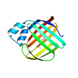
Crystal structure of monomer human cellular retinol binding protein II-Y60L
Descriptor: ACETATE ION, Retinol-binding protein 2
Authors:Assar, Z, Nossoni, Z, Wang, W, Gieger, J.H, Borhan, B.
Deposit date:2015-08-27
Release date:2016-09-21
Last modified:2024-03-06
Method:X-RAY DIFFRACTION (1.5 Å)
Cite:Domain-Swapped Dimers of Intracellular Lipid-Binding Proteins: Evidence for Ordered Folding Intermediates.
Structure, 24, 2016
4QYP
DownloadVisualize
BU of 4qyp by Molmil
The Crystal Structures of holo-wt human Cellular Retinol Binding protein II (hCRBPII) bound to Retinal
Descriptor: ACETATE ION, RETINAL, Retinol-binding protein 2
Authors:Nossoni, Z, Assar, Z, Yapici, I, Nosrati, M, Wang, W, Berbasova, T, Vasileiou, C, Borhan, B, Geiger, H.
Deposit date:2014-07-25
Release date:2014-12-10
Last modified:2023-09-20
Method:X-RAY DIFFRACTION (1.62 Å)
Cite:Structures of holo wild-type human cellular retinol-binding protein II (hCRBPII) bound to retinol and retinal.
Acta Crystallogr.,Sect.D, 70, 2014
4QYN
DownloadVisualize
BU of 4qyn by Molmil
The Crystal Structures of holo-wt human Cellular Retinol Binding protein II (hCRBPII) bound to Retinol
Descriptor: ACETATE ION, RETINOL, Retinol-binding protein 2
Authors:Nossoni, Z, Assar, Z, Yapici, I, Nosrati, M, Wang, W, Berbasova, T, Vasileiou, C, Borhan, B, Geiger, H.
Deposit date:2014-07-24
Release date:2014-12-31
Last modified:2024-02-28
Method:X-RAY DIFFRACTION (1.19 Å)
Cite:Structures of holo wild-type human cellular retinol-binding protein II (hCRBPII) bound to retinol and retinal.
Acta Crystallogr.,Sect.D, 70, 2014
4QZT
DownloadVisualize
BU of 4qzt by Molmil
Crystal Structure of wild type Human Cellular Retinol Binding Protein II (hCRBPII) bound to retinol at 7 KeV beam energy
Descriptor: ACETATE ION, RETINOL, Retinol-binding protein 2
Authors:Assar, Z, Geiger, J.H.
Deposit date:2014-07-28
Release date:2014-12-10
Last modified:2023-09-20
Method:X-RAY DIFFRACTION (1.9 Å)
Cite:Structures of holo wild-type human cellular retinol-binding protein II (hCRBPII) bound to retinol and retinal.
Acta Crystallogr.,Sect.D, 70, 2014
4QZU
DownloadVisualize
BU of 4qzu by Molmil
Crystal Structure of wild type Human Cellular Retinol Binding Protein II (hCRBPII) bound to retinol at 11 KeV beam energy
Descriptor: ACETATE ION, GLYCEROL, RETINOL, ...
Authors:Assar, Z, Geiger, J.H.
Deposit date:2014-07-28
Release date:2014-12-10
Last modified:2023-09-20
Method:X-RAY DIFFRACTION (1.5 Å)
Cite:Structures of holo wild-type human cellular retinol-binding protein II (hCRBPII) bound to retinol and retinal.
Acta Crystallogr.,Sect.D, 70, 2014
6E5E
DownloadVisualize
BU of 6e5e by Molmil
Crystal structure of the apo domain-swapped dimer Q108K:T51D mutant of human cellular retinol binding protein II
Descriptor: ACETATE ION, Retinol-binding protein 2
Authors:Ghanbarpour, A, Geiger, J.
Deposit date:2018-07-20
Release date:2019-10-16
Last modified:2023-10-11
Method:X-RAY DIFFRACTION (1.696 Å)
Cite:Engineering the hCRBPII Domain-Swapped Dimer into a New Class of Protein Switches.
J.Am.Chem.Soc., 141, 2019
6E5Q
DownloadVisualize
BU of 6e5q by Molmil
Crystal structure of the apo domain-swapped dimer Q108K:T51D:A28H mutant of human Cellular Retinol Binding Protein II
Descriptor: ACETATE ION, Retinol-binding protein 2
Authors:Ghanbarpour, A, Geiger, J.
Deposit date:2018-07-21
Release date:2019-10-16
Last modified:2023-10-11
Method:X-RAY DIFFRACTION (1.99 Å)
Cite:Engineering the hCRBPII Domain-Swapped Dimer into a New Class of Protein Switches.
J.Am.Chem.Soc., 141, 2019
6E7M
DownloadVisualize
BU of 6e7m by Molmil
Crystal structure of the holo retinal-bound domain-swapped dimer Q108K:T51D:A28C mutant of human Cellular Retinol Binding Protein II
Descriptor: ACETATE ION, GLYCEROL, RETINAL, ...
Authors:Ghanbarpour, A, Geiger, J.
Deposit date:2018-07-26
Release date:2019-10-16
Last modified:2023-10-11
Method:X-RAY DIFFRACTION (2.7 Å)
Cite:Engineering the hCRBPII Domain-Swapped Dimer into a New Class of Protein Switches.
J.Am.Chem.Soc., 141, 2019
6E5S
DownloadVisualize
BU of 6e5s by Molmil
Crystal structure of holo retinal-bound domain-swapped dimer Q108K:T51D mutant of human Cellular Retinol Binding Protein II
Descriptor: RETINAL, Retinol-binding protein 2
Authors:Nosrati, M, Ghanbarpour, A, Geiger, J.
Deposit date:2018-07-22
Release date:2019-07-24
Last modified:2023-10-11
Method:X-RAY DIFFRACTION (2.061 Å)
Cite:Engineering the hCRBPII Domain-Swapped Dimer into a New Class of Protein Switches.
J.Am.Chem.Soc., 141, 2019
6E51
DownloadVisualize
BU of 6e51 by Molmil
Crystal structure of the apo domain-swapped dimer Q108K:K40L:T51W mutant of human cellular retinol binding protein II
Descriptor: Retinol-binding protein 2
Authors:Ghanbarpour, A, Geiger, J.
Deposit date:2018-07-19
Release date:2019-10-16
Last modified:2023-10-11
Method:X-RAY DIFFRACTION (2.256 Å)
Cite:Engineering the hCRBPII Domain-Swapped Dimer into a New Class of Protein Switches.
J.Am.Chem.Soc., 141, 2019
6E50
DownloadVisualize
BU of 6e50 by Molmil
Crystal structure of the apo domain-swapped dimer Q108K:K40L:T51F mutant of human Cellular Retinol Binding Protein II
Descriptor: ACETATE ION, Retinol-binding protein 2
Authors:Ghanbarpour, A, Geiger, J.
Deposit date:2018-07-18
Release date:2019-10-16
Last modified:2023-10-11
Method:X-RAY DIFFRACTION (1.965 Å)
Cite:Engineering the hCRBPII Domain-Swapped Dimer into a New Class of Protein Switches.
J.Am.Chem.Soc., 141, 2019
6E6L
DownloadVisualize
BU of 6e6l by Molmil
Crystal structure of the holo retinal-bound domain-swapped dimer Q108K:K40L:T51F:Y60A mutant of human cellular retinol binding protein II
Descriptor: ACETATE ION, RETINAL, Retinol-binding protein 2
Authors:Ghanbarpour, A, Geiger, J.
Deposit date:2018-07-25
Release date:2019-10-16
Last modified:2023-10-11
Method:X-RAY DIFFRACTION (2.08 Å)
Cite:Engineering the hCRBPII Domain-Swapped Dimer into a New Class of Protein Switches.
J.Am.Chem.Soc., 141, 2019
6E5R
DownloadVisualize
BU of 6e5r by Molmil
Crystal structure of the apo domain-swapped dimer Q108K:T51D:A28C mutant of human Cellular Retinol Binding Protein II
Descriptor: ACETATE ION, GLYCEROL, Retinol-binding protein 2
Authors:Ghanbarpour, A, Geiger, J.
Deposit date:2018-07-22
Release date:2019-08-07
Last modified:2023-10-11
Method:X-RAY DIFFRACTION (2.592 Å)
Cite:Engineering the hCRBPII Domain-Swapped Dimer into a New Class of Protein Switches.
J.Am.Chem.Soc., 141, 2019
4YKO
DownloadVisualize
BU of 4yko by Molmil
Crystal structure of the R111K:Y134F:T54V:R132Q:P39Q:R59Y:A32Y:F3Q mutant of human Cellular Retinoic Acid Binding Protein II with Retinal at 1.58 angstrom resolution
Descriptor: Cellular retinoic acid-binding protein 2, RETINAL
Authors:Nosrati, M, Geiger, J.
Deposit date:2015-03-04
Release date:2016-03-09
Last modified:2023-09-27
Method:X-RAY DIFFRACTION (1.57 Å)
Cite:A Photoisomerizing Rhodopsin Mimic Observed at Atomic Resolution.
J.Am.Chem.Soc., 138, 2016
6MCV
DownloadVisualize
BU of 6mcv by Molmil
Crystal Structure of Holo Retinal-Bound Domain-Swapped Dimer of Wild Type Human Cellular Retinol Binding Protein II
Descriptor: RETINAL, Retinol-binding protein 2
Authors:Ghanbarpour, A, Geiger, J.
Deposit date:2018-09-02
Release date:2019-10-16
Last modified:2023-10-11
Method:X-RAY DIFFRACTION (3.3 Å)
Cite:Engineering the hCRBPII Domain-Swapped Dimer into a New Class of Protein Switches.
J.Am.Chem.Soc., 141, 2019
6MLB
DownloadVisualize
BU of 6mlb by Molmil
Crystal structure of the holo retinal-bound domain-swapped dimer Q108K:K40L:T51F mutant of human cellular retinol binding protein II
Descriptor: ACETATE ION, GLYCEROL, RETINAL, ...
Authors:Ghanbarpour, A, Geiger, J.
Deposit date:2018-09-27
Release date:2019-10-16
Last modified:2023-10-11
Method:X-RAY DIFFRACTION (2.15 Å)
Cite:Engineering the hCRBPII Domain-Swapped Dimer into a New Class of Protein Switches.
J.Am.Chem.Soc., 141, 2019
6MKV
DownloadVisualize
BU of 6mkv by Molmil
Crystal structure of Retinal-bound holo Q108K:K40L:T51W domain-swapped dimer of human cellular retinol binding protein 2
Descriptor: ACETATE ION, GLYCEROL, RETINAL, ...
Authors:Ghanbarpour, A, Geiger, J.
Deposit date:2018-09-26
Release date:2019-10-16
Last modified:2023-10-11
Method:X-RAY DIFFRACTION (2.107 Å)
Cite:Engineering the hCRBPII Domain-Swapped Dimer into a New Class of Protein Switches.
J.Am.Chem.Soc., 141, 2019
6MCU
DownloadVisualize
BU of 6mcu by Molmil
Crystal structure of the holo retinal-bound domain-swapped dimer Q108K:T51D:A28H mutant of human Cellular Retinol Binding Protein II
Descriptor: GLYCEROL, RETINAL, Retinol-binding protein 2
Authors:Ghanbarpour, A, Geiger, J.
Deposit date:2018-09-02
Release date:2019-10-16
Last modified:2023-10-11
Method:X-RAY DIFFRACTION (2.572 Å)
Cite:Engineering the hCRBPII Domain-Swapped Dimer into a New Class of Protein Switches.
J.Am.Chem.Soc., 141, 2019
<123


219140

PDB entries from 2024-05-01

PDB statisticsPDBj update infoContact PDBjnumon