4WUH
 
 | |
5HEV
 | |
4PDB
 | |
4WSZ
 | |
4WT0
 | |
4WUL
 | |
4WU4
 | |
3P9U
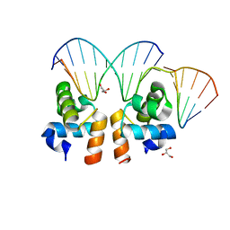
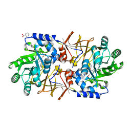
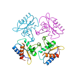
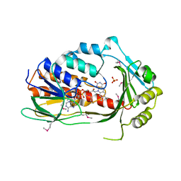 | | Crystal structure of TetX2 from Bacteroides thetaiotaomicron with substrate analogue | | 分子名称: | FLAVIN-ADENINE DINUCLEOTIDE, SULFATE ION, TetX2 protein | | 著者 | Walkiewicz, K, Davlieva, M, Sun, C, Lau, K, Shamoo, Y. | | 登録日 | 2010-10-18 | | 公開日 | 2011-04-27 | | 最終更新日 | 2017-11-08 | | 実験手法 | X-RAY DIFFRACTION (2.81 Å) | | 主引用文献 | Crystal structure of Bacteroides thetaiotaomicron TetX2: a tetracycline degrading monooxygenase at 2.8 A resolution.
Proteins, 79, 2011
|
|
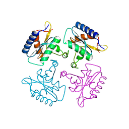
3S46
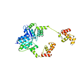
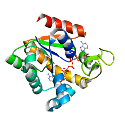
 | | The crystal structure of alanine racemase from streptococcus pneumoniae | | 分子名称: | Alanine racemase, BENZOIC ACID | | 著者 | Im, H, Sharpe, M.L, Strych, U, Davlieva, M, Krause, K.L. | | 登録日 | 2011-05-18 | | 公開日 | 2011-06-22 | | 最終更新日 | 2023-12-06 | | 実験手法 | X-RAY DIFFRACTION (2 Å) | | 主引用文献 | The crystal structure of alanine racemase from Streptococcus pneumoniae, a target for structure-based drug design.
BMC MICROBIOL., 11, 2011
|
|
3HA1
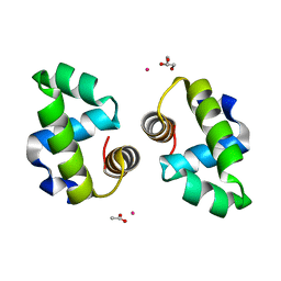
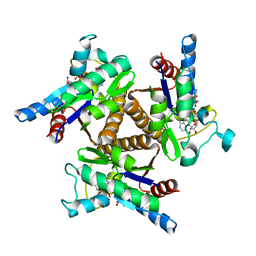
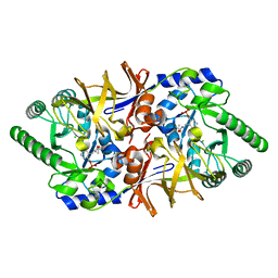 | | Alanine racemase from Bacillus Anthracis (Ames) | | 分子名称: | ACETATE ION, Alanine racemase, CHLORIDE ION | | 著者 | Counago, R.M, Davlieva, M, Strych, U, Hill, R.E, Krause, K.L. | | 登録日 | 2009-04-30 | | 公開日 | 2009-09-15 | | 最終更新日 | 2023-11-22 | | 実験手法 | X-RAY DIFFRACTION (1.95 Å) | | 主引用文献 | Biochemical and structural characterization of alanine racemase from Bacillus anthracis (Ames).
Bmc Struct.Biol., 9, 2009
|
|
6C0Z
 | |
6C84
 | |
6BWS
 | |
3FB4
 | |
3H86
 | |


