Loading
PDBj
MenuPDBj@FacebookPDBj@TwitterPDBj@YouTubewwPDB FoundationwwPDB
RCSB PDBPDBeBMRBAdv. SearchSearch help
Search by PDB author
7A33
DownloadVisualize
BU of 7a33 by Molmil
Crystal structure of the c-Src SH3 domain mutant S94A-T98D-V111L-N113S-T114S at pH 3.0
Descriptor: FORMIC ACID, Proto-oncogene tyrosine-protein kinase Src, SODIUM ION
Authors:Camara-Artigas, A, Plaza-Garrido, M, Salinas-Garcia, M.C.
Deposit date:2020-08-18
Release date:2021-08-25
Last modified:2024-01-31
Method:X-RAY DIFFRACTION (0.96 Å)
Cite:Crystal structure of the c-Src SH3 domain mutant S94A-T98D-V111L-N113S-T114S at pH 3.0
To be published
7A32
DownloadVisualize
BU of 7a32 by Molmil
Crystal structure of the c-Src SH3 domain mutant S94A-T98D-V111L-N113S-T114S at pH 7.0
Descriptor: GLYCEROL, Proto-oncogene tyrosine-protein kinase Src, SULFATE ION
Authors:Camara-Artigas, A, Plaza-Garrido, M, Salinas-Garcia, M.C.
Deposit date:2020-08-18
Release date:2021-08-25
Last modified:2024-01-31
Method:X-RAY DIFFRACTION (1.15 Å)
Cite:Crystal structure of the c-Src SH3 domain mutant S94A-T98D-V111L-N113S-T114S at pH 7.0
To be published
7A2T
DownloadVisualize
BU of 7a2t by Molmil
Crystal structure of the Fyn SH3 domain L112V-S114N-S115T-E121L-R123H mutant at pH 4.0
Descriptor: ACETATE ION, Tyrosine-protein kinase Fyn
Authors:Camara-Artigas, A, Plaza-Garrido, M, Salinas-Garcia, M.C.
Deposit date:2020-08-18
Release date:2021-08-25
Last modified:2024-01-31
Method:X-RAY DIFFRACTION (1.22 Å)
Cite:Crystal structure of the Fyn SH3 domain L112V-S114N-S115T-E121L-R123H mutant at pH 4.0
To be published
7A35
DownloadVisualize
BU of 7a35 by Molmil
Crystal structure of the c-Src SH3 domain mutant V111L-N113S-T114S at pH 7.0
Descriptor: Proto-oncogene tyrosine-protein kinase Src, SULFATE ION
Authors:Camara-Artigas, A, Plaza-Garrido, M, Salinas-Garcia, M.C.
Deposit date:2020-08-18
Release date:2021-08-25
Last modified:2024-01-31
Method:X-RAY DIFFRACTION (1.314 Å)
Cite:Crystal structure of the c-Src SH3 domain mutant V111L-N113S-T114S at pH 7.0
To be published
7A2U
DownloadVisualize
BU of 7a2u by Molmil
Crystal structure of the Fyn SH3 domain L112V-S114N-S115T at pH 4.5
Descriptor: ACETATE ION, Tyrosine-protein kinase Fyn
Authors:Camara-Artigas, A, Plaza-Garrido, M, Salinas-Garcia, M.C.
Deposit date:2020-08-18
Release date:2021-08-25
Last modified:2024-01-31
Method:X-RAY DIFFRACTION (1.7 Å)
Cite:Crystal structure of the Fyn SH3 domain L112V-S114N-S115T at pH 4.5
To be published
7A34
DownloadVisualize
BU of 7a34 by Molmil
Intertwined dimer of the c-Src SH3 domain mutant V111L-N113S-T114S
Descriptor: DI(HYDROXYETHYL)ETHER, Proto-oncogene tyrosine-protein kinase Src, TRIETHYLENE GLYCOL
Authors:Camara-Artigas, A, Plaza-Garrido, M, Salinas-Garcia, M.C.
Deposit date:2020-08-18
Release date:2021-08-25
Last modified:2024-01-31
Method:X-RAY DIFFRACTION (1.85 Å)
Cite:Intertwined dimer of the c-Src SH3 domain mutant V111L-N113S-T114S
To be published
7A2M
DownloadVisualize
BU of 7a2m by Molmil
Crystal structure of the Fyn SH3 domain A95S-D99T mutant in C21 space group
Descriptor: FORMIC ACID, SODIUM ION, SULFATE ION, ...
Authors:Camara-Artigas, A, Plaza-Garrido, M, Salinas-Garcia, M.C.
Deposit date:2020-08-18
Release date:2021-08-25
Last modified:2024-01-31
Method:X-RAY DIFFRACTION (1.5 Å)
Cite:Crystal structure of the Fyn SH3 domain A95S-D99T mutant in C21 space group
To be published
7A37
DownloadVisualize
BU of 7a37 by Molmil
Crystal structure of the c-Src SH3 domain mutant V111L-N113S-T114S in 3 M urea
Descriptor: Proto-oncogene tyrosine-protein kinase Src, UREA
Authors:Camara-Artigas, A, Plaza-Garrido, M, Salinas-Garcia, M.C.
Deposit date:2020-08-18
Release date:2021-08-25
Last modified:2024-01-31
Method:X-RAY DIFFRACTION (1.52 Å)
Cite:Crystal structure of the c-Src SH3 domain mutant V111L-N113S-T114S in 3 M urea
To be published
7A2S
DownloadVisualize
BU of 7a2s by Molmil
Crystal structure of the Fyn SH3 domain L112V-S114N-S115T-E121L-R123H mutant at pH 5.0
Descriptor: Tyrosine-protein kinase Fyn
Authors:Camara-Artigas, A, Plaza-Garrido, M, Salinas-Garcia, M.C.
Deposit date:2020-08-18
Release date:2021-08-25
Last modified:2024-01-31
Method:X-RAY DIFFRACTION (1.02 Å)
Cite:Crystal structure of the Fyn SH3 domain L112V-S114N-S115T-E121L-R123H mutant at pH 5.0
To Be Published
7A2K
DownloadVisualize
BU of 7a2k by Molmil
Crystal structure of the Fyn SH3 domain in space group P1 at pH 7.5
Descriptor: SODIUM ION, Tyrosine-protein kinase Fyn
Authors:Camara-Artigas, A, Plaza-Garrido, M, Salinas-Garcia, M.C.
Deposit date:2020-08-18
Release date:2021-08-25
Last modified:2024-01-31
Method:X-RAY DIFFRACTION (1.5 Å)
Cite:Crystal structure of the Fyn SH3 domain in space group P1 at pH 7.5
To be published
7A3A
DownloadVisualize
BU of 7a3a by Molmil
Intertwined dimer of the c-Src SH3 domain mutant V111L-N113S-T114S-Q128E at pH 6.0
Descriptor: 1,2-ETHANEDIOL, DI(HYDROXYETHYL)ETHER, Proto-oncogene tyrosine-protein kinase Src, ...
Authors:Camara-Artigas, A, Plaza-Garrido, M, Salinas-Garcia, M.C.
Deposit date:2020-08-18
Release date:2021-08-25
Last modified:2024-01-31
Method:X-RAY DIFFRACTION (1.8 Å)
Cite:Intertwined dimer of the c-Src SH3 domain mutant V111L-N113S-T114S-Q128E at pH 6.0
To be published
7A36
DownloadVisualize
BU of 7a36 by Molmil
Crystal structure of the c-Src SH3 domain mutant V111L-N113S-T114S in 7 M urea
Descriptor: Proto-oncogene tyrosine-protein kinase Src, SODIUM ION, UREA
Authors:Camara-Artigas, A, Plaza-Garrido, M, Salinas-Garcia, M.C.
Deposit date:2020-08-18
Release date:2021-08-25
Last modified:2024-01-31
Method:X-RAY DIFFRACTION (1.5 Å)
Cite:Crystal structure of the c-Src SH3 domain mutant V111L-N113S-T114S in 7 M urea
To be published
7A2N
DownloadVisualize
BU of 7a2n by Molmil
Crystal structure of the Fyn SH3 domain A95S-D99T mutant in space group P21
Descriptor: FORMIC ACID, SODIUM ION, Tyrosine-protein kinase Fyn
Authors:Camara-Artigas, A, Plaza-Garrido, M, Salinas-Garcia, M.C.
Deposit date:2020-08-18
Release date:2021-08-25
Last modified:2024-01-31
Method:X-RAY DIFFRACTION (1.4 Å)
Cite:Crystal structure of the Fyn SH3 domain A95S-D99T mutant in space group P21
To be published
7A3E
DownloadVisualize
BU of 7a3e by Molmil
Intertwined dimer of the c-Src SH3 domain mutant T126S
Descriptor: DI(HYDROXYETHYL)ETHER, Proto-oncogene tyrosine-protein kinase Src, SULFATE ION, ...
Authors:Camara-Artigas, A, Plaza-Garrido, M, Salinas-Garcia, M.C.
Deposit date:2020-08-18
Release date:2021-08-25
Last modified:2024-01-31
Method:X-RAY DIFFRACTION (1.52 Å)
Cite:Intertwined dimer of the c-Src SH3 domain mutant T126S
To be published
7A2W
DownloadVisualize
BU of 7a2w by Molmil
Crystal structure of the Fyn SH3 domain L112V-S114N-S115T-E121L-R123H mutant in complex with VSL12 at pH 3.0
Descriptor: CITRIC ACID, Tyrosine-protein kinase Fyn, VSL12
Authors:Camara-Artigas, A, Plaza-Garrido, M, Salinas-Garcia, M.C.
Deposit date:2020-08-18
Release date:2021-08-25
Last modified:2024-01-31
Method:X-RAY DIFFRACTION (0.99 Å)
Cite:Crystal structure of the Fyn SH3 domain L112V-S114N-S115T-E121L-R123H mutant in complex with VSL12 at pH 3.0
To be published
7A38
DownloadVisualize
BU of 7a38 by Molmil
Crystal structure of the c-Src SH3 domain mutant V111L-N113S-T114S-Q128E at pH 6.0
Descriptor: 2-(N-MORPHOLINO)-ETHANESULFONIC ACID, Proto-oncogene tyrosine-protein kinase Src, SULFATE ION
Authors:Camara-Artigas, A, Plaza-Garrido, M, Salinas-Garcia, M.C.
Deposit date:2020-08-18
Release date:2021-08-25
Last modified:2024-01-31
Method:X-RAY DIFFRACTION (1.62 Å)
Cite:Crystal structure of the c-Src SH3 domain mutant V111L-N113S-T114S-Q128E at pH 6.0
To be published
7NET
DownloadVisualize
BU of 7net by Molmil
Crystal structure of the v-Src SH3 domain W95R-I96T mutant
Descriptor: DI(HYDROXYETHYL)ETHER, SULFATE ION, TRIETHYLENE GLYCOL, ...
Authors:Camara-Artigas, A, Salinas-Garcia, M.C.
Deposit date:2021-02-04
Release date:2021-06-02
Last modified:2024-01-31
Method:X-RAY DIFFRACTION (1.5 Å)
Cite:The impact of oncogenic mutations of the viral Src kinase on the structure and stability of the SH3 domain.
Acta Crystallogr D Struct Biol, 77, 2021
7NES
DownloadVisualize
BU of 7nes by Molmil
Crystal structure of the v-Src SH3 domain N117D-V124L mutant
Descriptor: GLYCINE, v-Src SH3 domain
Authors:Camara-Artigas, A, Salinas-Garcia, M.C.
Deposit date:2021-02-04
Release date:2021-06-02
Last modified:2024-05-15
Method:X-RAY DIFFRACTION (1.35 Å)
Cite:The impact of oncogenic mutations of the viral Src kinase on the structure and stability of the SH3 domain.
Acta Crystallogr D Struct Biol, 77, 2021
7NER
DownloadVisualize
BU of 7ner by Molmil
Crystal structure of the v-Src SH3 domain Q128R mutant
Descriptor: SULFATE ION, TETRAETHYLENE GLYCOL, v-Src SH3 domain
Authors:Camara-Artigas, A, Salinas-Garcia, M.C.
Deposit date:2021-02-04
Release date:2021-06-02
Last modified:2024-01-31
Method:X-RAY DIFFRACTION (1.55 Å)
Cite:The impact of oncogenic mutations of the viral Src kinase on the structure and stability of the SH3 domain.
Acta Crystallogr D Struct Biol, 77, 2021
3UA6
DownloadVisualize
BU of 3ua6 by Molmil
Crystal Structure of the Human Fyn SH3 domain
Descriptor: FORMIC ACID, GLYCEROL, SODIUM ION, ...
Authors:Camara-Artigas, A, Martin-Garcia, J.M.
Deposit date:2011-10-21
Release date:2012-07-25
Last modified:2023-09-13
Method:X-RAY DIFFRACTION (1.85 Å)
Cite:The promiscuous binding of the Fyn SH3 domain to a peptide from the NS5A protein.
Acta Crystallogr.,Sect.D, 68, 2012
1Z9K
DownloadVisualize
BU of 1z9k by Molmil
Photosynthetic Reaction Center from Rhodobacter sphaeroides
Descriptor: BACTERIOCHLOROPHYLL A, BACTERIOPHEOPHYTIN A, FE (III) ION, ...
Authors:Camara-Artigas, A, Allen, J.P.
Deposit date:2005-04-02
Release date:2005-06-07
Last modified:2023-08-23
Method:X-RAY DIFFRACTION (4.6 Å)
Cite:Design of a Redox-Linked Active Metal Site: Manganese Bound to Bacterial Reaction Centers at a Site Resembling That of Photosystem II
Biochemistry, 44, 2005
6SYE
DownloadVisualize
BU of 6sye by Molmil
Crystal structure of orthorhombic lysozyme in presence of the dye bromophenol blue at pH 7.0
Descriptor: CHLORIDE ION, Lysozyme C, bromophenol blue
Authors:Camara-Artigas, A, Plaza-Garrido, M, Salinas-Garcia, M.C.
Deposit date:2019-09-27
Release date:2020-09-09
Last modified:2024-01-24
Method:X-RAY DIFFRACTION (0.97 Å)
Cite:Lysozyme crystals dyed with bromophenol blue: where has the dye gone?
Acta Crystallogr D Struct Biol, 76, 2020
6SYD
DownloadVisualize
BU of 6syd by Molmil
Crystal structure of the lysozyme in presence of bromophenol blue at pH 5.5
Descriptor: Lysozyme, SODIUM ION, bromophenol blue
Authors:Camara-Artigas, A, Plaza-Garrido, M, Salinas-Garcia, M.C.
Deposit date:2019-09-27
Release date:2020-09-09
Last modified:2024-01-24
Method:X-RAY DIFFRACTION (1.1 Å)
Cite:Lysozyme crystals dyed with bromophenol blue: where has the dye gone?
Acta Crystallogr D Struct Biol, 76, 2020
7ZR2
DownloadVisualize
BU of 7zr2 by Molmil
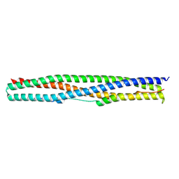
Crystal structure of a chimeric protein mimic of SARS-CoV-2 Spike HR1 in complex with HR2
Descriptor: Spike protein S2', Spike protein S2',Chimeric protein mimic of SARS-CoV-2 Spike HR1
Authors:Camara-Artigas, A, Gavira, J.A, Cano-Munoz, M, Polo-Megias, D, Conejero-Lara, F.
Deposit date:2022-05-03
Release date:2022-11-09
Last modified:2024-01-31
Method:X-RAY DIFFRACTION (1.45 Å)
Cite:Novel chimeric proteins mimicking SARS-CoV-2 spike epitopes with broad inhibitory activity.
Int.J.Biol.Macromol., 222, 2022
8AH5
DownloadVisualize
BU of 8ah5 by Molmil
Crystal Structure of the third PDZ domain of PSD-95 protein in the space group P212121 at pH 4.6
Descriptor: ACETATE ION, cDNA FLJ50577, highly similar to Discs large homolog 4
Authors:Camara-Artigas, A, Salinas Garcia, M.C.
Deposit date:2022-07-20
Release date:2023-03-08
Last modified:2024-02-07
Method:X-RAY DIFFRACTION (1.25 Å)
Cite:pH-Driven Polymorphic Behaviour of the Third PDZ Domain of PSD95: The Role of Electrostatic Interactions
Crystals, 2023

219869

PDB entries from 2024-05-15

PDB statisticsPDBj update infoContact PDBjnumon